

Intraoperative use of cone-beam computed tomography in a cadaveric ossified cochlea model
- PMID: 19393414
- PMCID: PMC2876243
- DOI: 10.1016/j.otohns.2008.12.046
Intraoperative use of cone-beam computed tomography in a cadaveric ossified cochlea model
Abstract
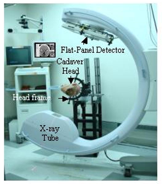
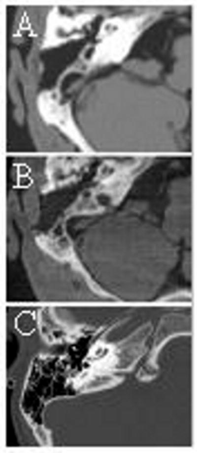
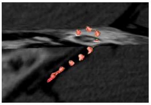
Objectives: To describe a cadaveric temporal bone model of labyrinthitis ossificans and investigate the utility of intraoperative cone-beam computed tomography (CBCT) in the facilitating cochlear implantation.
Design: Cadaveric temporal bone study.
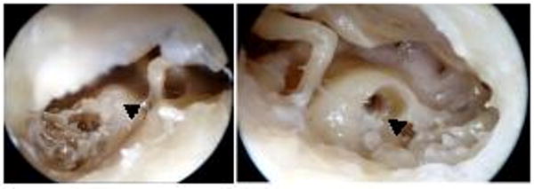
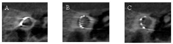
Methods: Five cadaveric heads had cement introduced into the 10 cochleas. CBCT and a conventional CT scan were compared to assess the extent of cochlear obliteration. The cement was drilled-out (under CBCT guidance, if required) and cochlear implant electrode arrays (from 3 different manufacturers) inserted.
Results: CBCT images demonstrated temporal bone anatomy and the extent of cochlear obliteration as clearly as conventional CT in all cases. Intraoperative CBCT guided drilling and facilitated electrode placement in two of five heads (3 of 10 ears). Streak-artifact from the electrodes of two devices partially obscured image clarity.
Conclusions: The obliterated cochlear model reproduced a disease-ossified cochlear both radiographically and surgically. CBCT is useful for intraoperative imaging to facilitate electrode array placement in the obliterated or congenitally abnormal cochlea.
Figures
Similar articles
-
Differences of radiological artefacts in cochlear implantation in temporal bone and complete head.Cochlear Implants Int. 2014 Mar;15(2):112-7. doi: 10.1179/1754762813Y.0000000035. Epub 2013 Nov 25. Cochlear Implants Int. 2014. PMID: 23938153
-
Imaging of the human cochlea using micro-computed tomography before and after cochlear implantation: comparison with cone-beam computed tomography.Eur Arch Otorhinolaryngol. 2023 Jul;280(7):3131-3140. doi: 10.1007/s00405-022-07811-y. Epub 2023 Jan 6. Eur Arch Otorhinolaryngol. 2023. PMID: 36604323
-
Cone beam CT for perioperative imaging in hearing preservation Cochlear implantation - a human cadaveric study.J Otolaryngol Head Neck Surg. 2019 Nov 21;48(1):65. doi: 10.1186/s40463-019-0388-x. J Otolaryngol Head Neck Surg. 2019. PMID: 31753027 Free PMC article.
-
Cone beam computed tomography and histological evaluations of a straight electrode array positioning in temporal bones.Acta Otolaryngol. 2017 Mar;137(3):229-234. doi: 10.1080/00016489.2016.1227477. Epub 2016 Sep 22. Acta Otolaryngol. 2017. PMID: 28225319
-
Optimal electrode diameter in relation to volume of the cochlea.Eur Ann Otorhinolaryngol Head Neck Dis. 2016 Jun;133 Suppl 1:S66-7. doi: 10.1016/j.anorl.2016.04.013. Epub 2016 May 27. Eur Ann Otorhinolaryngol Head Neck Dis. 2016. PMID: 27246746 Review.
Cited by
-
Cone-Beam CT with a Flat-Panel Detector: From Image Science to Image-Guided Surgery.Nucl Instrum Methods Phys Res A. 2011 Aug 21;648(S1):S241-S250. doi: 10.1016/j.nima.2010.11.088. Nucl Instrum Methods Phys Res A. 2011. PMID: 22942510 Free PMC article.
-
Overcoming Nonlinear Partial Volume Effects in Known-Component Reconstruction of Cochlear Implants.Proc SPIE Int Soc Opt Eng. 2013;8668:86681L. doi: 10.1117/12.2007945. Proc SPIE Int Soc Opt Eng. 2013. PMID: 24949189 Free PMC article.
-
[Progress of research on cone beam CT in cochlear implantation].Lin Chuang Er Bi Yan Hou Tou Jing Wai Ke Za Zhi. 2021 Jun;35(6):567-572. doi: 10.13201/j.issn.2096-7993.2021.06.019. Lin Chuang Er Bi Yan Hou Tou Jing Wai Ke Za Zhi. 2021. PMID: 34304522 Free PMC article. Review. Chinese.
-
An electromagnetic "Tracker-in-Table" configuration for X-ray fluoroscopy and cone-beam CT-guided surgery.Int J Comput Assist Radiol Surg. 2013 Jan;8(1):1-13. doi: 10.1007/s11548-012-0744-z. Epub 2012 May 15. Int J Comput Assist Radiol Surg. 2013. PMID: 22585463 Free PMC article.
-
Atlas-Based Segmentation of Temporal Bone Anatomy.Int J Comput Assist Radiol Surg. 2017 Nov;12(11):1937-1944. doi: 10.1007/s11548-017-1658-6. Epub 2017 Aug 29. Int J Comput Assist Radiol Surg. 2017. PMID: 28852952 Free PMC article.
References
-
- Labadie RF, Shah RJ, Harris SS, et al. Submillimetric target-registration error using a novel, non-invasive fiducial system for image-guided otologic surgery. Comput Aided Surg. 2004;9(4):145–153. - PubMed
-
- Cartellieri M, Vorbeck F. Endoscopic sinus surgery using intraoperative computed tomography imaging for updating a three-dimensional navigation system. Laryngoscope. 2000;110(2 Pt 1):292–296. - PubMed
-
- Fried MP, Hsu L, Topulos GP, et al. Image-guided surgery in a new magnetic resonance suite: preclinical considerations. Laryngoscope. 1996;106(4):411–417. - PubMed
-
- Siewerdsen JH, Moseley DJ, Burch S, et al. Volume CT with a Flat-Panel Detector on a Mobile C-Arm: Pre-Clinical Investigation in Guidance of Minimally Invasive Surgery. Med Phys. 2005;32(1):241–254. - PubMed
-
- Burch S, Bogaards A, Siewerdsen J, et al. Photodynamic therapy for the treatment of metastatic lesions in bone: studies in rat and porcine models. J Biomed Opt. 2005;10(3):034011. - PubMed
