. 2009 Jun 26;384(2):221-5.
doi: 10.1016/j.bbrc.2009.04.091. Epub 2009 Apr 23.

Peroxynitrite-induced p38 MAPK pro-apoptotic signaling in enterocytes

Affiliations

Peroxynitrite-induced p38 MAPK pro-apoptotic signaling in enterocytes

Yigit S Guner et al. Biochem Biophys Res Commun. .

Abstract

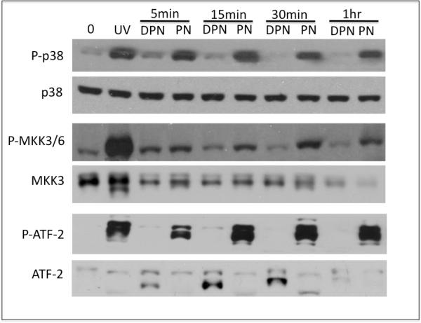
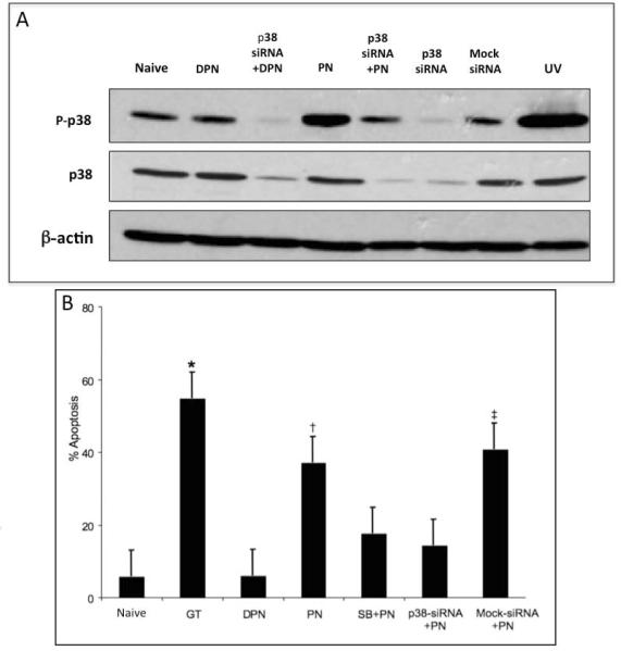
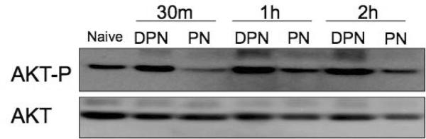
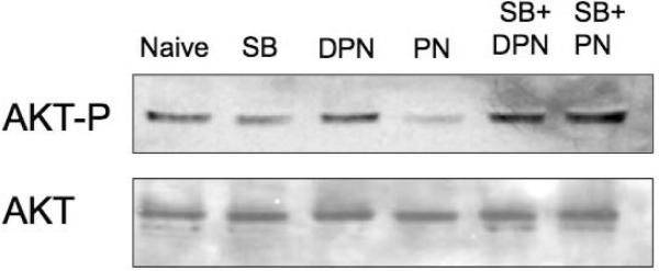
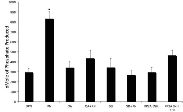
Enterocyte apoptosis in necrotizing enterocolitis is partly due to the elaboration of toxic intermediates of nitric oxide (NO), such as peroxynitrite (PN). Because p38 mitogen-activated protein kinase (MAPK) and serine-threonine kinase (AKT) are well-characterized pro- and anti-apoptotic mediators, respectively, we hypothesized that PN could induce enterocyte apoptosis via activation of p38 and deactivation of AKT. To test this hypothesis, the rat intestinal cell line, IEC-6, was treated with PN. PN caused phosphorylation of p38, its upstream activator, MKK3/6, and downstream effector, transcription factor ATF-2. PN-induced apoptosis was inhibited by the p38 inhibitor, SB202190, and by p38 siRNA. PN decreased AKT phosphorylation; this effect was abrogated by pre-treatment with SB202190 or p38 siRNA. PN exposure also increased the activity of the protein phosphatase 2A (PP2A). These data demonstrate that PN-mediated apoptosis depends on the p38 pathway and that p38 mediates deactivation of AKT survival pathways possibly by the involvement of PP2A.


Figures

Figure 1
Figure 1. PN increases phosphorylation of p38, MKK3/6 and ATF-2
IEC-6 cells were exposed to 50 M PN and DPN for the indicated times. Naïve (untreated cells) are indicated by 0min. Levels of phospho-p38, phospho-MKK3/6, phospho-ATF-2, total p38, total MMK3 and total ATF-2 were determined by Western blotting. UV, cells exposed to 50mJ/cm2 UVC and allowed to recover in growth medium for 1h (positive control for p38 activation). Data shown are representative of at least 3 independent experiments.
Figure 2
Figure 2. PN-induced apoptosis is prevented by p38 inhibition
(A) IEC-6 cells were transfected with p38 siRNA, or control (Mock) siRNA as indicated. 36h post transfection, cells were exposed to PBS, or 50 M PN, or equivalent concentration of DPN for 60min as indicated, and levels of phospho-p38, total p38, and -actin were determined. UV, cells exposed to 50 mJ/cm2 UVC and allowed to recover in growth medium for 1h (positive control for p38 activation). (B) IEC-6 cells were treated with PN or equivalent concentration of DPN for 1h, and allowed to recover for 6h. Percentage of apoptotic (sub-G1 DNA content) cells was determined by flow cytometry of 7-AAD stained cells were determined by flow cytometry. Cells were pre-treated with 10 M SB202190 (SB), or transfected with p38 siRNA or control siRNA 30 min or 48h prior to PN or DPN treatment, respectively, as indicated. GT, cells treated with 2.5 mol/L of gliotoxin for 6h. (*) p<0.001, gliotoxin treated cells vs. naïve DPN-treated, SB+PN-treated or p38 siRNA-transfected cells. (†) p<0.001, PN-treated vs. DPN-treated, SB+PN-treated and p38 siRNA-transfected PN-treated cells. (‡) p<0.001 control-siRNA-transfected vs.p38-siRNA-transfectedPN-treated cells. Data are depicted as the mean S.E. (n=6).
Figure 3
Figure 3. AKT phosphorylation in IEC-6 cells treated with PN
(A) Cells were treated with PN, or equivalent concentration of DPN for indicated time. Levels of phospho-AKT and total AKT were determined by Western blotting. (B) Levels of phospho-AKT and total AKT in cells untreated naive, or treated with PN or DPN for 1h, with or without pre-treatment with 10 M SB202190 (SB) as indicated. Data are representative of 3 independent experiments.
Figure 4
Figure 4. PN increases PP2A activity
IEC-6 cells were treated with PN (50 M) or equivalent concentration of DPN for 1 h. Cells were pre-treated with 10 M SB202190 (SB), 10 M okadaic acid (OA) or 100nM I1PP2A (PP2A INH) 30m prior to PN or DPN exposure. PP2A activity in cell lysates were measured. (*) PN exposure significantly elevated PP2A activity compared to all other variables (p<0.001, n=5). Data are shown as pmoles phosphate produced per mg protein in the form of mean S.E.

References

    1. Hackam DJ, Upperman JS, Grishin A, Ford HR. Disordered enterocyte signaling and intestinal barrier dysfunction in the pathogenesis of necrotizing enterocolitis. Semin Pediatr Surg. 2005;14:49–57. - PubMed
    1. Guner YS, Chokshi N, Petrosyan M, Upperman JS, Ford HR, Grikscheit TC. Necrotizing enterocolitis--bench to bedside: novel and emerging strategies. Semin Pediatr Surg. 2008;17:255–65. - PubMed
    1. Ford HR. Mechanism of nitric oxide-mediated intestinal barrier failure: insight into the pathogenesis of necrotizing enterocolitis. J Pediatr Surg. 2006;41:294–9. - PubMed
    1. Upperman JS, Potoka D, Grishin A, Hackam D, Zamora R, Ford HR. Mechanisms of nitric oxide-mediated intestinal barrier failure in necrotizing enterocolitis. Semin Pediatr Surg. 2005;14:159–66. - PubMed
    1. Nathan C. Nitric oxide as a secretory product of mammalian cells. Faseb J. 1992;6:3051–64. - PubMed

Publication types

MeSH terms

Substances

LinkOut - more resources