. 2009 Jul 1;331(1):26-37.
doi: 10.1016/j.ydbio.2009.04.018. Epub 2009 Apr 23.

Hedgehog signaling patterns mesoderm in the sea urchin

Affiliations

Hedgehog signaling patterns mesoderm in the sea urchin

Katherine D Walton et al. Dev Biol. .

Abstract

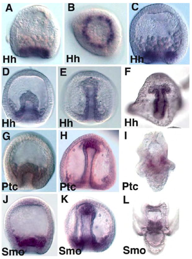
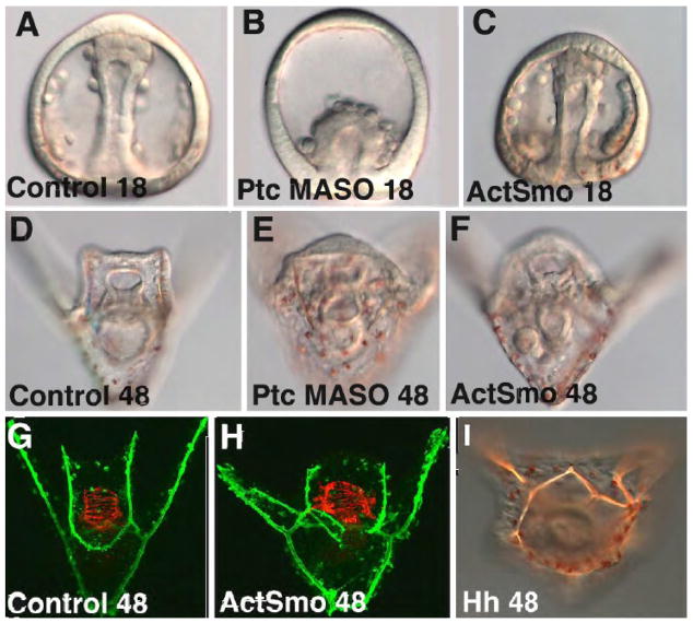
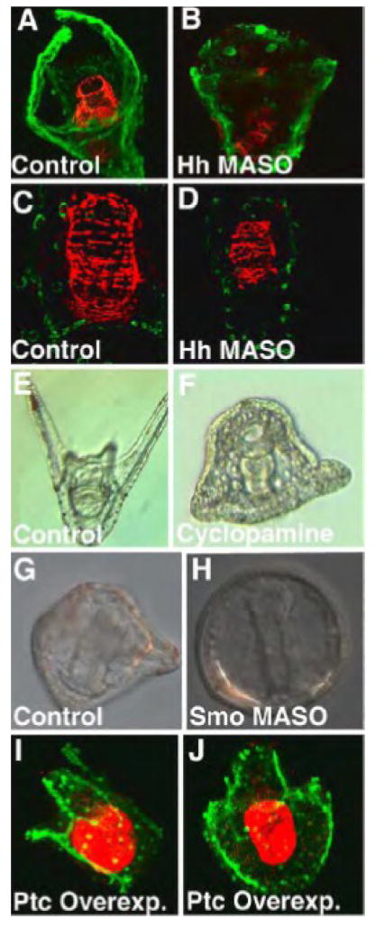
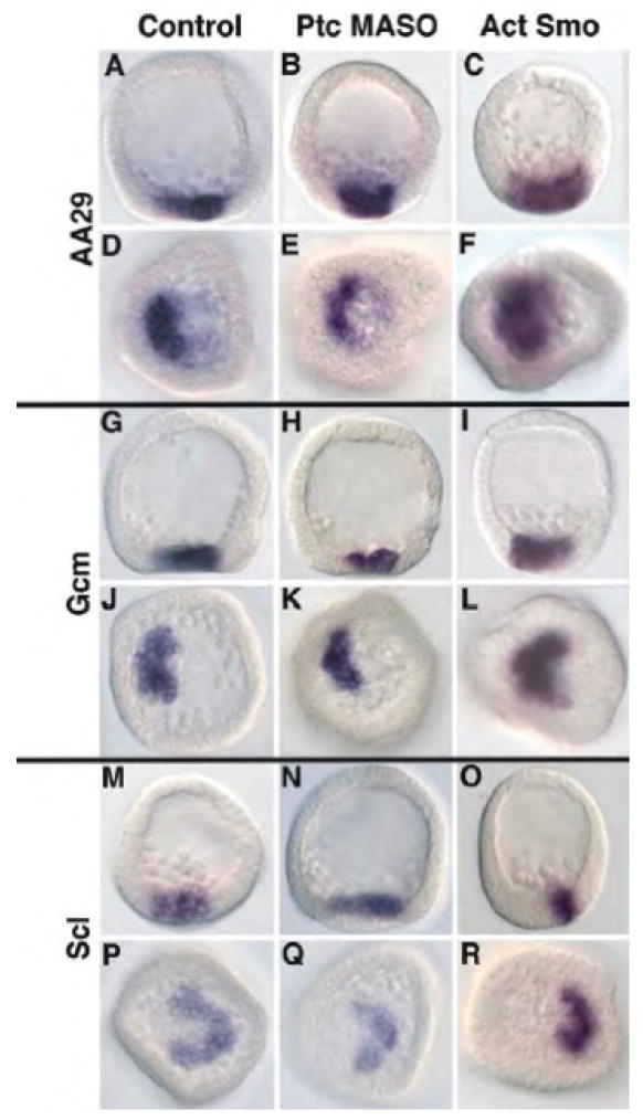
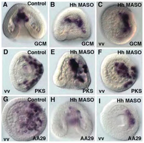
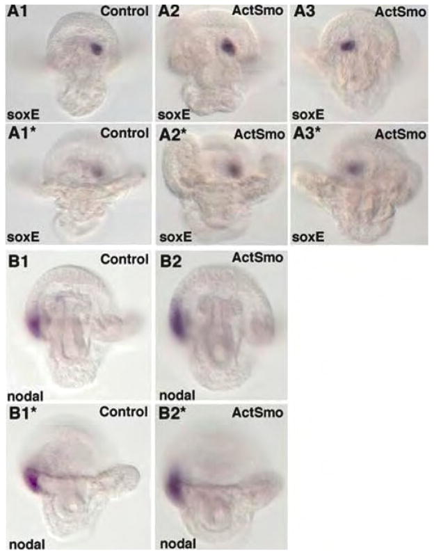
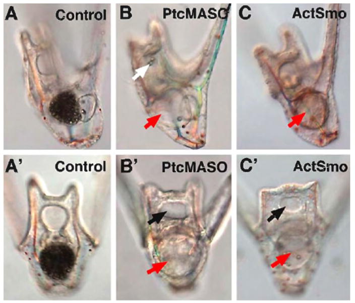
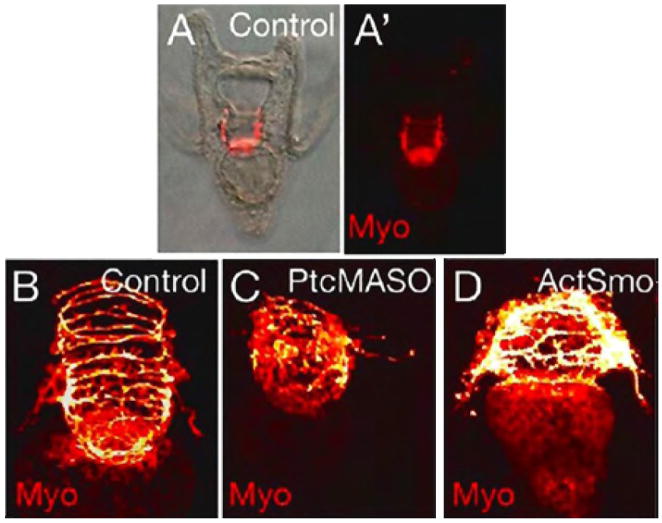
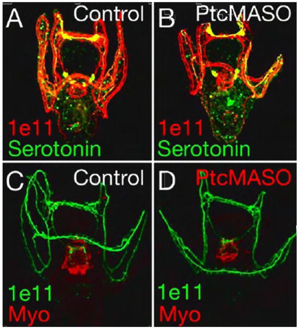
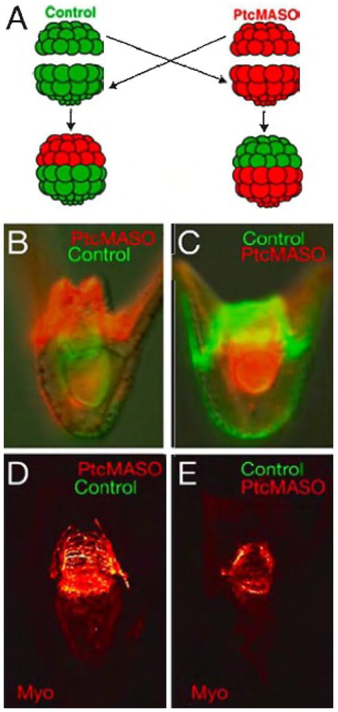
The Hedgehog (Hh) signaling pathway is essential for patterning many structures in vertebrates including the nervous system, chordamesoderm, limb and endodermal organs. In the sea urchin, a basal deuterostome, Hh signaling is shown to participate in organizing the mesoderm. At gastrulation the Hh ligand is expressed by the endoderm downstream of the Brachyury and FoxA transcription factors in the endomesoderm gene regulatory network. The co-receptors Patched (Ptc) and Smoothened (Smo) are expressed by the neighboring skeletogenic and non-skeletogenic mesoderm. Perturbations of Hh, Ptc and Smo cause embryos to develop with skeletal defects and inappropriate non-skeletogenic mesoderm patterning, although initial specification of mesoderm occurs without detectable abnormalities. Perturbations of the pathway caused late defects in skeletogenesis and in the non-skeletogenic mesoderm, including altered numbers of pigment and blastocoelar cells, randomized left-right asymmetry of coelomic pouches, and disorganized circumesophageal muscle causing an inability to swallow. Together the data support the requirement of Hh signaling in patterning each of the mesoderm subtypes in the sea urchin embryo.


Figures

Figure 1
Figure 1
Whole mount RNA in situ hybridization of Hh (A–F), Ptc (G–I) and Smo (J–L). In (A) and (B, ventral view), Hh expression is first detected at mid to late mesenchyme blastula stages as a single ring of cells in the endoderm. By early gastrula (C) the ring expands to a ring two – three cells wide, and during gastrulation (D,E) Hh expression is detected throughout the gut but not in the NSM at the tip of the archenteron. In pluteus larvae Hh expression spans the entire gut to the boundary of the coelomic pouches seen budding off the side of the foregut in (F). Ptc is first observed localized to the NSM at the beginning of gastrulation (G). At late gastrulation Ptc is still seen in the NSM and a new site in the PMCs surrounding the base of the archenteron (H). At the pluteus stage Ptc is expressed in the two coelomic pouches (I). Smo is first localized to the NSM at the beginning of gastrulation (J). At the end of gastrulation Smo is observed in the NSM at the tip of the archenteron (K), throughout the PMCs surrounding the NSM, and perhaps at a low level in the hindgut, though most of the staining observed in is in out-of-focus PMCs. At the pluteus larva stage Smo is observed in coelomic pouches (L).
Figure 2
Figure 2
Activation of the Hh pathway. The Hh pathway was activated by three treatments, injection of a Ptc MASO which blocked Ptc activity thereby allowing Smo to be active (B, E), injection of RNA construct that produced constitutively activated Smo (C,F,H), or over-expression of Hh (I). At 18 hr. the Ptc MASO (0.75 mM) delayed gastrulation (B) relative to controls (A), but development caught up and at 48 hr the phenotype was mild with an increased number of pigment cells (E), relative to controls (D). Activated Smo did not delay gastrulation at 0.63 pg/pl with ca. 1 pl injected per egg. However, at 48 hr there were an excess number of pigment cells (F), an abnormal skeletal pattern and abnormal circumesophageal musculature (H), relative to controls at 48 hr (D, G). At 48 hr, embryos expressing extra Hh (ca.1 pl at 0.58 pg/pl) displayed an abnormal skeleton (I).
Figure 3
Figure 3
Inhibition of the Hh pathway. The Hedgehog pathway was blocked by four different treatments. In (B and D) an Hh MASO (60 μM) caused skeletal (B) and circumesophageal muscle abnormalities (B,D) relative to controls (A,C). Treatment with Cyclopamine (6.25 μM) truncated skeletal development and reduced the number of pigment cells (F) relative to controls at 48 hr (E). A Smo MASO (375 μM) caused a truncated skeleton and reduced pigmentation (H) relative to controls at 22 hr (G). Over-expression of Ptc (0.5 pg/pl) caused an abnormal skeleton to develop (I,J). In (A–D and I, J), the skeleton is stained with an antibody to MSP130 (green). In (A–D) circumesophageal muscle is stained with anti-myosin (red). In (I,J) the midgut and hindgut is stained with an endoderm marker (red).
Figure 4
Figure 4
In situ analysis of non-skeletogenic mesoderm markers at the mesenchyme blastula stage. For each in situ imaged both a lateral view (A–C, G–I, M–O), and a vegetal view (D–F, J–L, P–R) are shown. (A,D,G,J,M,P) control embryos; (B,E,H,K,N,Q) embryos injected with Ptc MASO (0.75 mM); (C,F,I,L,O,R) embryos injected with Activated Smo (0.63 pg/pl). AA29 is a marker for all non-skeletogenic cells; GCM is an early activator of NSM and necessary for pigment cell specification; Scl, is a Marker activated prior to mesenchyme stage in NSM and necessary for blastocoelar cell specification. Both the Ptc MASO and Activated Smo had phenotypes at the pluteus stage but had no noticeable effect on expression of these three NSM markers at the mesenchyme blastula stage.
Figure 5
Figure 5
Hedgehog knockdowns do not affect expression of endomesoderm GRN components at late gastrula stage. GCM normally is expressed in the aboral NSM and in pigment cells at late gastrula (A). Embryos injected with Hh MASO at concentrations that affect late patterning nevertheless express GCM normally (B,C). PKS is a differentiation gene in the pigment cell lineage and pigment cells invade ectoderm on the aboral side (D). Hh MASO knockdowns do not alter PKS expression nor the ectodermal invasion (E,F). AA29 stains a number of NSM cells at late gastrula (G), and that staining pattern is not significantly altered in Hh MASO knockdowns (H,I). vv = ventral view.
Figure 6
Figure 6
Increased Hh signaling randomizes left-right asymmetry of the coelomic pouch expression of soxE, but not the ectodermal asymmetry of Nodal expression. RNA in situ hybridization with the normally left coelomic pouch marker, soxE (A), and the right side ectodermal marker, nodal (B) were examined at the pluteus larval stage. Images of larvae in (A1–A3) are focused on the soxE positive cells and B(1-B2) are focused on the nodal expressing cells. (A1*–A3*) are the same larvae as in (A1–A3) and (B1 and B2) are the same larvae as in (B1* and B2*), but the focus is on the anal arms to provide orientation of the markers within the larvae. A1–A1* is a control larva hybridized with soxE. A2– A2* is a larva with normal left sided expression of soxE. A3-A3* is a larva with right-sided expression of soxE. B1- B1* is a control larva, while B2, B2* is a larva injected with ActSmo mRNA that have been and probed with nodal.
Figure 7
Figure 7
Test of swallowing and peristalsis. The ability to swallow was examined by feeding latex beads for 1 hour to 2 day-old larvae. Control larvae have midguts filled with swallowed beads (A1 and A1′). Ptc MASO-injected larvae (0.75 mM) (A2 and A2′), and Act-Smo injected larvae (0.63 pg/pl) (A3 and A3′) fail to swallow the beads. Red arrows show midguts with very few beads in larvae with increased Hh signaling. Black arrows show that mouths do form in those larvae. White arrow indicates that beads enter the mouth of those embryos but those entering beads fail to be swallowed.
Figure 8
Figure 8
Myosin antibody staining shows circumesophageal muscle organization in 48 hr control embryos (A,A′,B). In 48 hr embryos injected with Ptc MASO (0.75 mM) (C), and in 48 hr embryos expressing ActSmo (0.63 pg/pl)(D), the circumesophageal muscle is patterned abnormally compared to the control. Images are captured by DIC (A), fluorescence microscopy (A′), and by confocal microscopy (B–D).
Figure 9
Figure 9
Patterning of serotonergic and non-serotonergic neurons throughout the larvae shown by confocal projections of fluorescent nerve markers. Anti-serotonin (green) shows a normal pattern of serotonergic neurons in both control (A) and Ptc MASO injected embryos (B). Anti-Synaptotagmin B shows all neurons in red in control embryos (A) and in Ptc MASO injected embryos (B) and in green in (C, D). Myosin antibody staining in red (C,D) shows a normal pattern of circumesophageal muscle in the pharynx (C) and an abnormal pattern in the Ptc MASO injected embryos (D).
Figure 10
Figure 10
Reciprocal transplantation of animal and vegetal halves from control and Ptc MASO injected embryos (0.75 mM). (A) shows a diagram of the experiment. A control animal half (green) is placed on a red Ptc MASO-injected vegetal half, and reciprocally, a red Ptc-MASO-injected animal half is placed on a green control vegetal half. 48 hr embryos in which the Ptc MASO was in the animal half (B,D) and reciprocally, in which the Ptc MASO was in the vegetal half (C,E). The fluorescent lineage tracers are shown in (B,C), and (D,E) show the circumesophageal muscle of the embryo immediately above them as seen by confocal microscopy. When the Ptc MASO is in the vegetal half the muscle patterning is abnormal (E), while if the Ptc MASO is in the animal half the muscle pattern is normal (D).

Similar articles

Cited by

References

    1. Ahlgren SC, Thakur V, Bronner-Fraser M. Sonic hedgehog rescues cranial neural crest from cell death induced by ethanol exposure. Proc Natl Acad Sci U S A. 2002;99:10476–10481. - PMC - PubMed
    1. Aihara M, Amemiya S. Left-right positioning of the adult rudiment in sea urchin larvae is directed by the right side. Development. 2001;128:4935–4948. - PubMed
    1. Armstrong N, Hardin J, McClay DR. Cell-cell interactions regulate skeleton formation in the sea urchin embryo. Development. 1993;119:833–840. - PubMed
    1. Armstrong N, McClay DR. Skeletal pattern is specified autonomously by the primary mesenchyme cells in sea urchin embryos. Dev Biol. 1994;162:329–338. - PubMed
    1. Aspock G, Kagoshima H, Niklaus G, Burglin TR. Caenorhabditis elegans has scores of hedgehog-related genes: sequence and expression analysis. Genome Res. 1999;9:909–923. - PubMed

Publication types

MeSH terms