. 2009 Jun 30;181(1):106-10.
doi: 10.1016/j.jneumeth.2009.04.012. Epub 2009 Apr 24.

A minimally invasive approach to long-term head fixation in behaving nonhuman primates

Affiliations

A minimally invasive approach to long-term head fixation in behaving nonhuman primates

T S Davis et al. J Neurosci Methods. .

Abstract

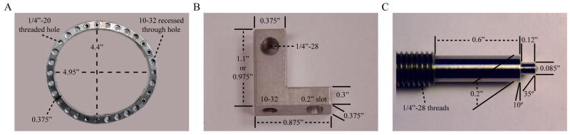
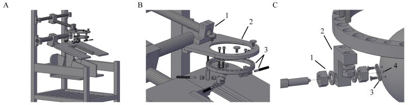
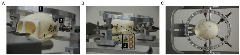
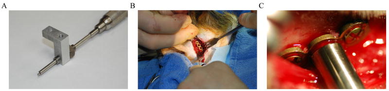
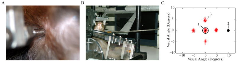
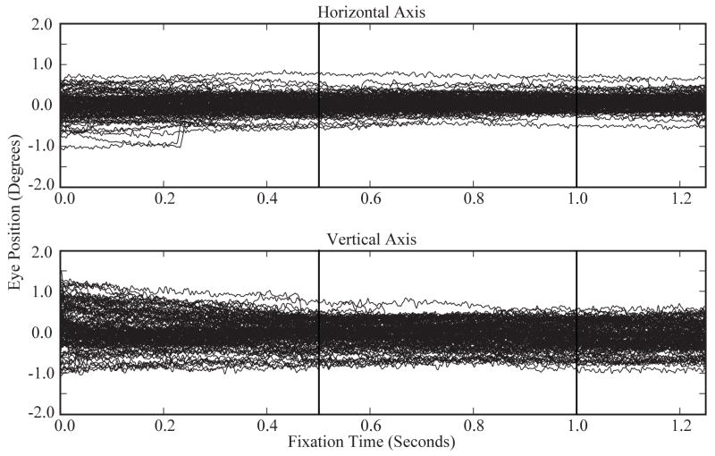
We have designed a device for long-term head fixation for use in behaving nonhuman primates that is robust yet minimally invasive and simple to use. This device is a modified version of the halo system that is used in humans for cervical traction and stabilization after spinal column injuries. This device consists of an aluminum halo with four titanium skull pins offset from the halo by aluminum posts. The titanium pins insert onto small segments of cranially reinforcing titanium plate, which are attached to the skull with titanium cortex screws. The surgery involves four scalp incisions, placement of the reinforcing plates, insertion of the pins for attachment of the halo, and incision closure. After the halo is attached, the animal's head can be fixed to a primate chair using a custom-built attachment arm that provides three degrees of adjustability for proper positioning during behavioral tasks. We have installed this device on two Macaque monkeys weighing 7 and 10kg. The halos have been in place on these animals for up to 8 months without signs of discomfort or loss of fixation. Using this method of head fixation, we have been able to track the animals' eye positions with an accuracy of less than two visual degrees while they perform behavioral tasks.


Figures

Figure 1
Figure 1
Device components with dimensions. A) Halo frame; B) Offset post; C) Modified titanium skull pin.
Figure 2
Figure 2
A) Overall view of primate chair with halo device. B) Exploded view of the attachment arm (1), attachment plate (2), and halo frame with posts and skull pins (3). C) Enlarged view showing insertion path of skull pin. The pin passes through stainless steel nuts and washers (1), an aluminum offset post (2), a titanium plate that is attached to the skull with titanium cortex screws (3), and into a predrilled hole in the skull surface (4).
Figure 3
Figure 3
Fixation device positioned on Macaque skull. A) Anterior view of device showing halo frame (1), offset post (2), and skull pin (3). The skull pin is placed in temporal bone superior and slightly anterior to the auditory canal. B) Posterior view of device showing post with skull pin and an enlarged image of the titanium reinforcement plate with cortex screws (4). The posterior pin is placed slightly superior to the superior nuchal line. C) Superior view of device showing pin position and angle with respect to cranial suture lines.
Figure 4
Figure 4
A) Drill guide used to align the drill bit during the attachment procedure. B) Reinforcement plate for the temporal pin with titanium cortex screws at each end. C) Reinforcement plate for the occipital pin with the pin threaded through the plate and into the predrilled hole in the skull surface. The predrilled hole was made using the drill guide.
Figure 5
Figure 5
A) Enlarged image of the right occipital pin site 5 months after surgery. B) Head-fixed animal performing a behavioral task requiring eye fixation. Eye positions were sampled at 1 kHz using the EyeLink 1000 infrared eye tracking system (SR Research, Ltd., Ontario, Canada). C) Graph of recorded eye positions in units of visual degrees. For each trial, the animal was required to fixate within an area of diameter 2.6° (1) while a stimulus (2) was presented and a response was acquired. If the animal looked outside of the defined area, the trial was aborted and no reward was received. A total of 118 trials were completed, each requiring a fixation duration of 1250 msec. Eye position data is plotted in red (3). Fixation positions were randomly alternated among five locations [(0,0); (5,0); (0,5); (−5,0); (0,−5)] during the task and are represented by the five clusters of eye position data. The central cluster of data follows a gaussian distribution with 2σ ≈ 0.8°.
Figure 6
Figure 6
Eye position data recorded at 1 kHz using the Cerebus data acquisition system (Blackrock Microsystems, Salt Lake City, UT). Plots show superimposed eye positions along the horizontal and vertical axis for 118 trials of the behavioral task described in Figure 5. Stimulus onset and offset are indicated by vertical lines. Individual tracings reveal small noise fluctuations on the order of minutes of visual angle. The smoothness of the tracings demonstrate the stability of this head fixation system while tracking eye movements with the EyeLink 1000 infrared eye tracking system (SR Research, Ltd., Ontario, Canada).

References

    1. Betelak KF, Margiotti EA, et al. The use of titanium implants and prosthodontic techniques in the preparation of non-human primates for long-term neuronal recording studies. J Neurosci Methods. 2001;112(1):9–20. - PubMed
    1. Dubowitz DJ, Chen DY, et al. Direct comparison of visual cortex activation in human and non-human primates using functional magnetic resonance imaging. J Neurosci Methods. 2001;107(1–2):71–80. - PubMed
    1. Evarts EV. Relation of pyramidal tract activity to force exerted during voluntary movement. J Neurophysiol. 1968;31(1):14–27. - PubMed
    1. Foeller P, Tychsen L. Eye movement training and recording in alert macaque monkeys: 1. Operant visual conditioning; 2. Magnetic search coil and head restraint surgical implantation; 3. Calibration and recording. Strabismus. 2002;10(1):5–22. - PubMed
    1. Friendlich AR. Primate head restrainer using a nonsurgical technique. J Appl Physiol. 1973;35(6):934–5. - PubMed

Publication types

LinkOut - more resources