Characterization of primary and restenotic atherosclerotic plaque from the superficial femoral artery: Potential role of Smad3 in regulation of SMC proliferation
- PMID: 19394554
- PMCID: PMC3302687
- DOI: 10.1016/j.jvs.2008.11.096
Characterization of primary and restenotic atherosclerotic plaque from the superficial femoral artery: Potential role of Smad3 in regulation of SMC proliferation
Abstract
Objective: To characterize and compare primary and restenotic lesions of the superficial femoral artery and analyze the contribution of TGF-beta/Smad3 signaling to the pathophysiology of peripheral artery occlusive disease.
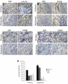
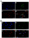
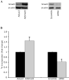
Methods and results: Immunohistochemical studies were performed on specimens retrieved from the superficial femoral artery of patients undergoing either atherectomy for primary atherosclerotic or recurrent disease after stenting and/or prior angioplasty. Immunohistochemical analysis revealed a significantly higher smooth muscle cell (SMC) content (alpha-actin+) and expression of Smad3 in restenotic lesions while primary lesions contained significantly more leukocytes (CD45+) and macrophages (CD68+). Further studies demonstrated colocalization of Smad3 with alpha-actin and PCNA, suggesting a role for Smad3 in the proliferation observed in restenotic lesions. To confirm a role for Smad3 in SMC proliferation, we both upregulated Smad3 via adenoviral mediated gene transfer (AdSmad3) and inhibited Smad3 through transfection with siRNA in human aortic SMCs, then assessed cell proliferation with tritiated thymidine. Overexpression of Smad3 enhanced whereas inhibition of Smad3 decreased cell proliferation.
Conclusion: Differences in cellular composition and cell proliferation in conjunction with the finding that Smad3 is expressed exclusively in restenotic disease suggest that TGF-beta, through Smad3 signaling, may play an essential role in SMC proliferation and the pathophysiology of restenosis in humans.
Figures


Similar articles
-
A crosstalk between TGF-β/Smad3 and Wnt/β-catenin pathways promotes vascular smooth muscle cell proliferation.Cell Signal. 2016 May;28(5):498-505. doi: 10.1016/j.cellsig.2016.02.011. Epub 2016 Feb 19. Cell Signal. 2016. PMID: 26912210 Free PMC article.
-
TGF-β/Smad3 inhibit vascular smooth muscle cell apoptosis through an autocrine signaling mechanism involving VEGF-A.Cell Death Dis. 2014 Jul 10;5(7):e1317. doi: 10.1038/cddis.2014.282. Cell Death Dis. 2014. PMID: 25010983 Free PMC article.
-
Arterial gene transfer of the TGF-beta signalling protein Smad3 induces adaptive remodelling following angioplasty: a role for CTGF.Cardiovasc Res. 2009 Nov 1;84(2):326-35. doi: 10.1093/cvr/cvp220. Epub 2009 Jul 1. Cardiovasc Res. 2009. PMID: 19570811 Free PMC article.
-
The role of Smad3-dependent TGF-beta signal in vascular response to injury.Trends Cardiovasc Med. 2006 Oct;16(7):240-5. doi: 10.1016/j.tcm.2006.04.005. Trends Cardiovasc Med. 2006. PMID: 16980181 Review.
-
Recanalization of infrainguinal vessels: silverhawk, laser, and the remote superficial femoral artery endarterectomy.Semin Vasc Surg. 2007 Mar;20(1):29-36. doi: 10.1053/j.semvascsurg.2007.02.002. Semin Vasc Surg. 2007. PMID: 17386361 Review.
Cited by
-
Protein kinase C delta mediates arterial injury responses through regulation of vascular smooth muscle cell apoptosis.Cardiovasc Res. 2010 Feb 1;85(3):434-43. doi: 10.1093/cvr/cvp328. Epub 2009 Oct 6. Cardiovasc Res. 2010. PMID: 19808702 Free PMC article.
-
Cell division cycle 7 mediates transforming growth factor-β-induced smooth muscle maturation through activation of myocardin gene transcription.J Biol Chem. 2013 Nov 29;288(48):34336-42. doi: 10.1074/jbc.M113.498238. Epub 2013 Oct 16. J Biol Chem. 2013. PMID: 24133205 Free PMC article.
-
FAM3A reshapes VSMC fate specification in abdominal aortic aneurysm by regulating KLF4 ubiquitination.Nat Commun. 2023 Sep 2;14(1):5360. doi: 10.1038/s41467-023-41177-x. Nat Commun. 2023. PMID: 37660071 Free PMC article.
-
Role of Smad3 signaling in the epithelial‑mesenchymal transition of the lens epithelium following injury.Int J Mol Med. 2018 Aug;42(2):851-860. doi: 10.3892/ijmm.2018.3662. Epub 2018 May 9. Int J Mol Med. 2018. PMID: 29750298 Free PMC article.
-
The role of Notch pathway in cardiovascular diseases.Glob Cardiol Sci Pract. 2013 Dec 30;2013(4):364-71. doi: 10.5339/gscp.2013.44. eCollection 2013. Glob Cardiol Sci Pract. 2013. PMID: 24749110 Free PMC article. Review.
References
-
- Schillinger M, Sabeti S, Loewe C, et al. Balloon Angioplasty versus Implantation of Nitinol Stents in the Superficial Femoral Artery. N Engl J Med. 2006;354:1879–88. - PubMed
-
- Schillinger M, Sabeti S, Dick P, et al. Sustained Benefit at 2 Years of Primary Femoropopliteal Stenting Compared With Balloon Angioplasty With Optional Stenting. Circulation. 2007;115:2745–9. - PubMed
-
- Bauriedel G, Schluckebier S, Hutter R, et al. Apoptosis in restenosis versus stable-angina atherosclerosis: implications for the pathogenesis of restenosis. Arteriosclerosis, thrombosis, and vascular biology. 1998;18(7):1132–9. - PubMed
-
- Nikol S, Murakami N, Pickering JG, et al. Differential expression of nonmuscle myosin II isoforms in human atherosclerotic plaque. Atherosclerosis. 1997;130:71–85. - PubMed
-
- Hao H, Gabbiani G, Bochaton-Piallat M-L. Arterial Smooth Muscle Cell Heterogeneity: Implications for Atherosclerosis and Restenosis Development. Arterioscler Thromb Vasc Biol. 2003;23:1510–20. - PubMed
Publication types
MeSH terms
Substances
Grants and funding
LinkOut - more resources
Full Text Sources
Medical
Research Materials
Miscellaneous

