Multisequence-imaging protocols to detect cortical lesions of patients with multiple sclerosis: observations from a post-mortem 3 Tesla imaging study
- PMID: 19394970
- PMCID: PMC3504465
- DOI: 10.1016/j.jns.2009.03.021
Multisequence-imaging protocols to detect cortical lesions of patients with multiple sclerosis: observations from a post-mortem 3 Tesla imaging study
Abstract
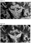
Neocortical lesions (NLs) are an important component of multiple sclerosis (MS) pathology and may account for part of the physical and cognitive disability. Visualizing NLs of patients with MS using magnetic resonance imaging (MRI) poses several significant challenges. We optimized the inversion time (TI) of T(1)-based magnetization-prepared rapid acquisition gradient-echo (MPRAGE) images by suppressing the signal of the lesions and enhancing their appearance as hypointensities, on the basis of the derived quantitative T(1) measurements. The latter were achieved by the means of 2D inversion recovery fast spin echo (IR-FSE), repeated using different inversion times (TI). Comparisons of detection of NLs by MPRAGE and dual echo T(2) weighted (T(2)W) and proton density (PD) W. Four coronal brain slices from a deceased MS patient and two coronal brain slices from two formerly healthy donors were imaged using a 3 Tesla magnet (3 T) equipped with a multi-channel coil. Based upon the averaged T1 values computed from the MS specimen as well as visual inspection, an optimal TI of 380 ms was selected for the MPRAGE image. No NLs were seen in the specimens of the two healthy donors. Of the 40 total NLs observed, 8 (20%) were visible in all three sequences employed. Three (7.5%) NLs were visible only in the PDW image and 5 (12.5%) were seen only in the T(2)W image. Four NLs (10%) had clearly unique conspicuity in the MPRAGE image. Of those, 3 were retrospectively scored in the PDW image (1 NL) or in the T2W image (2 NLs). We conclude that for the detection of MS-related NLs, high-resolution T(1)-based MPRAGE and T(2)-W images offer complementary information and the combination of the two image sequences is crucial for increasing the sensitivity of detecting MS-induced NLs.
Figures


References
-
- Zivadinov R, Stosic M, Cox JL, Ramasamy DP, Dwyer MG. The place of conventional MRI and newly emerging MRI techniques in monitoring different aspects of treatment outcome. J Neurol. 2008;255 (Suppl 1):61–74. - PubMed
-
- Boggild MD, Williams R, Haq N, Hawkins CP. Cortical plaques visualised by fluid-attenuated inversion recovery imaging in relapsing multiple sclerosis. Neuroradiology. 1996;38 (Suppl 1):10–13. - PubMed
-
- Filippi M, Yousry T, Baratti C, Horsfield MA, Mammi S, Becker C, et al. Quantitative assessment of MRI lesion load in multiple sclerosis. A comparison of conventional spin-echo with fast fluid-attenuated inversion recovery. Brain. 1996;119:1349–55. - PubMed
Publication types
MeSH terms
Grants and funding
LinkOut - more resources
Full Text Sources
Other Literature Sources
Medical

