. 2009 Aug;86(2):229-35.
doi: 10.1189/jlb.1208742. Epub 2009 Apr 24.

Hemopexin down-regulates LPS-induced proinflammatory cytokines from macrophages

Affiliations

Hemopexin down-regulates LPS-induced proinflammatory cytokines from macrophages

Xueya Liang et al. J Leukoc Biol. 2009 Aug.

Abstract

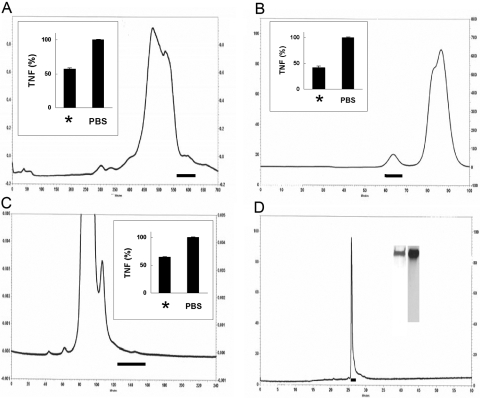
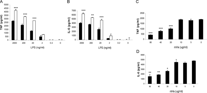
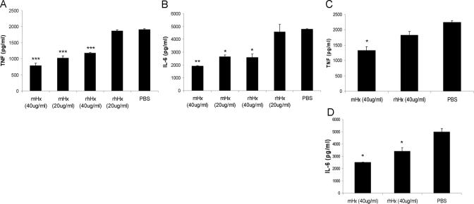
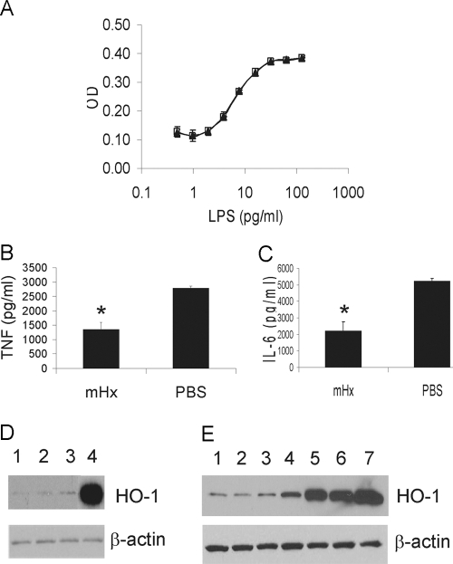
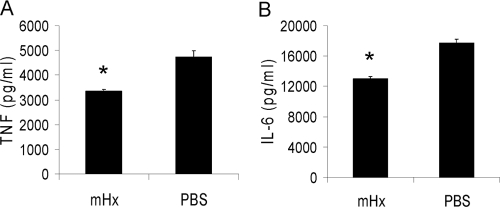
Detection of LPS in tissues is an integral component of innate immunity that acts to protect against invasion by Gram-negative bacteria. Plasma down-regulates LPS-induced cytokine production from macrophages, thereby limiting systemic inflammation in blood and distant tissues. To identify the protein(s) involved in this process, we used classical biochemical chromatographic techniques to identify fractions of mouse sera that suppress LPS-induced TNF from bone marrow-derived macrophages (BMDMs). Fractionation yielded microgram quantities of a protein that was identified by MS to be hemopexin (Hx). Mouse Hx purified on hemin-agarose beads and rhHx decreased the production of cytokines from BMDMs and peritoneal macrophages induced by LPS. Preincubation of LPS with Hx did not affect the activity of LPS on LAL, whereas preincubation of Hx with macrophages followed by washing resulted in decreased activity of these cells in response to LPS, suggesting that Hx acts on macrophages rather than LPS. Heme-free Hx did not stimulate HO-1 in the macrophages. Purified Hx also decreased TNF and IL-6 from macrophages induced by the synthetic TLR2 agonist Pam3Cys. Our data suggest that Hx, which is an acute-phase protein that increases during inflammation, limits TLR4 and TLR2 agonist-induced macrophage cytokine production directly through a mechanism distinct from HO-1.


Figures

Figure 1.
Figure 1.
Identification, purification, and purity of mHx. (A–C) Chromatograph of active fractions as described. Solid horizontal lines indicate material selected for activity in suppressing LPS-induced TNF from BMDMs. Insets show activity of pooled material from column; left bars are labeled with *, showing LPS-induced TNF in the presence of pooled material indicated by horizontal line, and right bars show LPS-induced TNF in the presence of PBS alone. (A) Chromatogram of 60–73% saturated ammonium sulfate precipitate of mouse serum chromatofocused on a 30-ml Tricorn 10/300 column in a decreasing pH 7 to 4 gradient. (B) Chromatogram of separation by anion exchange of material shown in A. (C) Chromatogram of a molecular sieving column of material shown in B. The material indicated was analyzed by MS and contained mHx. (D) Chromatogram on reverse-phase C4 column of 1 μg mHx purified on hemin-agarose as in Materials and Methods. Horizontal line shows material collected for final preparation. Inset shows SDS-PAGE and silver staining of this material (left lane) and Western blot developed with chicken anti-mHx IgY (right lane).
Figure 2.
Figure 2.
Effect of mHx on LPS-induced TNF and IL-6 production from BMDMs. Cells were washed three times with serum-free medium and then cultured overnight with 20 μg/ml mHx (solid bars) or PBS (open bars) and indicated concentrations of LPS (A and B) or 20 ng/ml LPS and indicated concentrations of mHx (C and D). Concentrations of TNF and IL-6 in the supernatants were determined by ELISA. The results represent mean ± se and are representative of more than five independent experiments. *, P < 0.05; **, P < 0.01; ***, P < 0.001, compared with cells treated with PBS.
Figure 3.
Figure 3.
rhHx suppresses LPS-induced TNF and IL-6 from macrophages. BMDMs (A and B) or peritoneal macrophages (C and D) were incubated overnight with 20 ng/ml LPS and the indicated concentrations (A and B) or 40 μg/ml (C and D) mHx and rhHx. Concentrations of TNF and IL-6 in the supernatants were determined by ELISA. The results represent mean ± se and are representative of three independent experiments. *, P < 0.05; **, P < 0.01; ***, P < 0.001, compared with cells treated with PBS.
Figure 4.
Figure 4.
Hx does not neutralize LPS as assessed by Limulus lysate actvity but acts on macrophages in a manner that does not up-regulate the expression of HO-1. (A) LAL assay of LPS preincubated with 2 μg/ml purified mHx (▴) or PBS (□). (B and C) BMDMs were preincubated with mHx (20 μg/ml) for 2 h, followed by extensive washes in serum-free medium. LPS (20 ng/ml) was then added to the culture and incubated overnight. Concentrations of TNF (B) and IL-6 (C) in the supernatants were determined by ELISA. The results represent mean ± se and are representative of five independent experiments. *, P < 0.05, compared with cells treated with PBS. (D and E) BMDMs were incubated overnight with: (D) Lane 1, PBS; Lane 2, mHx (40 μg/ml); Lane 3, rhHx (40 μg/ml); Lane 4, hemin chloride (3 μM), or (E) Lane 1, PBS; Lane 2, mHx (40 μg/ml, equal to 0.67 μM); Lane 3, mHx (200 μg/ml, equal to 3.35 μM); Lane 4, hemin-mHx complex (0.67 μM); Lane 5, hemin-mHx complex (3.35 μM); Lane 6, hemin-chloride (0.67 μM); Lane 7, hemin chloride (3 μM). Cells were then washed three times by PBS. Cell lysates were used for SDS-PAGE. Western blot was performed using anti-mouse HO-1 antibody, followed by stripping and reprobing by anti-β-actin antibody.
Figure 5.
Figure 5.
Effects of Hx on cytokine production from macrophages stimulated by Pam3Cys. BMDMs were incubated overnight with mHx (100 μg/ml) or PBS with Pam3Cys (200 ng/ml). Concentrations of TNF and IL-6 in the supernatants were determined by ELISA. The results represent mean ± se and are representative of more than five independent experiments. *, P < 0.05, compared with cells treated with PBS.

Comment in

References

    1. Raetz C R, Whitfield C. Lipopolysaccharide endotoxins. Annu Rev Biochem. 2002;71:635–700. - PMC - PubMed
    1. Akashi S, Shimazu R, Ogata H, Nagai Y, Takeda K, Kimoto M, Miyake K. Cutting edge: cell surface expression and lipopolysaccharide signaling via the Toll-like receptor 4-MD-2 complex on mouse peritoneal macrophages. J Immunol. 2000;164:3471–3475. - PubMed
    1. Takeda K, Akira S. Toll-like receptors in innate immunity. Int Immunol. 2005;17:1–14. - PubMed
    1. Bone R C, Balk R A, Cerra F B, Dellinger R P, Fein A M, Knaus W A, Schein R M H, Sibbald W J. Definitions for sepsis and organ failure and guidelines for the use of innovative therapies in sepsis. Chest. 1992;101:1644–1655. - PubMed
    1. Angus D C, Linde-Zwirble W T, Lidicker J, Clermont G, Carcillo J, Pinsky M R. Epidemiology of severe sepsis in the United States: analysis of incidence, outcome, and associated costs of care. Crit Care Med. 2001;29:1303–1310. - PubMed

Publication types

MeSH terms