. 2009 Jun;296(6):H1994-2006.
doi: 10.1152/ajpheart.00038.2009. Epub 2009 Apr 24.

beta-adrenergic regulation of a novel isoform of NCX: sequence and expression of shark heart NCX in human kidney cells

Affiliations

beta-adrenergic regulation of a novel isoform of NCX: sequence and expression of shark heart NCX in human kidney cells

Einsley Janowski et al. Am J Physiol Heart Circ Physiol. 2009 Jun.

Abstract

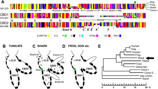
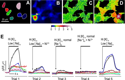
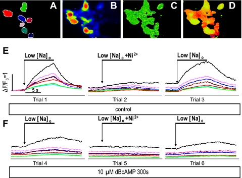
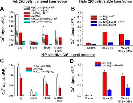
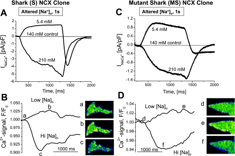
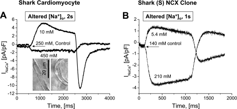
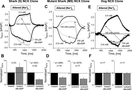
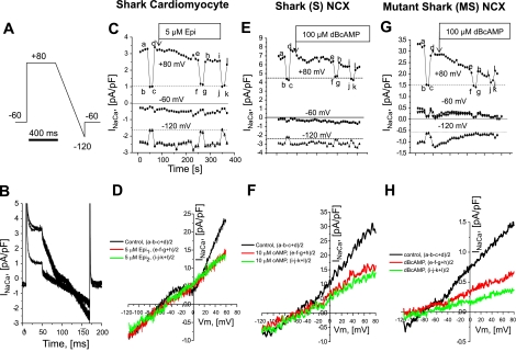
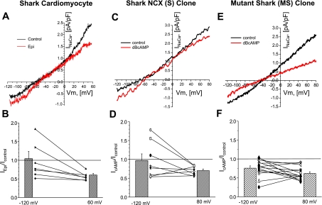
The function, regulation, and molecular structure of the cardiac Na(+)/Ca(2+) exchangers (NCXs) vary significantly among vertebrates. We previously reported that beta-adrenergic suppression of amphibian cardiac NCX1.1 is associated with specific molecular motifs. Here we investigated the bimodal, cAMP-dependent regulation of spiny dogfish shark (Squalus acanthias) cardiac NCX, exploring the effects of molecular structure, host cell environment, and ionic milieu. The shark cardiac NCX sequence (GenBank accession no. DQ 068478) revealed two novel proline/alanine-rich amino acid insertions. Wild-type and mutant shark NCXs were cloned and expressed in mammalian cells (HEK-293 and FlpIn-293), where their activities were measured as Ni(2+)-sensitive Ca(2+) fluxes (fluo 4) and membrane (Na(+)/Ca(2+) exchange) currents evoked by changes in extracellular Na(+) concentration and/or membrane potential. Regardless of Ca(2+) buffering, beta-adrenergic stimulation of cloned wild-type shark NCX consistently produced bimodal regulation (defined as differential regulation of Ca(2+)-efflux and -influx pathways), with suppression of the Ca(2+)-influx mode and either no change or enhancement of the Ca(2+)-efflux mode, closely resembling results from parallel experiments with native shark cardiomyocytes. In contrast, mutant shark NCX, with deletion of the novel region 2 insertion, produced equal suppression of the inward and outward currents and Ca(2+) fluxes, thereby abolishing the bimodal nature of the regulation. Control experiments with nontransfected and dog cardiac NCX-expressing cells showed no cAMP regulation. We conclude that bimodal beta-adrenergic regulation is retained in cloned shark NCX and is dependent on the shark's unique molecular motifs.


Figures

Fig. 1.
Fig. 1.
Amino acid sequence of shark cardiac NCX. A: graphic representation of a partial alignment of long cytoplasmic regulatory loops of cardiac Na+/Ca2+ exchangers (NCXs) from dog, frog, shark, and Ciona intestinalis (see Supplemental Fig. 2 for detailed alignment). B–D: major differences in alignment in tunicate, shark, and higher vertebrates. Labeled features include an α-catenin-like domain (α-CLD) with 4 putative α-helical segments (segments A–D) and Ca2+-binding domains, CBD1 and CBD2, each with 7 β-strands (a–g). Species-dependent linker sequences are shown in the A helix of α-CLD (shark region 1) and between β-strands f and g of Ca2+-binding domains: shark region 2 insert in CBD1 and the variably spliced exons C–F in CBD2. E: placement of shark NCX (arrow) in a phylogenetic tree (Clustal W2) of deuterostome NCXs rooted by NCX from the squid Loligo opalescens (GenBank accession no. U93214). Abscissa shows difference in amino acids (%) calculated for core regions of α-repeats and Ca2+-binding domains (see Supplemental Table 1). Vertebrate sequences are based on mRNA from cardiac tissue. GenBank accession nos. are as follows: NM021097 (human, Homo sapiens), M57523 (dog, Canis lupus familiaris), XM415002 (chicken, Gallus gallus), X90839/X90838 (frog, Xenopus laevis), DQ068478 (shark, Squalus acanthias), AAP37041 (tilapia, Oreochromis mossambicus), AAF06363 (trout, Oncorhynchus mykiss), and XP686228 (zebra fish, Danio rerio). Sea urchin is represented by a partial genomic construct (accession no. XM776256) that, similar to the modeled tunicate sequences [C. intestinalis (Ciona I) from AABS01000304 and C. savignyi (Ciona S) from AACT01059174/AACT01057381; see supplemental material in the online version of this article], showed homology with vertebrate NCXs.
Fig. 2.
Fig. 2.
Verification of transient transfection of shark NCX into nondialyzed HEK-293 cells by Ca2+ imaging. A: selected color-coded cellular regions corresponding to traces in E. B: average fluorescence intensity before activation. Image was used as divisor to generate ratiometrically normalized fluorescence images in C and D. Threshold for division was adjusted to generate dark areas around cells. C: normalized fluorescence (F/F0) image before activation shows noise in a single frame as green/blue mottle (F/F0 ≅ 1, see color scale). D: normalized fluorescence image at peak of activation (trial 1) shows individual cells as relatively uniform areas of orange or red. E: changes in fluorescence in selected cells during 5 trials with exposure to high extracellular K+ concentration (Hi [K]o, from 5.4 to 40 or 140 mM), with and without reduction in extracellular Na+ concentration {low [Na+]o, from 140 to 5 mM with tetraethylammonium chloride (TEA-Cl) substitution} and addition of 5 mM Ni2+. (Shark NCX was transiently expressed in nondialyzed HEK-293 cells stained with fluo 4-AM.)
Fig. 3.
Fig. 3.
Verification of stable transfection of shark NCX in nondialyzed FlpIn-293 cells (S cells). A–D (from trial 1) correspond to A–D in Fig. 2. E and F: bracketed measurements of changes in fluorescence in selected color-coded cells during 6 trials with exposure to reduced [Na+]o (low [Na+]o, from 140 to 5 mM with TEA-Cl substitution), with (trials 2 and 5) and without (trials 1, 3, 4, and 6) addition of 5 mM Ni2+, before (trials 1–3) and after (trials 4–6) application of 10 μM dibutyryl cAMP (DBcAMP) for 300 s.
Fig. 4.
Fig. 4.
Ni2+-sensitive Ca2+ signals (C and D) are of comparable magnitude for different NCX constructs and expression systems, but Ni2+-insensitive Ca2+ signals are smaller in stable FlpIn-293 cells (B) than in transiently expressing HEK-293 nondialyzed cells (A). Data are from experiments similar to those described in Figs. 2 and 3 legends. A and B: responses in the absence (solid bars) and presence (hatched bars) of 5 mM Ni2+ in HEK-293 cells transiently transfected with dog NCX, the empty vector (Sham), shark NCX, or mutant shark NCX (A) and in FlpIn-293 cells untransfected (Control) or stably transfected with shark (S cells) or mutant shark (MS cells) NCX (B). Ca2+-dependent fluorescence was activated by high [K]o and low [Na]o (white bars), normal [K]o and low [Na]o (red bars), high [K]o and normal [Na]o (teal bars), and low [Na]o after incubation with 100 μM DBcAMP (blue bars). Difference signals in C and D quantify Ni2+-sensitive changes in Ca2+-dependent fluorescence in the transiently (C) and stably (D) transfected cells.
Fig. 5.
Fig. 5.
Activation of Ca2+ fluxes by changes in [Na]o produces matching changes in Na+/Ca2+ exchange current (INaCa; A and C) and Ca2+ signals (B and D) in weakly Ca2+-buffered voltage-clamped cells with stable expression of shark NCX (S cells; A and B) and mutant shark NCX (MS cells; C and D). Ca2+-influx and -efflux modes were activated, respectively, by reduction of [Na+]o (from 140 to 5.4 mM, by TEA-Cl substitution) or elevation of [Na+]o (from 140 to 210 mM, by Na+ addition) during reduction of [Ca2+] (from 5 to 0.5 mM) for 1-s intervals. Insets (a–f): fluorescence images at the indicated times and [Na+]o. {Voltage-clamped FlpIn-293 cells were held at −60 mV and dialyzed with internal solution, where intracellular Ca2+ concentration ([Ca2+]i) was buffered at 100 nM by 0.1 mM BAPTA and 0.1 mM K5-fluo 4 and 0.02 Ca2+; ratiometric normalization of images and color scale as in Fig. 2.}
Fig. 6.
Fig. 6.
Comparison of shark NCX in its native environment (A) and in mammalian expression system (B). In the different ionic milieus, high Ca2+-buffering and Na+-dependent activation of outward and inward INaCa were achieved by slightly different means: in freshly dissociated shark ventricular cardiomyocytes, [Ca2+]i ∼200 nM (10 mM EGTA + 6 mM Ca2+), [Na+]o was reduced from 250 to 10 mM (by Cs+ substitution), and [Na+]o was elevated from 250 to 450 mM (by urea replacement); in FlpIn-293 cells with stable expression of shark NCX, [Ca2+]i ∼100 nM (0.1 mM BAPTA + 5 mM EGTA + 2.66 mM Ca2+), [Na+]o was reduced from 140 to 5.4 mM (by TEA-Cl substitution), and [Na+]o was elevated from 140 to 210 mM (by Na+ addition coupled with reduction of [Ca2+]o from 5 to 0.5 mM). Insets in A: midsection of a spindle-shaped shark cardiomyocyte (∼100 μm long, 5–10 μm diameter, 75 ± 5 pF membrane capacitance, n = 8) that was held in position by the patch pipette but bent when a flow of standard shark Ringer solution was applied via a rapid perfusion system. [Voltage-clamped dialyzed cells were held at −60 mV; Ca2+ current (ICa) was blocked by 10 μM nifedipine.]
Fig. 7.
Fig. 7.
Different modalities of cAMP-dependent regulation of shark (S cells; A and B: bimodal), mutant shark (MS cells; C and D: unimodal), and dog (E and F: no regulation) cardiac NCX. A, C, and E: [Na+]o-dependent activation of outward and inward INaCa in voltage-clamped stably expressing cells dialyzed with high concentrations of Ca2+ buffers. Dotted traces were recorded after 180 s of incubation with 100 μM DBcAMP. B, D, and F: fractional changes in inward (left) and outward (right) INaCa determined from pooled experiments with cells where [Ca2+]i = 100 nM was established by dialysis of titrated Ca2+ buffers in high (0.1 mM BAPTA + 5 mM EGTA + 2.66 mM Ca2+) or low (0.1 mM BAPTA, 0.1 mM K5-fluo 4, and 0.02 mM Ca2+) concentrations. Each graph shows response in the presence of DBcAMP (hatched bars) relative to response before incubation (≡1; solid bars). Values are means ± SE for experiments with n cells. P values are based on paired t-tests. {For voltage-clamped (−60 mV) dialyzed stable FlpIn-293 (A–D) and HEK-293 (E and F) cells, [Na+]o was reduced from 140 to 5.4 mM (by TEA-Cl substitution) and [Na+]o was elevated from 140 to 210 mM (by Na+ addition coupled with reduction of [Ca2+]o from 5 to 0.5 mM).}
Fig. 8.
Fig. 8.
cAMP-mediated regulation preferentially suppresses Ca2+-influx mode of NCX in shark ventricular myocytes (A–D) and S cells (E and F), whereas it suppresses influx and efflux modes of MS cells (G and H). A and B: ramp-clamp protocol used to measure membrane current before (a in B and C), during (b and c), and after (d) application of 5 mM Ni2+. C, E, and G: membrane current at +80, −60 (holding potential), and −120 mV recorded at 10-s intervals with 3 brief exposures to 5 mM Ni2+ and a long exposure to 5 μM epinephrine (Epi; C and D) or 100 μM DBcAMP (E–H). D, F, and H: current-voltage (I-V) relations for Ni2+-sensitive component of membrane current measured once before [control; (abc + d)/2] and twice after {red; [(efg + h)/2] and green [(ijk + l)/2] curves} β-adrenergic stimulation. Vm, membrane potential. [Voltage-clamped cells were dialyzed with high concentrations of Ca2+ buffers (see Fig. 6 legend).]
Fig. 9.
Fig. 9.
Bimodal β-adrenergic regulation of native (A and B) and cloned (C and D) shark NCX vs. unimodal suppression of mutant shark NCX (E and F). A and C: I-V relations of INaCa in high- and low-Ca2+-buffered cells, respectively, where epinephrine (A) or DBcAMP (C) decreased outward INaCa but increased inward INaCa. E: I-V relations of INaCa measured in a low-Ca2+-buffered mutant shark cell, where DBcAMP decreased outward and inward INaCa. B, D, and F: fractional changes in INaCa at Vm of −120 and +60 mV. Bars show average values of relative current [IEpi/IControl (B) and IDBcAMP/IControl (D and F)] based on individual experiments represented by symbols and connecting lines (n = 7 for shark ventricular myocytes, n = 8 for shark FlpIn-293 cells, and n = 14 for shark mutant FlpIn-293 cells). Closed symbols, strong Ca2+ buffering; open symbols, weak Ca2+ buffering.

Similar articles

Cited by

References

    1. Adachi-Akahane S, Cleemann L, Morad M. Cross-signaling between L-type Ca2+ channels and ryanodine receptors in rat ventricular myocytes. J Gen Physiol 108: 435–454, 1996. - PMC - PubMed
    1. Baker PF, Blaustein MP, Hodgkin AL, Steinhardt RA. The influence of calcium on sodium efflux in squid axons. J Physiol 200: 431–458, 1969. - PMC - PubMed
    1. Barry WH, Bridge JH. Intracellular calcium homeostasis in cardiac myocytes. Circulation 87: 1806–1815, 1993. - PubMed
    1. Bers DM, Bridge JH. Relaxation of rabbit ventricular muscle by Na-Ca exchange and sarcoplasmic reticulum calcium pump. Ryanodine and voltage sensitivity. Circ Res 65: 334–342, 1989. - PubMed
    1. Besserer GM, Ottolia M, Nicoll DA, Chaptal V, Cascio D, Philipson KD, Abramson J. The second Ca2+-binding domain of the Na+/Ca2+ exchanger is essential for regulation: crystal structures and mutational analysis. Proc Natl Acad Sci USA 104: 18467–18472, 2007. - PMC - PubMed

Publication types

MeSH terms

Associated data

LinkOut - more resources