. 2009 Jun;75(12):3851-8.
doi: 10.1128/AEM.00457-09. Epub 2009 Apr 24.

Utilization of mucus from the coral Acropora palmata by the pathogen Serratia marcescens and by environmental and coral commensal bacteria

Affiliations

Utilization of mucus from the coral Acropora palmata by the pathogen Serratia marcescens and by environmental and coral commensal bacteria

Cory J Krediet et al. Appl Environ Microbiol. 2009 Jun.

Abstract

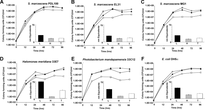
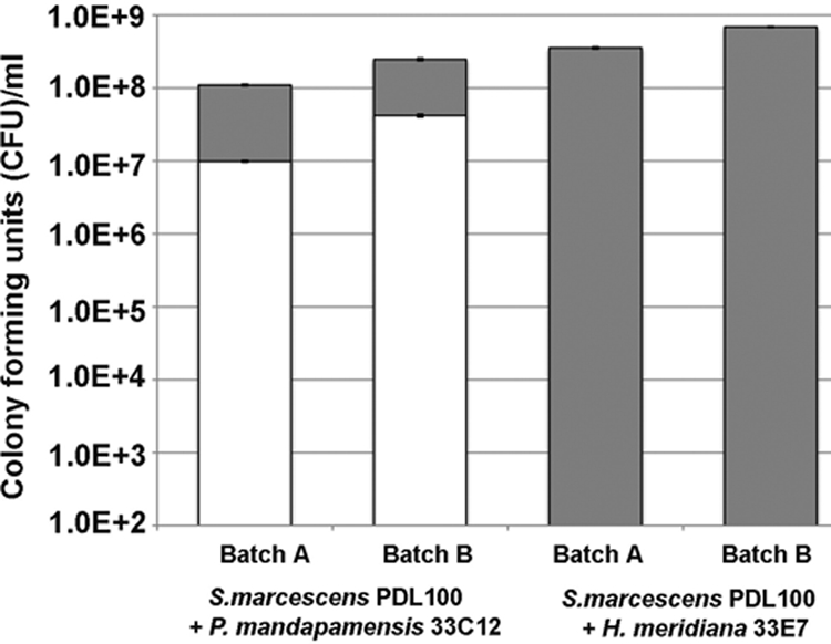
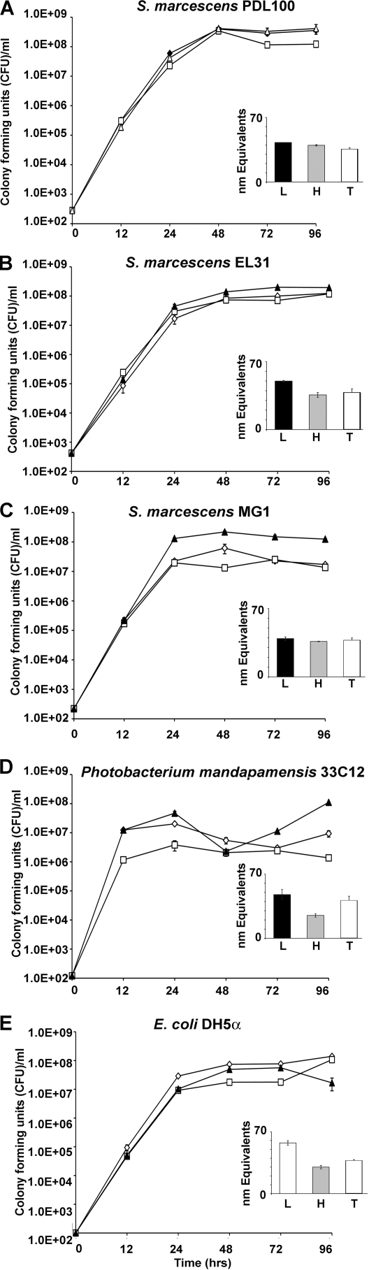
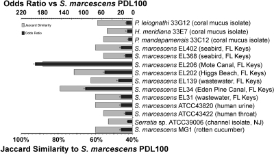
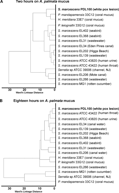
In recent years, diseases of corals caused by opportunistic pathogens have become widespread. How opportunistic pathogens establish on coral surfaces, interact with native microbiota, and cause disease is not yet clear. This study compared the utilization of coral mucus by coral-associated commensal bacteria ("Photobacterium mandapamensis" and Halomonas meridiana) and by opportunistic Serratia marcescens pathogens. S. marcescens PDL100 (a pathogen associated with white pox disease of Acroporid corals) grew to higher population densities on components of mucus from the host coral. In an in vitro coculture on mucus from Acropora palmata, S. marcescens PDL100 isolates outgrew coral isolates. The white pox pathogen did not differ from other bacteria in growth on mucus from a nonhost coral, Montastraea faveolata. The ability of S. marcescens to cause disease in acroporid corals may be due, at least in part, to the ability of strain PDL100 to build to higher population numbers within the mucus surface layer of its acroporid host. During growth on mucus from A. palmata, similar glycosidase activities were present in coral commensal bacteria, in S. marcescens PDL100, and in environmental and human isolates of S. marcescens. The temporal regulation of these activities during growth on mucus, however, was distinct in the isolates. During early stages of growth on mucus, enzymatic activities in S. marcescens PDL100 were most similar to those in coral commensals. After overnight incubation on mucus, enzymatic activities in a white pox pathogen were most similar to those in pathogenic Serratia strains isolated from human mucosal surfaces.


Figures

FIG. 1.
FIG. 1.
Bacterial growth on the mucus of Acropora palmata. Serratia marcescens, coral commensal bacteria, and E. coli DH5α were grown for 96 h in seawater containing approximately 60 mg ml−1 (wt/vol) total mucus (open diamonds) or its high-molecular-mass (>15 kDa; open squares) or low-molecular-mass (<15 kDa; closed triangles) fractions. (Insets) The extent to which the bacteria hydrolyzed substrates in mucus was examined using a BCA assay as described in Materials and Methods. Black bars indicate accumulation of reducing group equivalents in the low-molecular-mass fraction of mucus; gray bars indicate accumulation of reducing group equivalents in the high-molecular-mass fraction of mucus; and white bars indicate accumulation of reducing group equivalents in total mucus. (A) Growth of S. marcescens PDL100, a pathogen associated with white pox disease of A. palmata. PDL100 reached ∼2 × 10⁸ CFU ml−1 on all mucus treatments, which was significantly higher than the densities of all of the other strains (Tukey's HSD P value of <0.0025). (B) Growth of S. marcescens EL31 isolated from wastewater in the Florida Keys. (C) Growth of S. marcescens MG1 isolated from a rotten cucumber. (D) Growth of H. meridiana 33E7 isolated from A. palmata mucus. (E) Growth of P. mandapamensis 33C12 isolated from A. palmata mucus. (F) Growth of E. coli DH5α on mucus and its components. Graphs show the results of a representative experiment; error bars indicate standard errors from three parallel independent cultures.
FIG. 2.
FIG. 2.
Growth of S. marcescens PDL100 and coral commensals in an in vitro coculture on mucus. Starved washed cultures of S. marcescens PDL100 were coinoculated into sterilized mucus with either P. mandapamensis 33C12 or H. meridiana 33E7. Two batches of mucus samples harvested from different A. palmata colonies in the winter of 2007 were used in these assays. Initial inocula were ∼100 CFU ml−1 for all bacteria. After 96 h and 7 days, aliquots were dilution plated onto GASW and LB agar and then patched onto xylose lysine deoxycholate agar (Oxoid) to distinguish between S. marcescens and coral commensals. Growth of the coral commensals is indicated by white bars; growth of S. marcescens PDL100 is indicated by gray bars. Error bars indicate standard errors of three technical replications.
FIG. 3.
FIG. 3.
Bacterial growth on mucus of Montastraea faveolata. Isolates of Serratia marcescens, P. mandapamensis 33C12, and E. coli DH5α were grown on mucus from a nonhost coral, M. faveolata. Bacteria were seeded into seawater containing 60 mg ml−1 (wt/vol) total mucus (closed triangles) or high-molecular-mass (>15 kDa; open squares) or low-molecular-mass (<15 kDa; open diamonds) fractions. Growth was monitored by dilution plating. (Insets) The extent to which the bacteria hydrolyzed substrates in mucus was examined using a BCA assay as described in Materials and Methods. White bars indicate accumulation of reducing group equivalents in the low-molecular-mass fraction of mucus; gray bars indicate accumulation of reducing group equivalents in the high-molecular-mass fraction of mucus; and black bars indicate accumulation of reducing group equivalents in total mucus. (A) Growth of S. marcescens PDL100. (B) Growth of S. marcescens EL31. (C) Growth of S. marcescens MG1. (D) Growth of P. mandapamensis 33C12. (E) Growth of E. coli DH5α. Graphs show the results from a representative experiment; error bars indicate standard errors from three parallel independent cultures.
FIG. 4.
FIG. 4.
Metrics of correspondence between Biolog EcoPlate assays for S. marcescens PDL100, other S. marcescens isolates, and three coral commensals. The results of the Biolog assay were converted into binary data (see Table S2 in the supplemental material), and values for the strain PDL100 were compared with those for other S. marcescens strains, using the Jaccard similarity coefficient and the odds ratio. Isolates with similar metabolic capabilities have high Jaccard similarity and odds ratio. Statistical analyses were carried out using Statistica version 8.0 (StatSoft, Inc., Tulsa, OK) as described in Materials and Methods.
FIG. 5.
FIG. 5.
Clustering of strains based on enzymatic activities induced during growth on coral mucus. The hierarchical tree plot was constructed using Ward's linkage as the joining rule and Euclidean distance metric. (A) The hierarchical tree plot of enzymatic activities induced in S. marcescens isolates and coral commensals after 2 hours of incubation on the total mucus of A. palmata. (B) The hierarchical tree plot of enzymatic activities induced in S. marcescens isolates and coral commensals after 18 hours of incubation on the mucus of A. palmata. Statistical analyses were carried out using Statistica version 8.0 (StatSoft, Inc., Tulsa, OK) as described in Materials and Methods.

References

    1. Agresti, A. 1999. On logit confidence intervals for the odds ratio with small samples. Biometrics 55:597-602. - PubMed
    1. Brown, B. E., and J. C. Bythell. 2005. Perspectives on mucus secretion in reef corals. Mar. Ecol. Prog. Ser. 296:291-309.
    1. D'Aloia, M. A., T. A. Bailey, J. H. Samour, J. Naldo, and J. C. Howlett. 1996. Bacterial flora of captive houbara (Chlamydotis undulate), kori (Ardeotis kori) and rufous-crested (Eupodotis ruficrista) bustards. Avian Pathol. 3:459-468. - PubMed
    1. Ducklow, H. W., and R. Mitchell. 1979. Composition of mucus released by coral reef coelenterates. Limnol. Oceanogr. 21:706-714.
    1. Fabich, A. J., S. A. Jones, F. Z. Chowdhury, A. Cernosek, A. Anderson, D. Smalley, J. W. McHargue, G. A. Hightower, J. T. Smith, S. M. Autieri, M. P. Leatham, J. J. Lins, R. L. Allen, D. C. Laux, P. S. Cohen, and T. Conway. 2008. Comparison of carbon nutrition for pathogenic and commensal Escherichia coli strains in the mouse intestine. Infect. Immun. 76:1143-1152. - PMC - PubMed

Publication types

MeSH terms

LinkOut - more resources