. 2009 May;11(5):644-51.
doi: 10.1038/ncb1871. Epub 2009 Apr 26.

PP1-mediated dephosphorylation of phosphoproteins at mitotic exit is controlled by inhibitor-1 and PP1 phosphorylation

Affiliations

PP1-mediated dephosphorylation of phosphoproteins at mitotic exit is controlled by inhibitor-1 and PP1 phosphorylation

Judy Qiju Wu et al. Nat Cell Biol. 2009 May.

Abstract

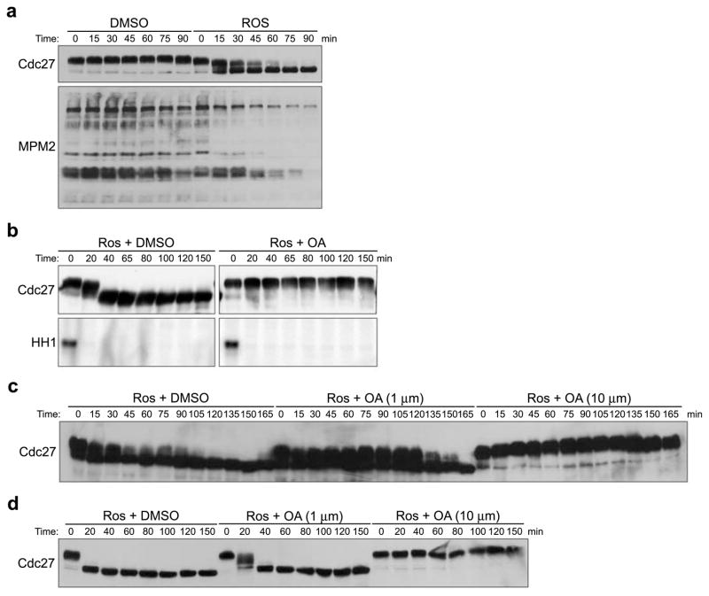
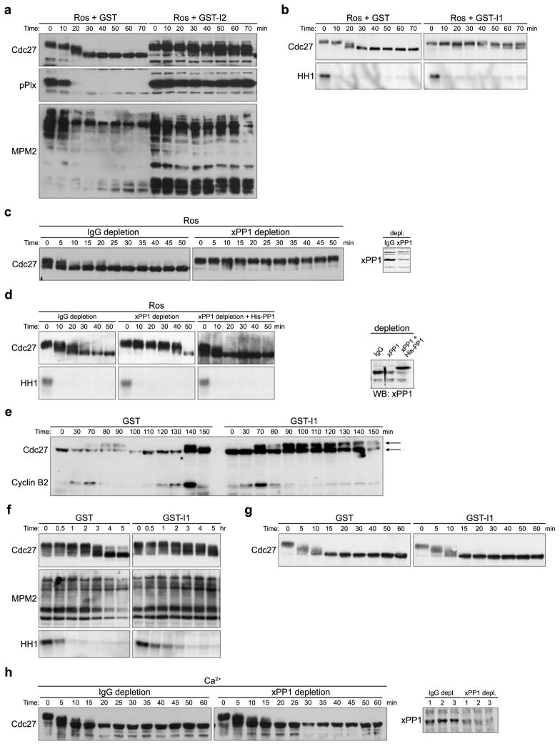
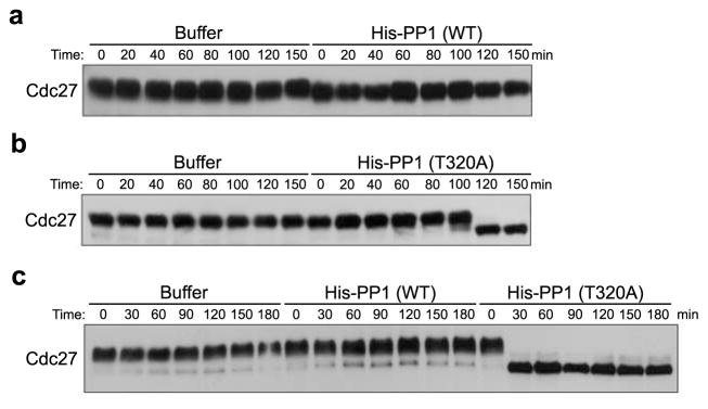
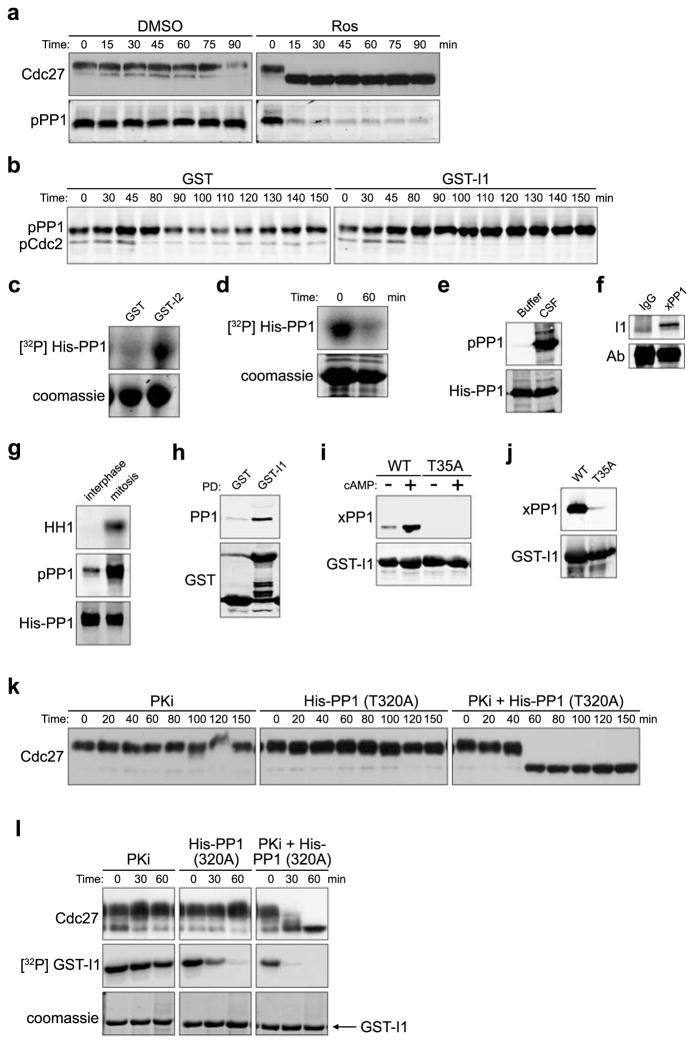
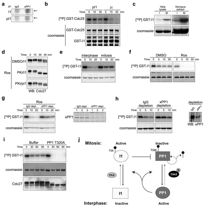
Loss of cell division cycle 2 (Cdc2, also known as Cdk1) activity after cyclin B degradation is necessary, but not sufficient, for mitotic exit. Proteins phosphorylated by Cdc2 and downstream mitotic kinases must be dephosphorylated. We report here that protein phosphatase-1 (PP1) is the main catalyst of mitotic phosphoprotein dephosphorylation. Suppression of PP1 during early mitosis is maintained through dual inhibition by Cdc2 phosphorylation and the binding of inhibitor-1. Protein kinase A (PKA) phosphorylates inhibitor-1, mediating binding to PP1. As Cdc2 levels drop after cyclin B degradation, auto-dephosphorylation of PP1 at its Cdc2 phosphorylation site (Thr 320) allows partial PP1 activation. This promotes PP1-regulated dephosphorylation at the activating site of inhibitor-1 (Thr 35) followed by dissociation of the inhibitor-1-PP1 complex and then full PP1 activation to promote mitotic exit. Thus, Cdc2 both phosphorylates multiple mitotic substrates and inhibits their PP1-mediated dephosphorylation.


Figures

Figure 1
Figure 1. Dephosphorylation of mitotic phosphoproteins by okadaic acid-sensitive phosphatase activity
A. Mitotic extracts were supplemented with DMSO or Roscovitine (Ros, 0.28mM). Aliquots were taken at the indicated times and immunoblotted with anti-Cdc27 or MPM2 antibodies. B. Ros (0.28mM) was added to mitotic extracts that had been pre-treated with DMSO or OA (10μM). Aliquots were withdrawn at the indicated times to immunoblot for Cdc27 and to measure Cdc2 kinase activity using Histone H1 (HH1) as an exogenous substrate. Full scan of Cdc27 blot and HH1 kinase assay is shown in S. Fig. 5A. C and D. Ros (0.28mM) was added to mitotic extracts (A) or CSF extracts (B) in the presence of either DMSO or different concentrations of OA. Samples were taken at the indicated times and blotted with anti-Cdc27 antibody.
Figure 2
Figure 2. PP1 is required for the dephosphorylation of substrates at mitotic, but not meiotic exit
A. Ros (0.28mM) was added to CSF extracts in the presence of GST or GST-I2. Aliquots were withdrawn at the indicated times and immunoblotted with anti-Cdc27, anti-pPlx, or MPM2 antibody. Full scans of Cdc27 and pPlx blots are shown in S. Fig. 5B. B. Ros (0.28mM) was added to CSF extracts in the presence of GST or GST-I1. Aliquots were withdrawn at the indicted times for Cdc27 immunoblotting and HH1 kinase assays. C. Left: PP1 was immunodepleted from mitotic extracts using anti-Xenopus PP1 antibodies. Rabbit IgG was used for mock depletion. Control or PP1-depleted extracts were treated with Ros (0.28mM). Aliquots were immunoblotted with anti-Cdc27 antibody. Right: Control and PP1-depleted extracts were immunoblotted with PP1 antibodies. D. Left: Same as Figure 2C except that CSF extract was used for PP1 depletion. PP1 depleted extract was supplemented with buffer or recombinant His-PP1 (0.5 μM). In addition to Cdc27 immunoblotting, HH1 kinase activity was assayed. Right: Extracts were also immunoblotted with PP1 antibodies. Full scan of PP1 blot is shown in S. Fig. 5C. E. GST or thio-phosphorylated GST-I1 was added to cycling extracts and aliquots were withdrawn at the indicated times and immunoblotted with anti-Cdc27 and anti-Cyclin B2 antibodies. Arrows indicate gel mobilities of Cdc27. Full scan of Cdc27 and Cyclin B2 blots are shown in S. Fig. 5D. F. HeLa cells were arrested in Mitosis with Nocodazole for 17 hours, and then allowed to resume cell cycle in fresh DMEM for 1 hour. Cell lysates were made and supplemented with GST or GST-I1. Aliquots were taken to immunoblot with MPM2 antibody and to measure HH1 kinase activity. G. Ca2+ was added to CSF extracts in the presence of GST or GST-I1. Aliquots were immunoblotted with anti-Cdc27 antibody. H. Left: PP1 antibodies were used for PP1 immunodepletion and rabbit IgG was used for control depletion. Calcium was added to the depleted CSF extracts. Aliquots were taken at the indicated times and blotted with anti-Cdc27 antibody. Right: Aliquots of depleted extracts were blotted with anti-PP1 antibody.
Figure 3
Figure 3. Cdc2 regulation of PP1 prevents premature substrate dephosphorylation in mitosis
A. Mitotic extracts were supplemented with rabbit His-PP1 catalytic subunit (4 μM) or buffer control. Aliquots were withdrawn at the indicated time points for Cdc27 immunoblotting. B. Mitotic extracts were supplemented with rabbit His-PP1T320A (4 μM) or buffer control. Aliquots were taken at the indicated time points for Cdc27 immunoblotting. C. CSF extracts were supplemented with rabbit His-PP1 wild type (WT), rabbit His-PP1T320A (4 μM) or buffer control. Samples were withdrawn at the indicated time points for Cdc27 immunoblotting.
Figure 4
Figure 4. PP1 autodephosphorylation and I1 dephosphorylation control PP1-regulated Cdc2 substrate dephosphorylation
A. CSF extracts treated with DMSO or Ros (0.28 mM) were immunoblotted for Cdc27 and PP1pT320. B. GST or thio-phosphorylated GST-I1 was added to cycling extracts and samples were immunoblotted with Cdc2pY15 (mitotic entry coincides with Y15 dephosphorylation) and PP1pT320 antibodies. Full scan in S. Fig. 5F. C. Ni-NTA agarose-bound His-PP1 was phosphorylated by Cdc2/Cyclin B in the presence of GST or GST-I2 and γ32P ATP for 30 min. His-PP1 phosphorylation was detected by SDS-PAGE/phosphorimager. Full scan in S. Fig. 5G. D. Ni-NTA agarose-bound His-PP1 was pre-phosphorylated with high concentrations of Cdc2/Cyclin B and γ32P ATP for 30min. Ros (0.28mM) was then added and reactions were either stopped with sample buffer immediately or were incubated in buffer for an additional 60 min before resolution by SDS-PAGE/phosphorimager. E. His-PP1 incubated with buffer or CSF extracts for 30 min, was retrieved and immunoblotted with anti-PP1pT320 or anti-His antibody. Full scan in S. Fig. 5H. F. Protein A Sepharose-bound Xenopus PP1 antibody or control IgG were incubated with mitotic extracts for 1hr. Immunoprecipitates were blotted with anti-I1 antibody. Full scan in S. Fig. 5I. G. His-PP1 protein was dipped into interphase or mitotic extracts for 10 min, washed and immunoblotted with anti-PP1pT320 or anti-His antibody. HH1 phosphorylation is shown. H. GST or GST-I1 incubated with nocodazole-arrested HeLa lysates were retrieved and immunoblotted for GST or PP1. I. Glutathione Sepharose-bound GST-I1 (WT) or GST-I1T35A incubated in interphase extracts +/− cAMP for 1 hour were washed and immunoblotted with anti-Xenopus PP1 or anti-GST antibodies. Full scan in S. Fig. 5J. J. Glutathione Sepharose-bound GST-I1 (WT) or GST-I1T35A were incubated in mitotic extracts for 1 hour, washed, and immunoblotted with anti-Xenopus PP1 or anti-GST antibodies. K. CSF extracts incubated with PKA specific inhibitor (PKi) H89 (0.2 mM), rabbit His-PP1T320A (2 μM) or PKI together with His-PP1T320 were immunoblotted with anti-Cdc27 antibody. L. Glutathione Sepharose-bound GST-I1 was prephosphorylated by PKA and γ32P ATP, washed and added to CSF extracts. Extracts were treated as in 4K except GST-I1 was also retrieved and visualized by SDS-PAGE/phosphorimager.
Figure 5
Figure 5. I1 Cell cycle regulation
A. Protein A Sepharose-bound Xenopus PP1 antibody or control IgG were incubated in mitotic extracts for 1hr. Immunoprecipitates resolved by SDS-PAGE were immunoblotted for I1 or p-I1 (pT35). B. Glutathione Sepharose-bound GST-Cdc25 pre-phosphorylated with Chk1 and γ32P ATP were dipped into phosphatase buffer in the presence of His-PP1 and either GST-I1 or T35 thio-phosphorylated GST-I1. Aliquots were resolved by SDS-PAGE/phosphorimager. C. PKA was immunoprecipitated from interphase (I) or mitotic (M) HeLa cell lysate (Left) or Xenopus egg extracts (Right). Immunoprecipitates were incubated with GST-I1 and γ32P ATP for 20 min and resolved by SDS-PAGE/phosphorimager. D. CSF extracts pretreated with DMSO or PKi (0.2 mM) for 15 min were supplemented with GST-I1 that had been either pre-thiophosphorylated or not. Samples were then immunoblotted for Cdc27. E. Glutathione Sepharose-bound GST-I1 was prephosphorylated by PKA and γ32P ATP and dipped in interphase or mitotic extracts, retrieved at the indicated times and resolved by SDS-PAGE/phosphorimager. F. Glutathione Sepharose-bound GST-I1 was prephosphorylated as in 5E and then dipped in CSF extracts or Ros-pretreated CSF extracts and processed as in 5E. G.(Left): Glutathione Sepharose-bound GST-I1 was prephosphorylated as in 5E and dipped into control or PP1 depleted CSF extracts in the presence of Ros. Aliquots were processed as in 5E. (Right): Aliquots of control or PP1-depleted extracts were immunoblotted for PP1. H. (Left) Anti-PP1 or IgG-depleted CSF extracts were treated with Ca2+ for 20 min. Glutathione Sepharose-bound GST-I1 prephosphorylated as in 5E was dipped into the Ca2+ treated extracts. Aliquots were resolved by SDS-PAGE/phosphorimager. (Right) IgG- or PP1-depleted extracts were immunoblotted for PP1. I. Glutathione Sepharose –bound GST-I1 prephosphorylated as in 5E was dipped into CSF extracts supplemented with Buffer or His-PP1T320A (4 μM), and processed as in 5E. Samples were also immunoblotted for Cdc27. J. PP1 is inhibited both by Cdc2/Cyclin B and by phosphorylated I1 in mitosis. During mitotic exit, Cdc2 inactivation allows PP1 auto-dephosphorylation on T320 to prevail, and at the same time, active PP1 dephosphorylates I1 at T35, resulting in full activation of PP1.

References

    1. Lew DJ, Kornbluth S. Regulatory roles of cyclin dependent kinase phosphorylation in cell cycle control. Curr Opin Cell Biol. 1996;8:795–804. - PubMed
    1. Azzam R, et al. Phosphorylation by cyclin B-Cdk underlies release of mitotic exit activator Cdc14 from the nucleolus. Science. 2004;305:516–519. - PubMed
    1. Amon A. A decade of Cdc14--a personal perspective. Delivered on 9 July 2007 at the 32nd FEBS Congress in Vienna, Austria. FEBS J. 2008;275:5774–5784. - PMC - PubMed
    1. Queralt E, Uhlmann F. Cdk-counteracting phosphatases unlock mitotic exit. Curr Opin Cell Biol. 2008;20:661–668. - PMC - PubMed
    1. Trautmann S, McCollum D. Cell cycle: new functions for Cdc14 family phosphatases. Curr Biol. 2002;12:R733–735. - PubMed

Publication types

MeSH terms

Substances