

Comment
doi: 10.1038/ng.371.
Epub 2009 Apr 26.
Cooperativity of TMPRSS2-ERG with PI3-kinase pathway activation in prostate oncogenesis
Affiliations
- PMID: 19396167
- PMCID: PMC2898503
- DOI: 10.1038/ng.371
Item in Clipboard
Comment
Cooperativity of TMPRSS2-ERG with PI3-kinase pathway activation in prostate oncogenesis
Nat Genet.
2009 May.
Abstract
The TMPRSS2-ERG fusion, present in approximately 50% of prostate cancers, is less common in prostatic intraepithelial neoplasia (PIN), raising questions about whether TMPRSS2-ERG contributes to disease initiation. We identified the translational start site of a common TMPRSS2-ERG fusion and showed that transgenic TMPRSS2-ERG mice develop PIN, but only in the context of PI3-kinase pathway activation. TMPRSS2-ERG-positive human tumors are also enriched for PTEN loss, suggesting cooperation in prostate tumorigenesis.
Figures
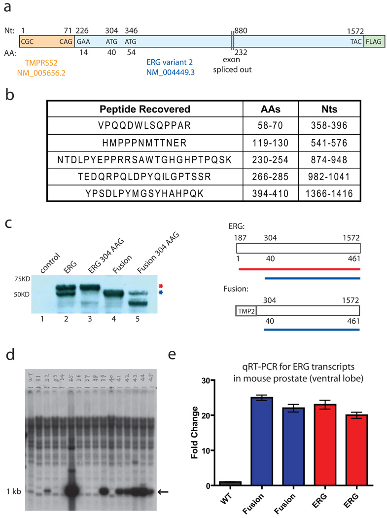
TMPRSS2-ERG fusion transcript produces a truncated ERG protein product. a, Diagram of TMPRSS2-ERG variant 2 construct. b, Tryptic peptides recovered from mass spectrometry performed on immunoprecipitated, gel-resolved protein from the fusion construct. c, ERG immunoblot of transiently transfected vector control, full length ERG, full length ERG with codon 304 mutation, fusion, or fusion with the codon 304 mutation. A diagram showing the protein products derived from the non-mutated constructs is shown on the right. d, Southern blot showing founder animals derived from blastocyst injections of ARR2PB-TMPRSS2-ERG construct. DNA was cut with BamHI and probed with a BamHI fragment that encodes the first 1050bp of the fusion. e, qRT-PCR using primers for the C-terminus of ERG on ventral prostates from a wildtype mouse, two TMPRSS2-ERG mice, and two full-length ERG transgenic mice. All Ct values were normalized to the Ct values of Nkx3.1 and fold change was calculated relative to the wildtype animal.
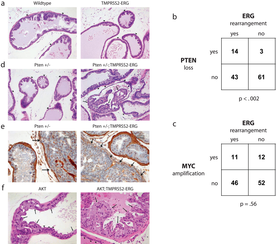
TMPRSS2-ERG fusion and PI3K pathway activation are cooperative events in prostate cancer. All pictures show the ventral prostate lobe. a, No PIN is noted in either wild type or TMPRSS2-ERG animals at 12 months of age (H&E, 200X). b, The proportion of human prostate cancers that show PTEN genomic loss, ERG rearrangement, both, or neither are shown (n = 121). PTEN loss was defined as having reduced copy number by RAE (heterozygous or homozygous loss; FDR q-value<6.93×10⁻⁸). ERG rearrangement was defined as the consequence of interstitial deletion by aCGH (q-value<6.93×10⁻⁸) and increased expression of ERG downstream of common translocation breakpoints (exons 5–8) relative to early ERG exons (exons 1–2) [>2 s.d. above the mean of normal samples (n=29) by exon expression array] The significance of the association between PTEN loss and ERG rearrangement is p-value<0.002 as determined by Fisher's exact test (right tail). c, The proportion of human prostate cancers that show MYC genomic amplification, ERG rearrangement, both, or neither are shown (n = 121). MYC amplification was defined as having increased copy number (q-value<1.2×10⁻⁷). ERG rearrangement and the significance of association (p-value=0.56) was defined as in Fig 2b. d, Normal prostate in Pten +/− and high grade PIN in Pten +/−; TMPRSS2-ERG mice at 6 months of age (H&E, 200X). Arrows indicate PIN and arrowheads show normal epithelium for comparison. e, Immunohistochemistry for smooth muscle actin in a Pten +/− and a Pten +/−; TMPRSS2-ERG mouse (400X). Compare disrupted and absent smooth muscle stroma around areas of possible microinvasion from a Pten +/−; TMPRSS2-ERG mouse (right column, arrows) to minimally attenuated smooth muscle sheath around area of high grade PIN in Pten +/− (left column, arrows) and to intact stroma of normal glands in both animals. f, Low grade PIN in AKT transgenic mice and high grade PIN in AKT; TMPRSS2-ERG double transgenic animals at 11 months of age (H&E, 400X). Arrows indicate PIN and arrowheads show normal epithelium for comparison.
Comment in
-
TMPRSS2-ERG and PTEN loss in prostate cancer.Nat Genet. 2009 May;41(5):509-10. doi: 10.1038/ng0509-509. Nat Genet. 2009. PMID: 19399032
Comment on
-
Aberrant ERG expression cooperates with loss of PTEN to promote cancer progression in the prostate.Nat Genet. 2009 May;41(5):619-24. doi: 10.1038/ng.370. Epub 2009 Apr 26. Nat Genet. 2009. PMID: 19396168 Free PMC article.
References
Publication types
MeSH terms
Substances
Grants and funding
LinkOut - more resources
Full Text Sources
Other Literature Sources
Medical
Molecular Biology Databases
Research Materials
