. 2009 May;16(5):468-76.
doi: 10.1038/nsmb.1603. Epub 2009 Apr 26.

A conserved structural motif mediates formation of the periplasmic rings in the type III secretion system

Affiliations

A conserved structural motif mediates formation of the periplasmic rings in the type III secretion system

Thomas Spreter et al. Nat Struct Mol Biol. 2009 May.

Abstract

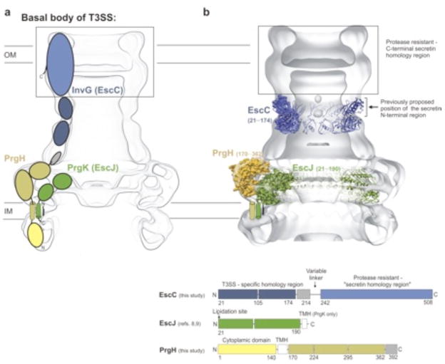
The type III secretion system (T3SS) is a macromolecular 'injectisome' that allows bacterial pathogens to transport virulence proteins into the eukaryotic host cell. This macromolecular complex is composed of connected ring-like structures that span both bacterial membranes. The crystal structures of the periplasmic domain of the outer membrane secretin EscC and the inner membrane protein PrgH reveal the conservation of a modular fold among the three proteins that form the outer membrane and inner membrane rings of the T3SS. This leads to the hypothesis that this conserved fold provides a common ring-building motif that allows for the assembly of the variably sized outer membrane and inner membrane rings characteristic of the T3SS. Using an integrated structural and experimental approach, we generated ring models for the periplasmic domain of EscC and placed them in the context of the assembled T3SS, providing evidence for direct interaction between the outer membrane and inner membrane ring components and an unprecedented span of the outer membrane secretin.


Figures

Figure 1
Figure 1
Basal body of the T3SS. (a) Representation of the basal body of the T3SS and the components that assemble the complex (the basal body contains only the integral T3SS membrane proteins and lacks the internal stalk and extending needle of the assembled T3SS needle complex (NC)) –. InvG/EscC and PrgK/EscJ are the respective orthologs from S. typhimurium and EPEC –. A third prominent component of the basal body has been most well characterized in S. typhimurium (PrgH) and is thought to interact with the inner membrane ring PrgK –. The coloring corresponds to the schematic in the bottom panel, illustrating the domain organization of EscC (InvG), EscJ (PrgK) and PrgH. (b) Crystal structures of T3SS basal body components EscC (21–174), PrgH (170–362) and EscJ (21–190) and their proposed position in the S. typhimurium T3SS EM map, as described here (EM accession codes emd_1224 and emd_1214 5, 21). The box in black indicates the approximate dimensions of the C-terminal protease resistant - “secretin homology region” as determined by EM reconstructions of the secretin PulD and the black arrow below highlights the previously proposed position for the secretin N-terminal region and the secretin N-termus , . The docked models are equivalent to the models presented in Figures 2 and 4.
Figure 2
Figure 2
EscC, PrgH and EscJ share a common fold for the assembly of the T3SS multi-ring basal body. (a) Ribbon representation of the EscC (21–174) crystal structure, highlighting the two domain fold and the connecting hinge region. (b) Ribbon representation of the EscJ crystal structure (pdb accession code 1yj7 8). The top shows monomeric EscJ and the ring structure on the bottom illustrates the assembled 24mer oligomeric structure of EscJ, which has been previously described , . (c) Ribbon representation of the crystal structure of monomeric PrgH (170–362). (d) Superposition of both the outer membrane secretin EscC (21–174) (left panel) and the inner membrane protein PrgH (170–362) (right panel) to EscJ (pdb accession code 1yj7). Highlighted in orange and green are the conserved folds shared among the three proteins. The coloring scheme corresponds to the structures of EscC, EscJ and PrgH in panels (a) to (c). The schematic illustrates the integrated approach, based on the crystal structures and the observed conservation, of molecular modeling, experimental validation and docking into the EM density maps of the S. typhimurium basal body (EM accession codes emd_1224 and emd_1100) , . PrgH (170–362) was manually docked into the EM maps. The previously published EscJ structure and the EscC (21–174) models are automatically docked as described in the Methods.
Figure 3
Figure 3
EscC (21–174) and the T3SS specific N-terminal secretin region within the T3SS needle complex (NC). (a) Negative Stain EM of isolated InvG complexes from S. typhimurium. A raw image and representative class averages showing the top and side view of the complex (scale bar represents 50 nm). (b) Comparison of the side view of isolated InvG to the docked EscC ring-models and the previously published EM reconstruction of the T3SS NC . The docked EscC (21–174) ring-models represent the ensemble of final models, which fulfill all structural and biochemical constraints and show the highest correlation to the periplasmic region of the EM density maps (EM accession codes emd_1224 and emd_1100). The docked models in orange and blue (both 12mers) are localized directly adjacent to the C-terminal protease resistant core with a notable match to the bilobal structure observed in the EM map and which also encompasses the periplasmic region previously suggested to contain the N-terminal domain as described above. Also shown is a slightly larger ring-model in green with 14fold symmetry that localizes more towards the IM rings, but for which less biochemical support currently exists. The orange and blue models are favored by existing literature and have thus been used as the representative docked EscC model shown in Figs. 1, 2 and 4.
Figure 4
Figure 4
EscC (21–174) and the T3SS specific N-terminal secretin region within the T3SS needle complex (NC). (a) Representative EscC model in the EM density maps of the S. typhimurium basal body and the fully assembled T3SS NC containing in addition the stalk and needle, which are built up by the proteins InvJ, PrgJ, PrgI (EM accession codes emd_1224; emd_1100 , ; the docked EscC model is equivalent to the model shown in orange in Figures 3b). The blue bar highlights the important region that undergoes considerable structural rearrangement during the NC assembly process , . (b) Effects of EscC mutations on type III secretion in EPEC. Shown are the Tir/EspB secretion profiles in secretion assays of escC-deficient EPEC strains complemented with the EscC mutants. The proteins were detected with respective monoclonal antibodies. (c) Effects of InvG mutation on type III secretion in Salmonella. Secreted proteins were separated by SDS-PAGE and stained with Coomassie Blue from wildtype and chromosomal InvG (E106P) S. typhimurium strains. Western-blots of secreted proteins were performed with respective monoclonal antibodies. Needle Complexes (NC) were isolated, separated on SDS-PAGE and stained with Coomassie (molecular weight reference shown in kDa). Levels of InvG in NC were detected with a polyclonal antibody.
Figure 5
Figure 5
Illustration of conformational sampling during protein-protein docking simulations. (a) Conformational sampling of the hinge region (residue 102–108) of EscC. The position of the α-carbon of residue 162 in a random selection of docking models after a hinge perturbation protocol is shown in red spheres. The yellow spheres indicate a subset of docking models with more than 300 contacts across an interface and green the 10% lowest energy subset of the yellow spheres. The wheat colored domain 2 correspond to the orientation of the model best fitting the experimental data and the magenta sphere is the α-carbon of residue 162 of this model. When a smaller sized hinge-perturbation is used spheres clusters around the magenta sphere, which roughly coincides with the position of the residue 162 in the crystal structure. In order to pick out the best models out of the green ensemble (and correspondingly in other simulations), geometric information and biotinylation data was used as a filter, see Figure 6. (b) Symmetrical docking of the N-terminal EscC domain (21–101) into 12mer ring models. The plot shows the Rosetta full atom energy vs backbone rmsd for three consecutive subunits relative the model selected as best fitting with the experimental data.
Figure 6
Figure 6
Illustration of the evaluation process of the ring-models. The predicted models (Fig. 5) are further discriminated by experimentally derived data of accessible and buried lysine residues, identified by limited biotinylation (Supplementary Fig. 3). The models in panels a – f show a representative subset of models to illustrate the process in detail. Solvent accessible residues are rendered in green and buried in yellow spheres. In addition we utilized site-directed mutagenesis data on two negatively charged patches of EscC that show defects in type III secretion upon mutation (residues in orange spheres) (Supplementary Fig. 4). The illustrated process identifies a subset of models that all satisfy both the modeling/structural constraints and the experimental data (the models a, b, c, d and e are representative for the identified subset; the model f can be excluded because it does not meet the experimental data). This ensemble of validated models was further evaluated by docking to the EM density maps of the T3SS basal body and NC from S. typhimurium , . This process led to a set of final models that fulfil all structural/modelling, experimental and docking constrains. In the representative subset above only models a and b show considerable complementarity to the periplasmic regions of the EM density maps and satisfy all imposed criteria (Supplementary Fig. 8). These two models and their docking positions are equivalent to the models described in Figure 3b.

Comment in

Similar articles

Cited by

References

    1. Ghosh P. Process of protein transport by the type III secretion system. Microbiol Mol Biol Rev. 2004;68:771–795. - PMC - PubMed
    1. Kimbrough TG, Miller SI. Contribution of Salmonella typhimurium type III secretion components to needle complex formation. Proc Natl Acad Sci U S A. 2000;97:11008–11013. - PMC - PubMed
    1. Sukhan A, Kubori T, Wilson J, Galan JE. Genetic analysis of assembly of the Salmonella enterica serovar Typhimurium type III secretion-associated needle complex. J Bacteriol. 2001;183:1159–1167. - PMC - PubMed
    1. Kimbrough TG, Miller SI. Assembly of the type III secretion needle complex of Salmonella typhimurium. Microbes Infect. 2002;4:75–82. - PubMed
    1. Marlovits TC, et al. Structural insights into the assembly of the type III secretion needle complex. Science. 2004;306:1040–1042. - PMC - PubMed

Publication types

MeSH terms