. 2009 May;16(5):499-508.
doi: 10.1038/nsmb.1593. Epub 2009 Apr 26.

Structure of the RAG1 nonamer binding domain with DNA reveals a dimer that mediates DNA synapsis

Affiliations

Structure of the RAG1 nonamer binding domain with DNA reveals a dimer that mediates DNA synapsis

Fang Fang Yin et al. Nat Struct Mol Biol. 2009 May.

Abstract

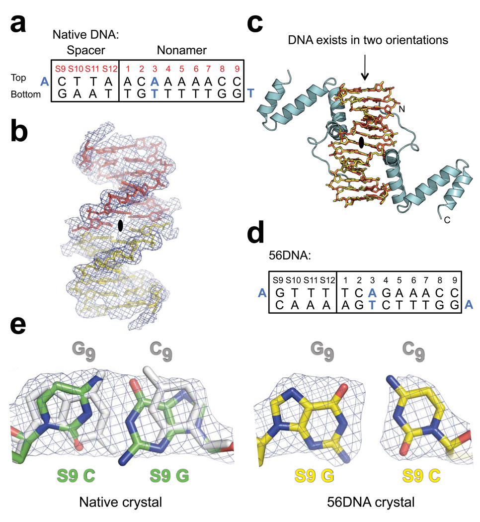
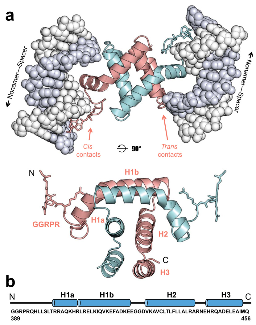
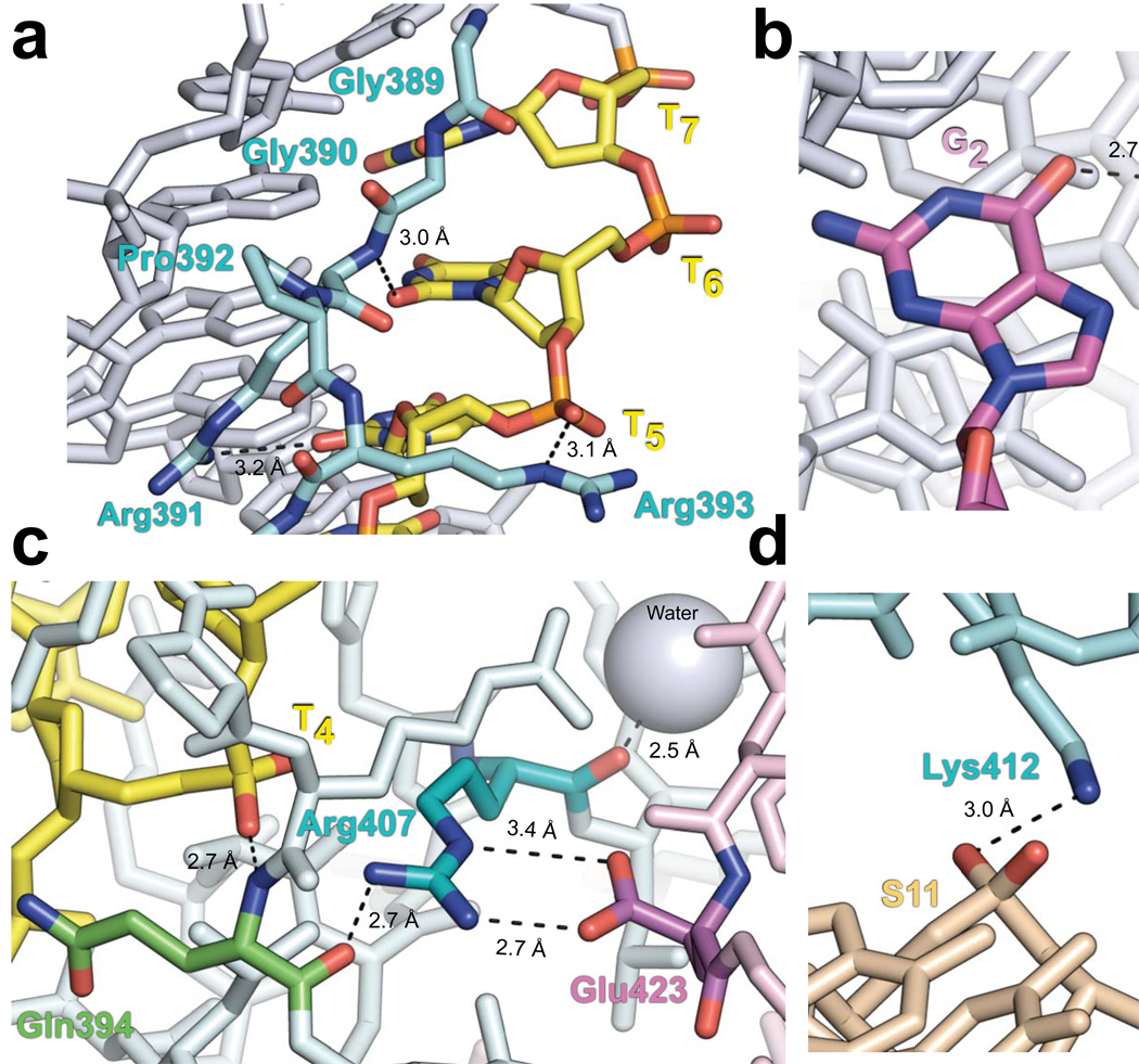
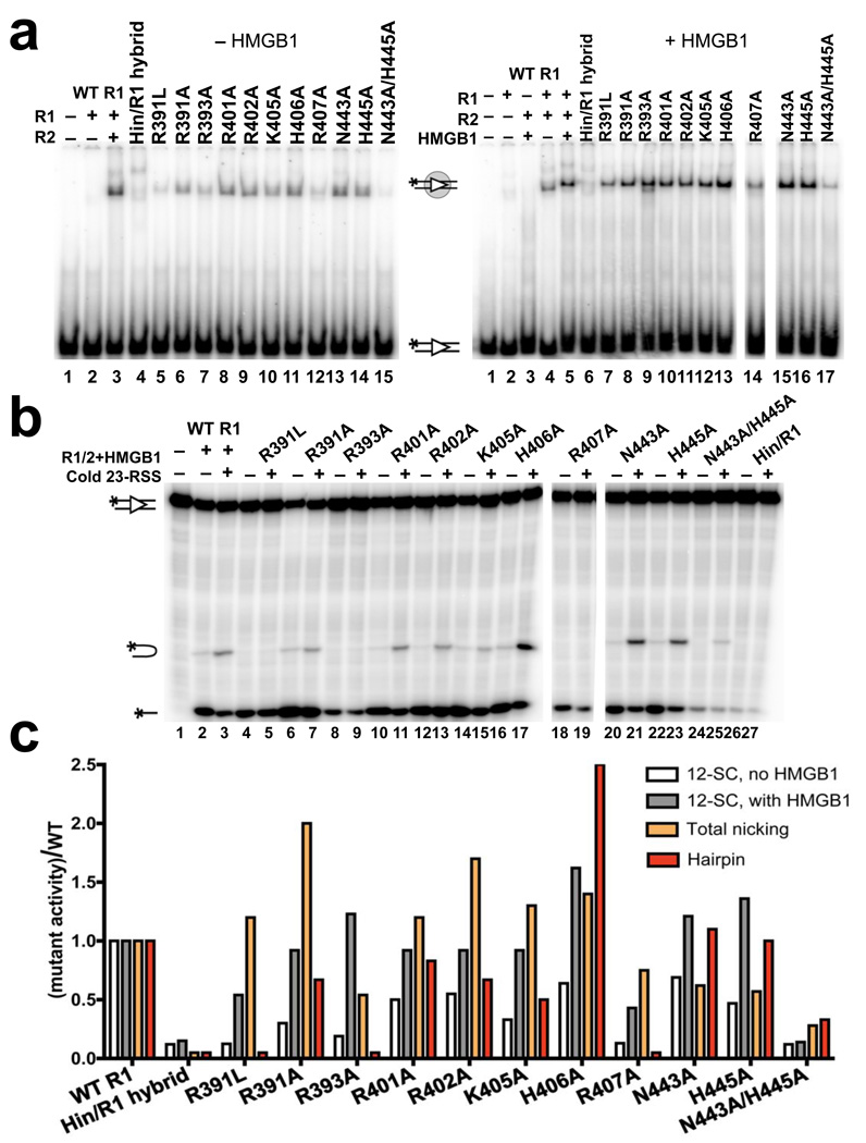
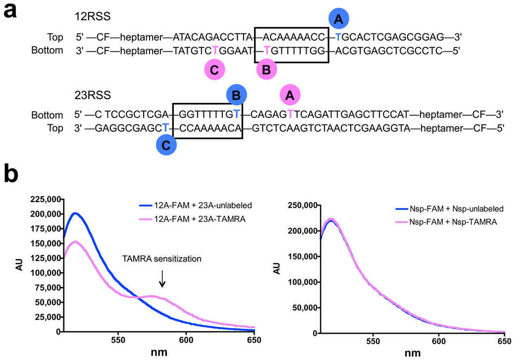
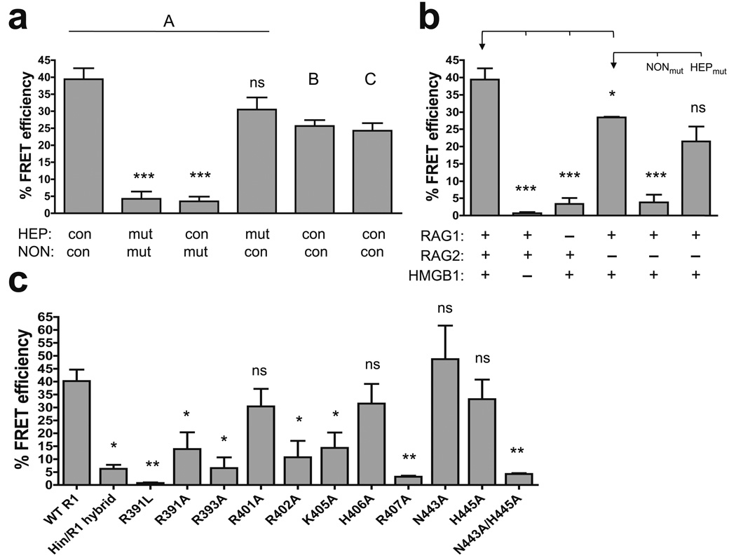
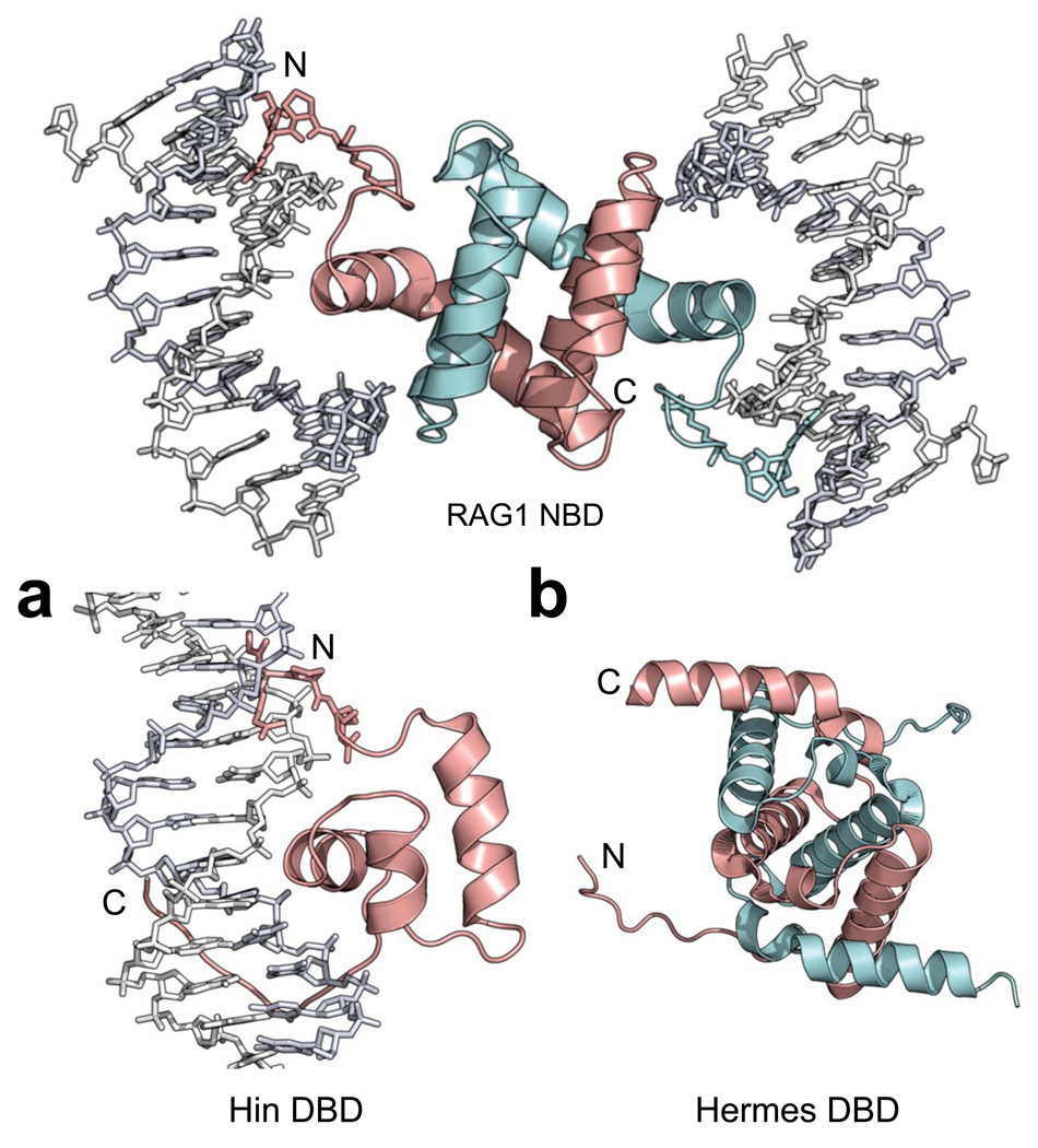
The products of recombination-activating genes RAG1 and RAG2 mediate the assembly of antigen receptor genes during lymphocyte development in a process known as V(D)J recombination. Lack of structural information for the RAG proteins has hindered mechanistic studies of this reaction. We report here the crystal structure of an essential DNA binding domain of the RAG1 catalytic core bound to its nonamer DNA recognition motif. The RAG1 nonamer binding domain (NBD) forms a tightly interwoven dimer that binds and synapses two nonamer elements, with each NBD making contact with both DNA molecules. Biochemical and biophysical experiments confirm that the two nonamers are in close proximity in the RAG1/2-DNA synaptic complex and demonstrate the functional importance of the protein-DNA contacts revealed in the structure. These findings reveal a previously unsuspected function for the NBD in DNA synapsis and have implications for the regulation of DNA binding and cleavage by RAG1 and RAG2.


Figures

Figure 1
Figure 1
Schematic of RAG-mediated DNA binding and cleavage. RAG1/RAG2/HMGB1 (shaded oval) bind to either a 12RSS or 23RSS (gray or white triangles) to form the SC and then capture a second, complementary RSS to form the PC, within which DNA cleavage is completed. DNA cleavage occurs in two steps, nicking and hairpin formation (inset). Nicking can occur in the SC or PC, while hairpin formation is restricted to the PC. The DNA sequence elements that make up the RSS DNA substrate are shown at bottom.
Figure 2
Figure 2
Electron density maps of DNA in the native and 56DNA crystals. (a) Sequence of the DNA used in native crystals is shown with the consensus nonamer (numbered 1–9) and four base pairs from the consensus 12RSS spacer (numbered S9-S12). Bases coinciding with the crystallographic 2-fold symmetry axis are blue. (b) Solvent-flattened experimental electron density map (mesh) derived from heavy-atom phasing of the native crystal shows continuous electron density extending from the base pair at nonamer position 3 across the 2-fold symmetry axis (black oval; the axis is perpendicular to the page). Two halves of a single 14-mer DNA duplex minus the overhang bases are shown as yellow and red stick models. (c) Application of the crystallographic 2-fold symmetry operator generates a model consistent with the DNA existing in two possible orientations (overlap of yellow and red stick models), each at half occupancy, and the NBD (cyan) bound to the two A•T-rich sites in the DNA. The termini of one NBD are indicated with N and C. Crystals containing a DNA with additional symmetrized bases, 56DNA (d), in complex with the NBD were also obtained. (e) 2Fo-Fc omit electron density maps (mesh) of bases at S9, which were generated by performing simulated annealing refinement without the DNA coordinates, are shown for both the native (green) and 56DNA (yellow) crystals. The corresponding symmetry-mate bases at nonamer position 9 (subscript) are colored gray in both models, but are not visible in the case of 56DNA due to perfect overlap with the S9 base pair. Nitrogen and oxygen atoms are indicated with blue and red, respectively.
Figure 3
Figure 3
Structure of the RAG1 NBD-DNA complex. (a) The NBD homodimer (subunits colored cyan and pink with the GGRPR motif shown as stick model) binds to two DNA molecules (gray space-filling model) and makes both cis and trans DNA contacts as indicated for the pink subunit. The lower panel shows the NBD dimer alone, rotated 90° from the view in the top panel. (b) The sequence of the NBD in the model (murine RAG1 residues 389–456) is shown below the corresponding secondary structures, with loops and helices represented as lines and blue cylinders, respectively.
Figure 4
Figure 4
Important protein-DNA and protein-protein contacts in the NBD-DNA crystal structure. Nitrogen, oxygen, and phosphate atoms are indicated with blue, red, and orange, respectively. Dashed lines represent bonds. Subscript denotes the nonamer position. (a) Structure of the GGRPR motif (cyan) in the minor groove of the nonamer is shown with thymines colored yellow, while all other DNA bases are gray. The Arg391 side chain amino group and main chain amide are hydrogen-bonded to the O2 atoms of T5 and T6, respectively. The Arg393 ε-imino group is hydrogen-bonded to the phosphate of T5. (b) Base-specific interaction between the Arg402 side chain (cyan) and the O6 atom of G2 (magenta). (c) The Arg407 guanidino group (cyan) is hydrogen-bonded to the carbonyl of Gln394 (green) in the same subunit (light cyan) and forms a salt bridge with Glu423 (magenta) of the other NBD subunit (light pink). The main chain amide of Gln394 is hydrogen-bonded to the phosphate of T4 (yellow). Additionally, the carbonyl of Arg407 is hydrogen-bonded to a nearby water molecule (gray sphere). (d) In the spacer, the side chains of Lys405 and Lys412 (cyan) form hydrogen bonds with the DNA backbone at positions S10 and S11 (beige), respectively. See also Supplementary Fig. 2.
Figure 5
Figure 5
NBD-DNA contacts are important for DNA binding and hairpin formation. (a) Gel shift analysis of RAG1/2 binding to a 32P-labeled consensus 12RSS substrate in the presence (right gel) or absence (left gel) of HMGB1 in a binding buffer containing 5 mM CaCl2. Proteins were added as indicated above the lanes. All reactions with mutant RAG1 proteins contained RAG2, and the positions of the free and bound substrates are indicated with diagrams. (b) DNA cleavage assay was performed with 32P-labeled consensus 12RSS and a 5-fold molar excess of unlabeled consensus 23RSS in the presence of 1.5 mM MgCl2. Proteins and 23RSS substrate were added as indicated above the lanes. All reactions with mutant RAG1 proteins contained RAG2 and HMGB1. The positions of the nicked product, hairpin product, and input substrate are indicated with diagrams. (c) Quantitation of binding and cleavage results from panels (a) and (b), with the activity of the mutants normalized to that of wild-type RAG1, whose activity was arbitrarily set to one. Similar results were obtained in repeat experiments (data not shown). See also Supplementary Fig. 3.
Figure 6
Figure 6
Detection of nonamer synapsis using FRET. (a) Diagram showing the positions of fluorophore labeling in the A, B, and C pairs of 12/23 RSS FRET substrates. TAMRA (pink circle); FAM (blue circle); CF, coding flank. (b) Representative corrected emission spectra obtained with the A substrate pair (left panel) or nonspecific (nsp) substrates, which are identical to the A substrates except that the nonamer and heptamer elements have been mutated in both the 12 and 23 RSSs (right panel). In each case, the FAM-labeled substrate was incubated with a 4-fold molar excess of either a TAMRA-labeled (pink spectrum) or unlabeled (blue, control spectrum) partner substrate together with RAG1/RAG2/HMGB1 in a buffer containing 5 mM MgCl2. Background intrinsic TAMRA fluorescence has been subtracted from both pink spectra. Donor quenching and TAMRA sensitization are observed only with the consensus A substrates.
Figure 7
Figure 7
FRET analysis of the DNA and protein requirements for nonamer synapsis. Average FRET efficiencies of at least two experiments are plotted with error bars indicating the s.e.m. *, P < 0.05; **, P < 0.01; ***, P < 0.001; ns, not significant (unpaired two-tailed t-test). (a) Energy transfer was assessed with the A, B, and C substrate pairs (as indicated above the bars), and with A substrates in which the heptamer (HEP) and/or nonamer (NON) were mutated in both the 12 and 23 RSSs, as indicated below the bars. mut, mutant; con, consensus. Statistical comparisons were made with the data obtained with the consensus A substrate pair. (b) Energy transfer was assessed using various combinations of RAG1, RAG2, and HMGB1, as indicated below the bars, with the A substrate pair, or the A substrate pair with mutant nonamer (NONmut), or mutant heptamer (HEPmut) sequences. Statistical comparisons (brackets) were made with the data indicated with an arrow. (c) Energy transfer was assessed with the A substrate pair using WT and mutant RAG1 core proteins, as indicated below the bars. Statistical comparisons were made with the data obtained with WT RAG1 core.
Figure 8
Figure 8
Structural comparisons of the RAG1 NBD with the DNA-binding domains (DBDs) of Hin and Hermes. The NBD dimer (subunits colored cyan and pink) with the GGRPR motif (stick model) is shown. (a) The Hin DBD (pink) is a monomer that uses a HTH motif to interact with DNA in the major groove, while a GGRPR motif (stick model) and a C-terminal tail interact with the minor grooves. Hin PDB: 1HCR (b) The Hermes DBD (residues 76–159) also has an intertwined dimer structure (subunits colored cyan and pink). Hermes PDB: 2BW3.

References

    1. Schatz DG, Oettinger MA, Baltimore D. The V(D)J recombination activating gene, RAG-1. Cell. 1989;59:1035–1048. - PubMed
    1. Oettinger MA, Schatz DG, Gorka C, Baltimore D. RAG-1 and RAG-2, adjacent genes that synergistically activate V(D)J recombination. Science. 1990;248:1517–1523. - PubMed
    1. Sawchuk DJ, et al. V(D)J recombination: modulation of RAG1 and RAG2 cleavage activity on 12/23 substrates by whole cell extract and DNA-bending proteins. J. Exp. Med. 1997;185:2025–2032. - PMC - PubMed
    1. van Gent DC, Hiom K, Paull TT, Gellert M. Stimulation of V(D)J cleavage by high mobility group proteins. EMBO J. 1997;16:2665–2670. - PMC - PubMed
    1. Tonegawa S. Somatic generation of antibody diversity. Nature. 1983;302:575–581. - PubMed

Publication types

LinkOut - more resources