. 2009 Jun;5(6):397-9.
doi: 10.1038/nchembio.167. Epub 2009 Apr 26.

FTIR analysis of GPCR activation using azido probes

Affiliations

FTIR analysis of GPCR activation using azido probes

Shixin Ye et al. Nat Chem Biol. 2009 Jun.

Abstract

We demonstrate the site-directed incorporation of an IR-active amino acid, p-azido-L-phenylalanine (azidoF, 1), into the G protein-coupled receptor rhodopsin using amber codon suppression technology. The antisymmetric stretch vibration of the azido group absorbs at approximately 2,100 cm(-1) in a clear spectral window and is sensitive to its electrostatic environment. We used FTIR difference spectroscopy to monitor the azido probe and show that the electrostatic environments of specific interhelical networks change during receptor activation.


Figures
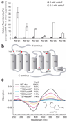
Figure 1
Figure 1
Non-natural amino acid mutagenesis. (a) HEK 293T cells were transfected with plasmids carrying the genes for firefly luciferase with an amber mutation (FLuc.Y70am), a suppressor tRNA (Bst-Yam), and one of six mutant synthetases (azidoF RS-V1 to RS-V6) at a time. Cells were cultured in the absence and in the presence of azidoF. The average FLuc activities were determined from the chemiluminescence intensities (from three independent experiments). The amber stop codon suppression efficiencies of the mutant synthetases were described as relative FLuc intensities compared to wild-type E. coli Tyr-RS in suppression activity (%). Error bars show s.d. (b) Positions in rhodopsin subjected to site-specific incorporation of azidoF are indicated as red dots in a schematic secondary structure plot. (c) Functional expression and expression yield were determined in light-induced UV-visible difference spectra (dark minus photoproduct) of purified wild-type rhodopsin and azidoF rhodopsin mutants. The structure of azidoF is indicated.
Figure 2
Figure 2
FTIR spectroscopy on azidoF rhodopsin mutants. (a) The light-induced FTIR difference spectrum (Meta II photoproduct minus dark state) of Val250azidoF mutant (blue) reveals the characteristic band pattern below 1,800 cm−1 that reflects conformational changes of the protein during light-dependent receptor activation, similar to that in wild-type rhodopsin (gray). At 2,100 cm−1, the antisymmetric stretch (νas) of the azido label is observed (the absorption peak of Asp83 in the dark state is marked for comparison). (b) The broad featureless bands underlying the difference spectrum of the wild type were subtracted from the Val250azidoF difference spectrum (top row). The resulting double difference spectrum Val250azidoF minus wild type (second row) reveals in the first approximation a downshift of the azido νas during activation of Val250azidoF. In the analogous spectrum of the Val227azidoF mutant, on the other hand, a clear upshift is observed, while no substantial changes are observed in the spectrum of the Tyr102azidoF mutant.
Figure 3
Figure 3
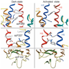
Structural models of rhodopsin activation. Structural models of rhodopsin azidoF mutants derived from the dark state and in an activated conformation of the ligand-free opsin state were obtained by modeling and molecular dynamic equilibration (cytoplasmic side points upwards). Receptor activation leads to rearrangement of a charged cluster at the H3/H6 interface and formation of a new charged cluster at the H5/H6 interface in the vicinity of positions 250 and 227. The environment of position 102, on the other hand, is not hypothesized to be affected by receptor activation. The results of the simulations are consistent with the shifts of the azido stretching bands in the Meta II difference spectra of the three azidoF rhodopsin mutants shown in Figure 2b.

References

    1. Pierce KL, Premont RT, Lefkowitz RJ. Nat. Rev. Mol. Cell Biol. 2002;3:639–650. - PubMed
    1. Huber T, Menon S, Sakmar TP. Biochemistry. 2008;47:11013–11023. - PubMed
    1. Kobilka B, Schertler GFX. Trends Pharmacol. Sci. 2008;29:79–83. - PubMed
    1. Hanson MA, Stevens RC. Structure. 2009;17:8–14. - PMC - PubMed
    1. Mahalingam M, Martinez-Mayorga K, Brown MF, Vogel R. Proc. Natl. Acad. Sci. USA. 2008;105:17795–17800. - PMC - PubMed

Publication types

MeSH terms