. 2009 Aug;13(8B):2622-2633.
doi: 10.1111/j.1582-4934.2008.00643.x. Epub 2008 Dec 29.

Laminin isoforms in human embryonic stem cells: synthesis, receptor usage and growth support

Affiliations

Laminin isoforms in human embryonic stem cells: synthesis, receptor usage and growth support

Sanna Vuoristo et al. J Cell Mol Med. 2009 Aug.

Abstract

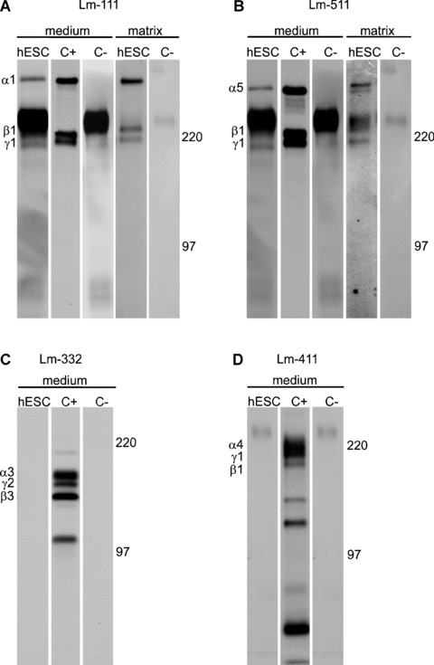
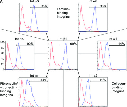
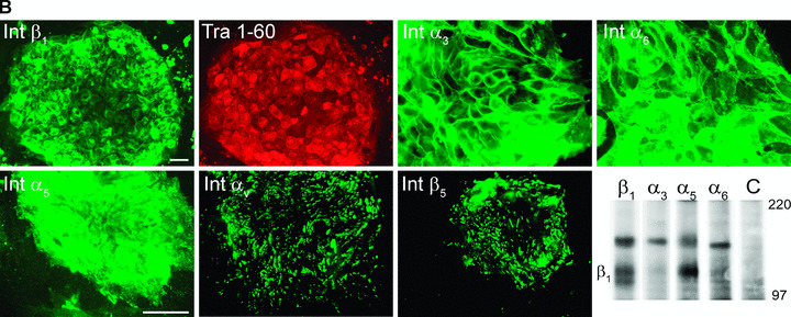
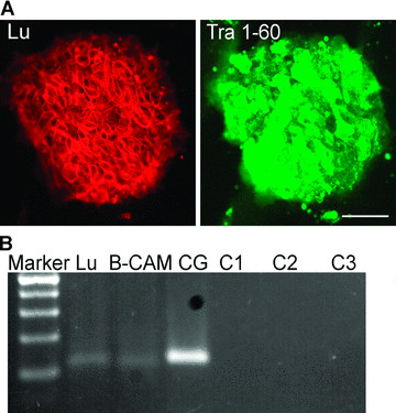
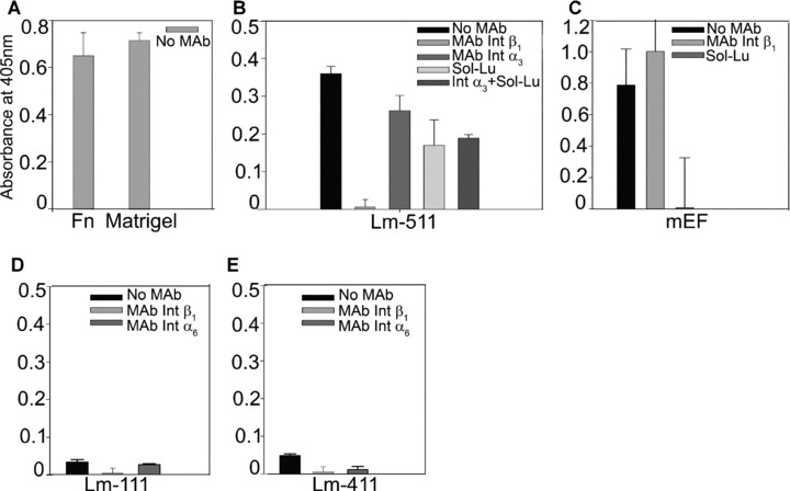
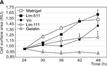
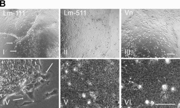
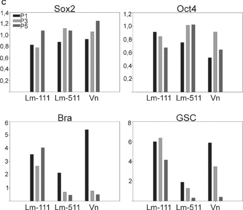
To reveal the functional intrinsic niche of human embryonic stem cells (hESC) we examined the production of basement membrane (BM) proteins and the presence of their receptors in feeder-free cell culture conditions. In addition, we investigated binding of hESCs to purified human BM proteins and identified the receptors mediating these contacts. Also, we tested whether purified human laminin (Lm) isoforms have a role in hESC self-renewal and growth in short-term cultures. The results show that hESCs synthesize Lm alpha(1) and Lm alpha(5) chains together with Lm beta(1) and gamma(1) chains suggesting the production of Lms-111 and -511 into the culture medium and deposits on cells. hESCs contain functionally important integrin (Int) subunits, Int beta(1), alpha(3), alpha(6), alpha(5), beta(5) and alpha(V), as well as the Lm alpha(5) receptor, Lutheran (Lu) glycoprotein and its truncated form, basal cell adhesion molecule (B-CAM). In cell adhesion experiments, Int beta(1) was crucial for adhesion to most of the purified human BM proteins. Lu/B-CAM mediated adhesion to Lm-511 together with Int alpha(3)beta(1), and was essential for the adhesion of hESCs to embryonic feeder cells. Adhesion to Lm-411 was mediated by Int alpha(6)beta(1). Lm-511 supported hESC growth in defined medium equally well as Matrigel. These results provide consequential information of the biological role of BM in hESCs, warranting further investigation of BM biology of human pluripotent stem cells.


Figures

Figure 1
Figure 1
Production of laminin (Lm) chains by hESCs. hESCs were metabolically labelled with radioactive methionine followed by the immunoprecipitation of Lm chains. (a) and (b) hESCs synthesized and secreted chains of Lm‐111 (α1β1γ1) and Lm‐511 (α5β1γ1) into culture medium and matrix. (c) and (d) hESCs did not produce Lm‐332 or Lm‐411. C+; positive control, C–; negative control without antibody.
Figure 2
Figure 2
hESCs contain laminin (Lm‐), fibronectin‐ and vitronectin‐binding integrins. (a) Flow cytometry analysis showed that hESCs contained Int β1, α3, α6, α5 and αV subunits. Int β1 subunit can bind different Lms by pairing with Int α3 and α6 subunits, fibronectin with Int α5 subunit, and vitronectin with Int αV subunit. Functions of fibronectin and vitronectin receptors may overlap. hESCs did not contain collagen binding Integrins α1 and α2. (b) hESCs were double‐stained by monoclonal antibodies against integrin subunits and Tra1–60 antigen. Tra1–60‐positive hESCs showed immunoreactivity for Int β1, α3, α6 and α5 subunits in a cell surface‐confined manner (Tra1–60 shown only for Int β1). Int αV and β5 subunits were seen as a pattern resembling point adhesions. Scale bar, 50 μm. Immunoprecipitation confirmed production of Int β1, α3, α5 and α6 subunits.
Figure 2
Figure 2
hESCs contain laminin (Lm‐), fibronectin‐ and vitronectin‐binding integrins. (a) Flow cytometry analysis showed that hESCs contained Int β1, α3, α6, α5 and αV subunits. Int β1 subunit can bind different Lms by pairing with Int α3 and α6 subunits, fibronectin with Int α5 subunit, and vitronectin with Int αV subunit. Functions of fibronectin and vitronectin receptors may overlap. hESCs did not contain collagen binding Integrins α1 and α2. (b) hESCs were double‐stained by monoclonal antibodies against integrin subunits and Tra1–60 antigen. Tra1–60‐positive hESCs showed immunoreactivity for Int β1, α3, α6 and α5 subunits in a cell surface‐confined manner (Tra1–60 shown only for Int β1). Int αV and β5 subunits were seen as a pattern resembling point adhesions. Scale bar, 50 μm. Immunoprecipitation confirmed production of Int β1, α3, α5 and α6 subunits.
Figure 3
Figure 3
Undifferentiated hESCs contain a known Lm α5 receptor, Lutheran/B‐CAM. (a) Antibody against Lutheran/B‐CAM intensively stained Tra1–60‐positive hESCs. Scale bar, 50 μm. (b) Both mRNA variants of the same gene, 115 bp for Lutheran and 109 bp for B‐CAM, were expressed by hESCs. Negative controls; C1 for Lutheran, C2 for B‐CAM, C3 for Cyclophilin G.
Figure 4
Figure 4
hESC adhesion to BM proteins was measured by quantitative adhesion experiments. Monoclonal antibodies (MAb) directed against various receptors, or recombinant Lutheran (Sol‐Lu) were used to determine functional receptors in hESCs. (a) hESCs adhered potently to positive control substrata, Matrigel and fibronectin. (b) Prominent hESC adhesion to purified human Lm‐511 was mediated via Int β1, α3 and Sol‐Lu (P < 0.001). (c) Sol‐Lu, but not MAb against Int β1, totally blocked adhesion to mEFs. (d) and (e) Adhesion to Lm‐111 or to Lm‐411 was poor. MAb against Int β1 prevented (P < 0.005) adhesion to Lm‐111. MAbs against Int β1 and α6 blocked adhesion to Lm‐411 (P < 0.001; P < 0.005).
Figure 5
Figure 5
Individual laminin isoforms have distinct effects on the hESC self‐renewal. (a) Proliferation was measured in a continuous live‐cell imaging system. The area of hESC colonies in relative units is shown in function of time (hours). Results are presented as the mean ± S.E.M. for three culture wells. The growth rate was highest on Matrigel and on purified human Lm‐511. In contrast, proliferation on Lm‐111 was low. (b) hESCs cultured on purified human Lm‐111 (I and IV), Lm‐511 (II and V) or Vn (III and VI) in defined medium, StemPro, at passage 5. hESCs tended to differentiate on human Lm‐111 (I, IV; arrows) whereas typical characteristics of undifferentiated cells were present on Lm‐511 and on Vn. Scale bars, 100 μm. (c) qPCR analysis of hESCs cultured on BM proteins in StemPro, at passage 1, 3 and 5. The relative expression levels of the pluripotency marker genes, Sox2 and Oct4 sustained, while the expression of the differentiation markers GSC and Bra decreased during culture on Lm‐511 or Vn. In contrast, differentiation occurred when the hESCs were cultured on Lm‐111, as shown by the presence of GSC and Bra and a decreasing level of Oct4. Data are presented as the mean expression level relative to cells grown on Matrigel, determined in duplicate.
Figure 5
Figure 5
Individual laminin isoforms have distinct effects on the hESC self‐renewal. (a) Proliferation was measured in a continuous live‐cell imaging system. The area of hESC colonies in relative units is shown in function of time (hours). Results are presented as the mean ± S.E.M. for three culture wells. The growth rate was highest on Matrigel and on purified human Lm‐511. In contrast, proliferation on Lm‐111 was low. (b) hESCs cultured on purified human Lm‐111 (I and IV), Lm‐511 (II and V) or Vn (III and VI) in defined medium, StemPro, at passage 5. hESCs tended to differentiate on human Lm‐111 (I, IV; arrows) whereas typical characteristics of undifferentiated cells were present on Lm‐511 and on Vn. Scale bars, 100 μm. (c) qPCR analysis of hESCs cultured on BM proteins in StemPro, at passage 1, 3 and 5. The relative expression levels of the pluripotency marker genes, Sox2 and Oct4 sustained, while the expression of the differentiation markers GSC and Bra decreased during culture on Lm‐511 or Vn. In contrast, differentiation occurred when the hESCs were cultured on Lm‐111, as shown by the presence of GSC and Bra and a decreasing level of Oct4. Data are presented as the mean expression level relative to cells grown on Matrigel, determined in duplicate.

Similar articles

Cited by

References

    1. Kalluri R. Basement membranes: structure, assembly and role in tumour angiogenesis. Nat Rev Cancer . 2003; 3: 422–33. - PubMed
    1. Scheele S, Nystrom A, Durbeej M, et al . Laminin isoforms in development and disease. J Mol Med . 2007; 85: 825–36. - PubMed
    1. Miner JH, Yurchenco PD. Laminin functions in tissue morphogenesis. Ann Rev Cell Dev Biol . 2004; 20: 255–84. - PubMed
    1. Ekblom P. Receptors for laminins during epithelial morphogenesis. Curr Opin Cell Biol . 1996; 8: 700–6. - PubMed
    1. Gustafsson E, Fassler R. Insights into extracellular matrix functions from mutant mouse models. Exp Cell Res . 2000; 261: 52–68. - PubMed

Publication types