. 2009 Apr 27:7:17.
doi: 10.1186/1741-7007-7-17.

The calcium-sensing receptor regulates parathyroid hormone gene expression in transfected HEK293 cells

Affiliations

The calcium-sensing receptor regulates parathyroid hormone gene expression in transfected HEK293 cells

Hillel Galitzer et al. BMC Biol. .

Abstract

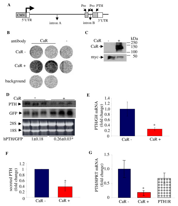
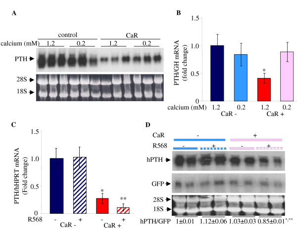
Background: The parathyroid calcium receptor determines parathyroid hormone secretion and the response of parathyroid hormone gene expression to serum Ca2+ in the parathyroid gland. Serum Ca2+ regulates parathyroid hormone gene expression in vivo post-transcriptionally affecting parathyroid hormone mRNA stability through the interaction of trans-acting proteins to a defined cis element in the parathyroid hormone mRNA 3'-untranslated region. These parathyroid hormone mRNA binding proteins include AUF1 which stabilizes and KSRP which destabilizes the parathyroid hormone mRNA. There is no parathyroid cell line; therefore, we developed a parathyroid engineered cell using expression vectors for the full-length human parathyroid hormone gene and the human calcium receptor.

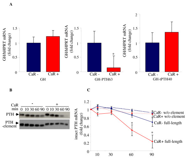
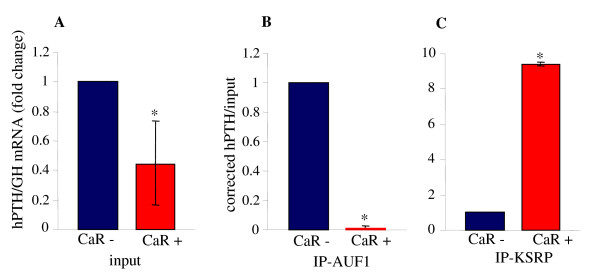
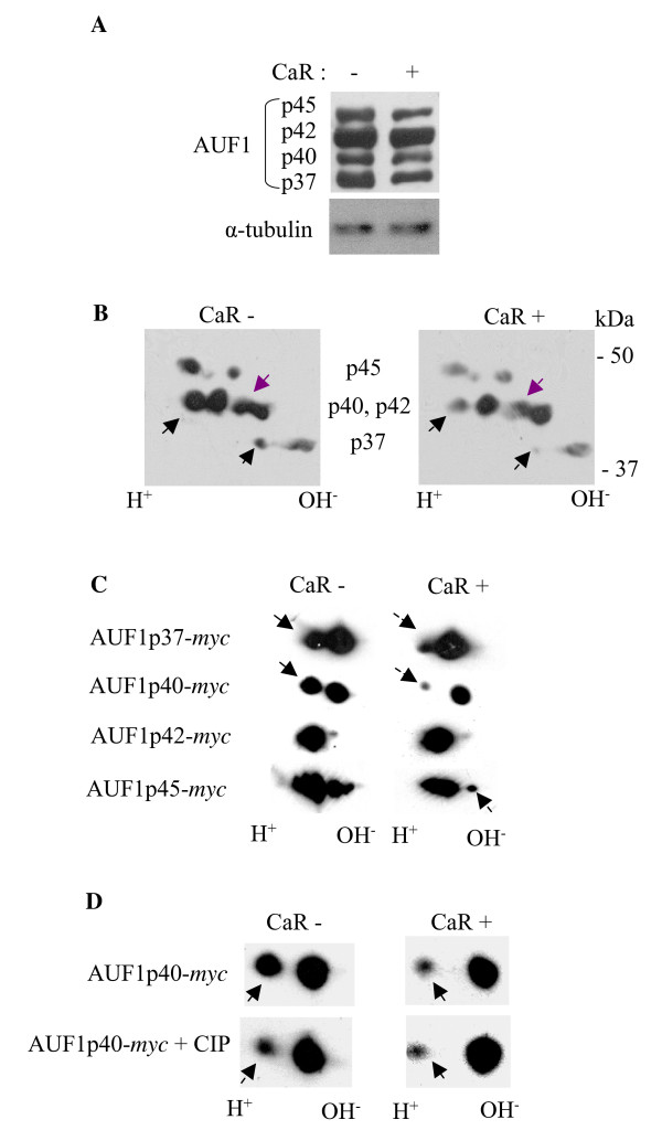
Results: Co-transfection of the human calcium receptor and the human parathyroid hormone plasmid into HEK293 cells decreased parathyroid hormone mRNA levels and secreted parathyroid hormone compared with cells that do not express the calcium receptor. The decreased parathyroid hormone mRNA correlated with decreased parathyroid hormone mRNA stability in vitro, which was dependent upon the 3'-UTR cis element. Moreover, parathyroid hormone gene expression was regulated by Ca2+ and the calcimimetic R568, in cells co-transfected with the calcium receptor but not in cells without the calcium receptor. RNA immunoprecipitation analysis in calcium receptor-transfected cells showed increased KSRP-parathyroid hormone mRNA binding and decreased binding to AUF1. The calcium receptor led to post-translational modifications in AUF1 as occurs in the parathyroid in vivo after activation of the calcium receptor.

Conclusion: The expression of the calcium receptor is sufficient to confer the regulation of parathyroid hormone gene expression to these heterologous cells. The calcium receptor decreases parathyroid hormone gene expression in these engineered cells through the parathyroid hormone mRNA 3'-UTR cis element and the balanced interactions of the trans-acting factors KSRP and AUF1 with parathyroid hormone mRNA, as in vivo in the parathyroid. This is the first demonstration that the calcium receptor can regulate parathyroid hormone gene expression in heterologous cells.


Figures

Figure 1
Figure 1
Over-expression of the CaR decreases PTH mRNA levels in co-transfected HEK293 cells. HEK293 cells were transiently co-transfected in triplicate with expression plasmids for the hPTH gene and control GFP or GH and either the CaR (CaR+) or empty vector (CaR-). A. Schematic representation of the human PTH expression plasmid used for transient transfections. The boxes show the CMV promoter (grey) and the PTH exons with the untranslated regions (UTRs) (white) and coding regions (diagonal lines). The arrows show the pre, pro and mature PTH. B. Immunohistochemistry on whole cells using an intact cell enzyme-linked immunoassay for the cell surface expression of the CaR. Untransfected (background), CaR (+) or control plasmid (-) transfected cells were analyzed using a CaR antibody or IgG (-). C. Immunoblot analysis of extracts from HEK293 cells co-transfected with expression plasmids for the CaR and myc-AUF1 as control plasmid, using anti-CaR or myc antibodies. D-G. Effect of CaR on PTH expression. D. Northern blot for hPTH and co-transfected GFP with the CaR (+) or an empty vector (-). Ethidium bromide staining of the membrane is shown as a loading control. E. qPCR for PTH and co-transfected GH mRNA levels from cells with (red) and without (blue) the CaR. F. Secreted PTH from cells as above 1 h after an incubation in fresh medium, 1 mM Ca2+. G. qRT-PCR for PTH mRNA levels from cells without and with either the CaR or the PTH1R (checkered). Data in D-G are expressed as fold change (mean ± SE) (n = 3). *, P < 0.01, CaR: control.
Figure 2
Figure 2
The CaR decreases PTH mRNA levels and stability through the PTH mRNA 3'-UTR cis element. A. Effect of CaR over-expression on reporter GH mRNA containing the PTH mRNA ARE. qRT-PCR for GH and control HPRT mRNA levels in cells transfected with either control (CaR-) or CaR (CaR+) and expression plasmids for GH (left panel), GH containing the PTH mRNA 63 nt ARE (GH-PTH63) (middle panel) or GH containing a 40 nt truncated ARE (GH-tPTH40) (right panel). Results are fold changes compared with cells without the CaR expressed as mean ± SE of three experiments. *, P < 0.05. B. Effect of CaR on PTH mRNA decay. Representative IVDA of transcripts for the full-length hPTH mRNA and the PTH mRNA with an internal deletion of the 3'-UTR ARE, incubated with extracts from cells expressing either the CaR or empty vector. C. Quantification of the amount of intact transcripts remaining with time related to time 0 (mean ± SE, in three repeat experiments; *, P < 0.05). Blue square, full length PTH mRNA without CaR (CaR-) and red square, with CaR (CaR+); blue triangle, PTH w/o ARE without CaR (CaR-) and red triangle, with the CaR, (CaR+).
Figure 3
Figure 3
Expression of the CaR in HEK293 cells decreases AUF1 and increases KSRP interaction with the PTH mRNA. RNA immunoprecipitation (RIP) analysis of extracts from cells transiently transfected with expression plasmids for PTH and GH as control and either the CaR plasmid or empty vector. Immunoprecipitation was performed using antibodies for AUF1, KSRP or control IgG. Input (A) and immunoprecipitiated (B, C) samples were analyzed by qPCR for PTH and GH mRNA. Results are presented as PTH mRNA corrected for GH mRNA. PTH mRNA in the immunoprecipitated samples was corrected for PTH mRNA in the input. The results are mean ± SE of three repeat experiments. *, P < 0.05 compared with cells transfected with empty vector (CaR-).
Figure 4
Figure 4
AUF1 is post-translationally modified in HEK293 cells transfected with CaR. A. 1D gels for AUF1 and α-tubulin as a loading control. B. 2D gel analysis for endogenous AUF1 in extracts from cells transiently transfected with the CaR (CaR+) plasmid or empty vector (CaR-). Molecular weight markers are shown on the right and the four AUF1 isoforms are indicated. C. 2D gel analysis of extracts from cells transiently transfected with the CaR plasmid or empty vector and the myc-AUF1 isoforms p37, p40, p42 or p45 separately. D. 2D analysis of extracts from cells transiently transfected with the CaR plasmid or empty vector and myc-AUF1p40 without and after treatment with a non-specific phosphatase (CIP). The results all represent one of two repeat experiments with similar results.
Figure 5
Figure 5
Expression of the CaR decreases PTH mRNA levels and confers responsivity to a low [Ca2+]o and the calcimimetic R568. A. Representative Northern blot analysis for PTH mRNA in cells transiently transfected in triplicate with hPTH and either the CaR plasmid or empty vector. After transfection cells were grown in 1.2 or 0.2 mM Ca2+ medium for an additional 48 h. Ethidium bromide staining of the membrane is shown as loading control. B. qRT PCR for PTH corrected for co-transfected GH mRNAs for cells treated as in A. C. qPCR for PTH and HPRT mRNAs from cells expressing the hPTH gene and the CaR (CaR+) or empty vector (CaR-) in cells grown in 1.2 mM Ca2+ supplemented with either R568 or vehicle. The results in A-C represent one of three repeat experiments performed in triplicate with similar results. D. Representative Northern blot for PTH and GFP mRNA levels in cells transfected with PTH and GFP and either CaR (CaR+) or empty vector (CaR-). The cells were grown in 0.2 mM calcium supplemented with R568 or vehicle. Quantification of the results is shown below the gel. The results are presented as mean ± SE of two repeat experiments performed in triplicate. *, P < 0.05, CaR+:CaR-; **, P < 0.05, CaR+, treated:CaR+, untreated.

References

    1. Brown EM, Gamba G, Riccardi D, Lombardi M, Butters R, Kifor O, Sun A, Hediger MA, Lytton J, Hebert J. Cloning and characterization of an extracellular Ca2+-sensing receptor from bovine parathyroid. Nature. 1993;366:575–580. doi: 10.1038/366575a0. - DOI - PubMed
    1. Silver J, Naveh-Many T, Kronenberg HM. Parathyroid hormone: molecular biology. In: Bilezikian JB, Raisz LG, Rodan GA, editor. Principles of bone biology. San Diego: Academic Press; 2002. pp. 407–422.
    1. Rodriguez M, Nemeth E, Martin D. The calcium-sensing receptor: a key factor in the pathogenesis of secondary hyperparathyroidism. Am J Physiol Renal Physiol. 2005;288:F253–F264. doi: 10.1152/ajprenal.00302.2004. - DOI - PubMed
    1. Hofer AM, Brown EM. Extracellular calcium sensing and signalling. Nat Rev Mol Cell Biol. 2003;4:530–538. doi: 10.1038/nrm1154. - DOI - PubMed
    1. Nemeth EF, Steffey ME, Hammerland LG, Hung BC, Van Wagenen BC, DelMar EG, Balandrin MF. Calcimimetics with potent and selective activity on the parathyroid calcium receptor. Proc Natl Acad Sci USA. 1998;95:4040–4045. doi: 10.1073/pnas.95.7.4040. - DOI - PMC - PubMed

Publication types

MeSH terms

LinkOut - more resources