. 2009 Apr 27;11(1):11.
doi: 10.1186/1532-429X-11-11.

Cell tracking and therapy evaluation of bone marrow monocytes and stromal cells using SPECT and CMR in a canine model of myocardial infarction

Affiliations

Cell tracking and therapy evaluation of bone marrow monocytes and stromal cells using SPECT and CMR in a canine model of myocardial infarction

Gerald Wisenberg et al. J Cardiovasc Magn Reson. .

Abstract

Background: The clinical application of stem cell therapy for myocardial infarction will require the development of methods to monitor treatment and pre-clinical assessment in a large animal model, to determine its effectiveness and the optimum cell population, route of delivery, timing, and flow milieu.

Objectives: To establish a model for a) in vivo tracking to monitor cell engraftment after autologous transplantation and b) concurrent measurement of infarct evolution and remodeling.

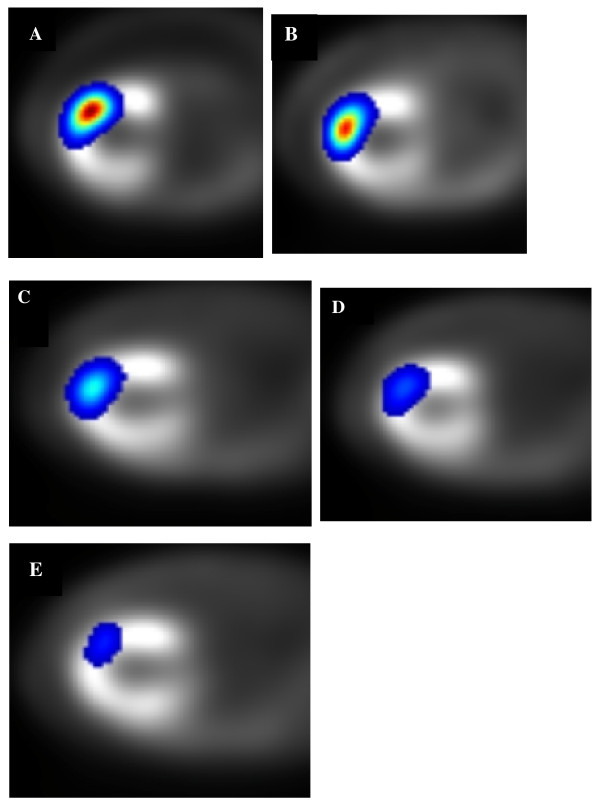
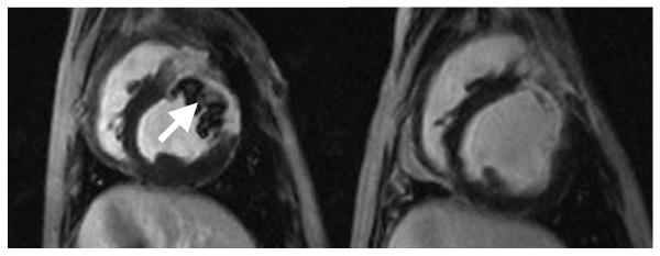
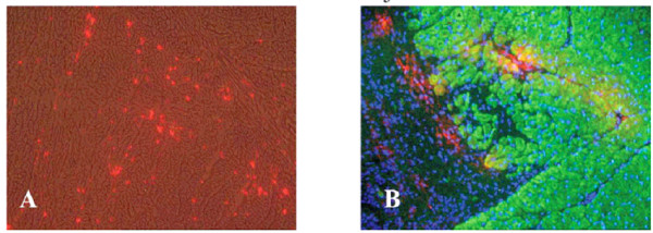
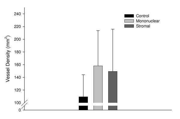
Methods: We evaluated 22 dogs (8 sham controls, 7 treated with autologous bone marrow monocytes, and 7 with stromal cells) using both imaging of 111Indium-tropolone labeled cells and late gadolinium enhancement CMR for up to12 weeks after a 3 hour coronary occlusion. Hearts were also examined using immunohistochemistry for capillary density and presence of PKH26 labeled cells.

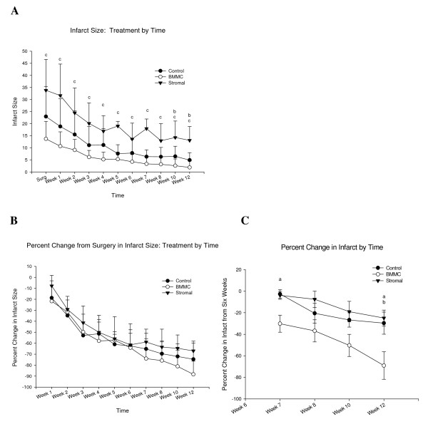
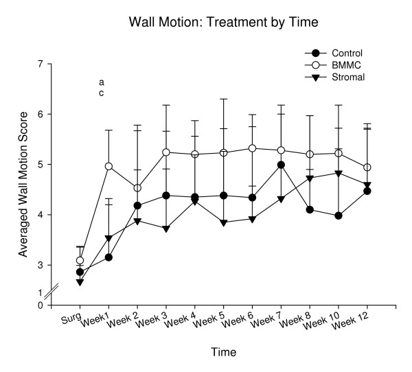
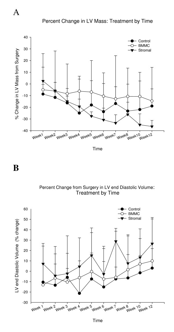
Results: In vivo Indium imaging demonstrated an effective biological clearance half-life from the injection site of ~5 days. CMR demonstrated a pattern of progressive infarct shrinkage over 12 weeks, ranging from 67-88% of baseline values with monocytes producing a significant treatment effect. Relative infarct shrinkage was similar through to 6 weeks in all groups, following which the treatment effect was manifest. There was a trend towards an increase in capillary density with cell treatment.

Conclusion: This multi-modality approach will allow determination of the success and persistence of engraftment, and a correlation of this with infarct size shrinkage, regional function, and left ventricular remodeling. There were overall no major treatment effects with this particular model of transplantation immediately post-infarct.


Figures

Figure 1
Figure 1
Serial Transaxial SPECT images following Indium labeling and intravenous Tc-99m MIBI. Panels A-E respectively are fused images of Tc-MIBI and 111In-labeled stromal cells in a dog at day 0, 4, 7, 10 and 14.
Figure 2
Figure 2
Absolute and relative changes in infarct size over time. There was a progressive decline in both absolute (A) and relative (B) CMR measured infarct size, in comparison to baseline, for controls, and both treatment groups. Values are means ± SE. One way analyses of variance showed that significant group differences were observed in relative infarct size changes. Posthoc Tukey Tests showed significant paired group differences at 12 weeks when the 6 week time point was used as the reference point for further change. a-Control vs. BMMC p = 0.046, b-Stromal vs. BMMC p = 0.032,
Figure 3
Figure 3
Changes in regional wall motion scores. Although there was a progressive improvement in regional function in the infarct and peri-infarct areas by almost 2 wall motion scores, there was no difference between treatments at 12 weeks. Separate one way analyses of variance showed that significant group differences were observed at week one only, F(2,16) = 8.08, p < .01. Posthoc Tukey Tests showed significant paired group differences between a = Controls and BMMC, and c = Stromal and BMMC.
Figure 4
Figure 4
Changes in left ventricular volume and enddiastolic volumes. A The stromal cell animals had a significant decline in left ventricular volume (total mass) in comparison to both controls, and BMMC from 8 weeks through 12 weeks: b- stromal different from controls c- stromal different from BMMC. There were small increases in endiastolic volume over time but there were no differences between treatments. The first data point on the graph is at the first week following surgery relative to the volume on the day of surgery.
Figure 5
Figure 5
Late gadolinium enhanced end diastolic images from one of the stromal cell animals on the day of infarction, left, and at 12 weeks, right. The initial image demonstrates a large infarct with a central area of no enhancement. Such a pattern is often seen with extensive microvascular injury leading to "no reflow" and failure of delivery of tracer to the central zone of infarction. The 12 week image demonstrates a considerable reduction in the extent of infarction, and thinning of the infarct area, with loss of this no reflow effect, and cavitary dilatation.
Figure 6
Figure 6
Identification and characterization of PKH26+ (fluorescent) labeled donor cells in damaged myocardium 3 weeks following direct injection of Bone Marrow Mononuclear cells (BMMCs) into myocardium. A) Overlay of PKH26 fluorescence (red) on 160× bright field image of unfixed cryostat section reveals bright red foci, which represent engrafted PKH26+ donor BMMCs. B) Overlay of beta cardiac myosin heavy chain immunofluoresence (IF = green) with PHK26 fluoresence (red) and Hoesch 33258 fluoresence (blue) reveals red donor cells at the interface between scar (left) and myocardium (right). Yellow MyHC+/PKH26 + BMMCs derived cardiomyocytes can also be observed. (Mag 160×).
Figure 7
Figure 7
Bar graph illustrating the vessel density (vessels/mm2) in the peri-infarct region of each experimental group. A total of five random fields of view (400×) bordering the infarct scar had vessel structures counted for two injection sites for each animal in each group. The number of capillaries for each animal was averaged and calculated per square millimeters and used to calculate the vessel density for each group +/- SD (control n = 6, mononuclear n = 5, stromal n = 7). Of the three groups, the mononuclear and stromal stem cell treated animals showed approximately a 1/3 increase in the density of blood vessels within the peri-infarcted region compared to control animals that received an infarct but no cells, but was not significant due to the small number of animals per group (F 2,15 = 1.30; p = 30).

Similar articles

Cited by

References

    1. Chiu RC, Zibaitis A, Kao RL. Cellular cardiomyoplasty: myocardial regeneration with satellite cell implantation. Ann Thorac Surg . 1995;60(1):12–18. - PubMed
    1. Atkins BZ, Lewis CW, Kraus WE, Hutcheson KA, Glower DD, Taylor DA. Intracardiac transplantation of skeletal myoblasts yields two populations of striated cells in situ. Ann Thorac Surg. 1999;67:124–9. doi: 10.1016/S0003-4975(98)01197-7. - DOI - PubMed
    1. Wang JS, Shum-Tim D, Chedrawy E, Chiu RC. The coronary delivery of marrow stromal cells for myocardial regeneration: Pathophysiological and therapeutic implications. J Thor Cardiovasc Surg. 2001;122:699–705. doi: 10.1067/mtc.2001.116317. - DOI - PubMed
    1. Condorelli G, Borello U, De Angelis L, Latronico M, Sirabella D, Coletta M, Galli R, Balconi G, Follenzi A, Frati G, Cusella De Angelis MG, Gioglio L, Amuchastegui S, Adorini L, Naldini L, Vescovi A, Dejana E, Cossu G. Cardiomyocytes induce endothelial cells to trans-differentiate into cardiac muscle: Implications for myocardium regeneration. Proc Natl Academ Sci USA. 2001;98:10733–10738. doi: 10.1073/pnas.191217898. - DOI - PMC - PubMed
    1. Jackson KA, Majka SM, Wang H, Pocius J, Hartley CJ, Majesky MW, Entman ML, Michael LH, Hirschi KK, Goodell MA. Regeneration of ischemic cardiac muscle and vascular endothelium by adult stem cells. J Clin Invest. 2001;107:1395–1402. doi: 10.1172/JCI12150. - DOI - PMC - PubMed

Publication types

MeSH terms