. 2009 Nov;41(11):2232-9.
doi: 10.1016/j.biocel.2009.04.019. Epub 2009 May 4.

The ion channel activity of the SARS-coronavirus 3a protein is linked to its pro-apoptotic function

Affiliations

The ion channel activity of the SARS-coronavirus 3a protein is linked to its pro-apoptotic function

Chak-Ming Chan et al. Int J Biochem Cell Biol. 2009 Nov.

Abstract

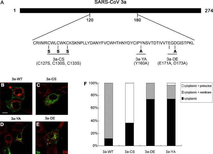
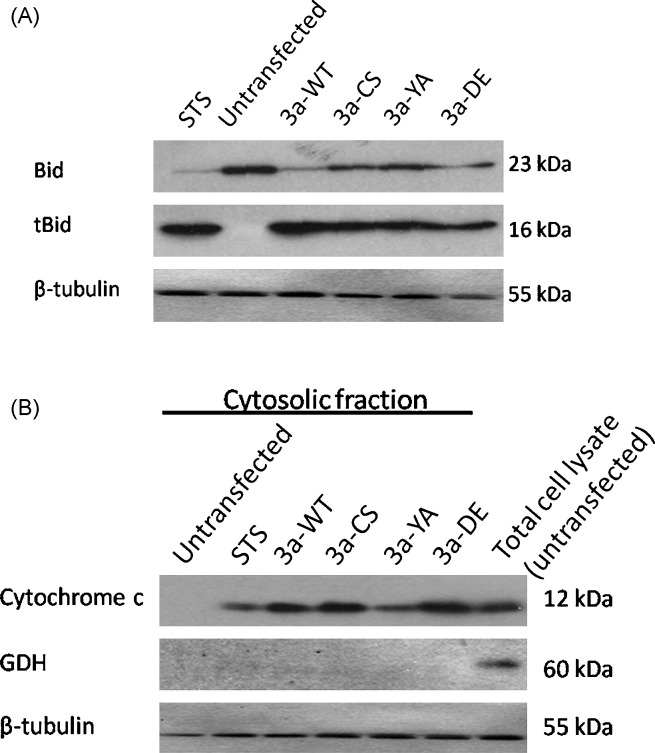
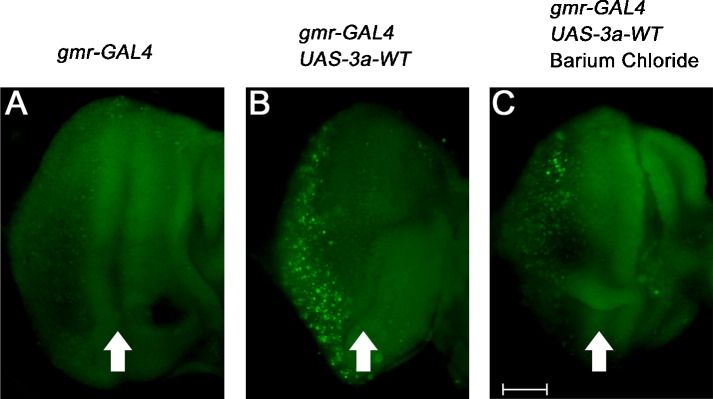
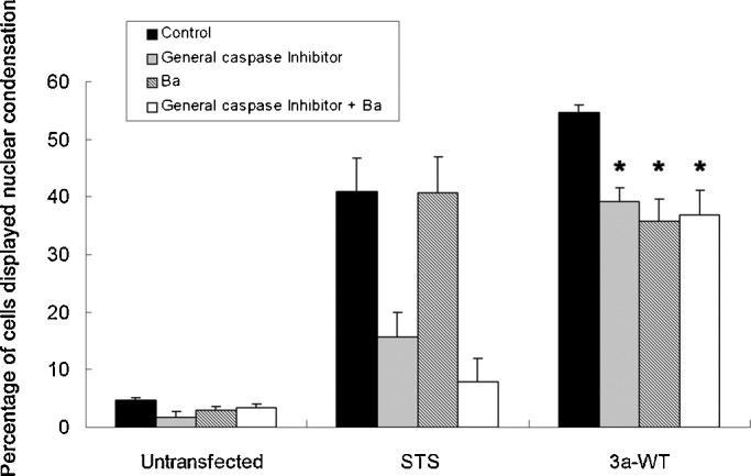
The severe acute respiratory syndrome-coronavirus (SARS-CoV) caused an outbreak of atypical pneumonia in 2003. The SARS-CoV viral genome encodes several proteins which have no homology to proteins in any other coronaviruses, and a number of these proteins have been implicated in viral cytopathies. One such protein is 3a, which is also known as X1, ORF3 and U274. 3a expression is detected in both SARS-CoV infected cultured cells and patients. Among the different functions identified, 3a is a capable of inducing apoptosis. We previously showed that caspase pathways are involved in 3a-induced apoptosis. In this study, we attempted to find out protein domains on 3a that are essential for its pro-apoptotic function. Protein sequence analysis reveals that 3a possesses three major protein signatures, the cysteine-rich, Yxx phi and diacidic domains. We showed that 3a proteins carrying respective mutations in these protein domains exhibit reduced pro-apoptotic activities, indicating the importance of these domains on 3a's pro-apoptotic function. It was previously reported that 3a possesses potassium ion channel activity. We further demonstrated that the blockade of 3a's potassium channel activity abolished caspase-dependent apoptosis. This report provides the first evidence that ion channel activity of 3a is required for its pro-apoptotic function. As ion channel activity has been reported to regulate apoptosis in different pathologic conditions, finding ways to modulate the ion channel activity may offer a new direction toward the inhibition of apoptosis triggered by SARS-CoV.


Figures

Fig. 1
Fig. 1
Subcellular localization of wild type and mutant SARS-CoV 3a proteins in Vero E6 cells. (A) Mutagenesis scheme of this study. (B–E) Subcellular localization of wild type and mutant 3a proteins in Vero E6 cells. Wild type 3a protein (3a-WT) displayed plasma membrane and punctate cytoplasmic staining pattern (B). The cysteine-rich domain mutant protein 3a-CS lost the plasma membrane localization, and concentrated in the cytoplasm and the perinuclear region (C). Both 3a-YA (D) and 3a-DE (E) mutant proteins showed cytoplasmic localization and fractional plasma membrane localization was retained. Different degrees of protein aggregation were also observed in 3a-YA and 3a-DE mutant proteins (D and E). ER-Tracker™ Red was used as a counter-stain to label the endoplasmic reticulum (shown in red). Scale bar represents 16 μm. (F) Quantification of subcellular distribution of 3a-WT and mutant 3a proteins. (For interpretation of the references to color in this figure legend, the reader is referred to the web version of the article.)
Fig. 2
Fig. 2
Wild type and mutant SARS-CoV 3a proteins possess caspase activities and cause nuclear condensation in Vero E6 cells. (A) Expression of wild type 3a protein (3a-WT) induced nuclear condensation (arrows) in Vero E6 cells. Scale bar represents 16 μm. (B) Vero E6 cells transfected with 3a-WT construct showed nuclear condensation while cells transfected with the 3a triple mutant (3a-CS-YA-DE) construct showed a much reduced level of nuclear condensation. At least 100 cells were counted in each experiment. *p < 0.05, 3a-CS-YA-DE triple mutant versus 3a-WT control. (C) Vero E6 cells transfected with 3a-WT and mutant 3a (3a-CS, 3a-YA and 3a-DE) constructs displayed different degrees of nuclear condensation. Nuclear condensation induced by 3a-WT, but not mutant 3a proteins, was inhibited by caspase-8 inhibitor II (z-IETD-fmk) and caspase-9 inhibitor I (z-LEHD-fmk). #p < 0.05, caspase inhibitor-treated 3a-WT cells versus 3a-WT control; *p < 0.05, 3a mutant-expressing cells versus 3a-WT-expressing cells. At least 100 cells were counted in each experiment. (D) General caspase inhibitor (z-VAD-fmk) significantly reduced nuclear condensation induced by 3a-WT protein in Vero E6 cells. At least 100 cells were counted in each experiment. *p < 0.05, caspase inhibitor-treated 3a-WT cells versus 3a-WT control. (E and F) Caspase-8 and -9 activities were detected in Vero E6 cells transfected with 3a-WT construct but these activities were significantly reduced in mutant 3a-expressing cells. *p < 0.05, 3a mutant-expressing cells versus 3a-WT-expressing cells.
Fig. 3
Fig. 3
3a induces Bid truncation and mitochondrial cytochrome C release. (A) Truncation of Bid was observed when untransfected HEK293 cells were treated with staurosporine (STS). Similar to STS treatment, HEK293 cells transfected with 3a-WT, -CS, -YA, and -DE constructs all showed Bid truncation. Bid: uncleaved Bid (23 kDa); tBid: truncated Bid (16 kDa). (B) Upon STS treatment, cytochrome c was detected in the cytosolic fraction of untransfected HEK293 cells. Cytosolic cytochrome c was also detected in cells transfected with 3a-WT, -CS, -YA and -DE constructs. Glutamate dehydrogenase (GDH), a mitochondrial marker, was used to demonstrate the cytosolic fractions were free of mitochondrial contamination. Staurosporine (1 μM) was used to induce apoptosis, and β-tubulin was used as loading control.
Fig. 4
Fig. 4
In vivo expression of wild type and mutant 3a disrupt adult eye structures in Drosophila. (A–E) Light microscopic examination of adult fly eyes. When compared to the gmr-GAL4 driver alone control (A), expression of the 3a-WT protein in eye tissues resulted in rough-eye phenotype (B) as characterized by loss of regularity of the adult external eye structure. Expression of mutant 3a proteins (3a-CS, 3a-YA and 3a-DE) showed minimal dominant external eye phenotype (C–E). (F–J) Scanning electron microscopic examination of adult fly eyes. Expression of 3a-WT caused severe loss of sensory bristles (G) when compared to the gmr-GAL4 control (F); while the expression of 3a-CS, 3a-YA and 3a-DE mutants caused less severe loss of sensory bristles (H–J). Scale bar represents 20 μm.
Fig. 5
Fig. 5
In vivo expression of wild type and mutant 3a induce apoptosis in Drosophila. Acridine orange (AO) staining was performed on third instar larval eye imaginal discs to label apoptotic cells. (A) The gmr-GAL4 control showed low levels of AO-stained cells. (B) 3a-WT expression induced apoptosis as indicated by an increase in the number of AO-stained cells. The 3a-CS (C), 3a-YA (D) and 3a-DE (E) mutants also induced apoptosis but to a less extent when compared to 3a-WT (B) as indicated by the relatively less number of AO-stained cells detected. Arrows indicate morphogenetic furrows. Scale bar represents 50 μm.
Fig. 6
Fig. 6
Potassium ion channel blockers suppress 3a-WT-induced apoptosis. (A–E) Ion channel properties of 3a. The 3a-WT (B), -YA (C), -DE (D) but not 3a-CS (E) proteins display ion channel activity. The IV relationship of mock transfected HEK293 cells (A), and cells transfected with cDNA encoding for 3a-WT-EGFP (B), 3a-YA-EGFP (C) and 3a-DE-EGFP (D) and 3a-CS-EGFP (E) were measured by whole cell patch clamping. Whole cell currents were recorded under voltage steps before (diamond) and 5 min after (square) 10 mM barium chloride (Ba) application. (F) Vero E6 cells transfected with 3a-WT and 3a-CS constructs were treated with potassium ion channel blockers 4-aminopyridine (AP) or barium chloride (Ba). AP or Ba treatment alone only induced minimal nuclear condensation on untransfected cells. Nuclear condensation induced by 3a-WT expression was significantly suppressed by potassium channel blockers (AP and Ba), while such treatments did not result in any significant inhibitory effect on 3a-CS-expressing cells. Untransfected Vero E6 cells treated with 1 μM staurosporine (STS) for 8 h were used as a control because neither AP nor Ba exerted any suppressive effect on STS-induced cell death. Results were plotted as percentage of cells showed nuclear condensation and expressed as means + S.E.M. of three independent experiments. At least 100 cells were counted in each experiment. *p < 0.05, AP- or Ba-treated 3a-WT-expressing cells versus 3a-WT control.
Fig. 7
Fig. 7
Barium chloride treatment suppresses 3a-WT-induced apoptosis in Drosophila. Acridine orange (AO) staining in third instar larval eye imaginal discs was performed to identify apoptotic cells in flies. (A) The gmr-GAL4 control showed low levels of AO-stained cells in third instar imaginal eye discs. (B) 3a-WT expression induced apoptosis as indicated by the increase in the number of AO-stained cells. (C) Feeding 3a-WT-expressing transgenic animals with barium chloride (Ba) reduced the number of AO-stained cells. Arrows indicate morphogenetic furrows. Scale bar represents 50 μm.
Fig. 8
Fig. 8
Pro-apoptotic and ion channel activities of 3a are linked. Vero E6 cells, either untransfected or transfected with 3a-WT construct, were treated with general caspase inhibitor (z-VAD-fmk) and/or Ba. z-VAD-fmk and/or barium chloride (Ba) treatment alone only induced minimal nuclear condensation on untransfected cells. Cells transfected with 3a-WT construct displayed nuclear condensation. z-VAD-fmk and/or Ba treatment significantly reduced nuclear condensation induced by 3a-WT expression in Vero E6 cells. Untransfected Vero E6 cells treated with 1 μM staurosporine (STS) for 8 h were used as control, only z-VAD-fmk but not Ba was able to suppress STS-induced nuclear condensation. All results were plotted as percentage of cells that showed nuclear condensation, and were expressed as means + S.E.M. of three independent experiments. At least 100 cells were counted in each experiment. *p < 0.05, Ba and/or caspase inhibitor-treated 3a-WT-expressing cells versus 3a-WT control.

References

    1. Bordi L., Castilletti C., Falasca L., Ciccosanti F., Calcaterra S., Rozera G. Bcl-2 inhibits the caspase-dependent apoptosis induced by SARS-CoV without affecting virus replication kinetics. Arch Virol. 2006;151:369–377. - PMC - PubMed
    1. Burg E.D., Remillard C.V., Yuan J.X. K+ channels in apoptosis. J Membr Biol. 2006;209:3–20. - PubMed
    1. Chan W.S., Wu C., Chow S.C., Cheung T., To K.F., Leung W.K. Coronaviral hypothetical and structural proteins were found in the intestinal surface enterocytes and pneumocytes of severe acute respiratory syndrome (SARS) Mod Pathol. 2005;18:1432–1439. - PMC - PubMed
    1. Chan C.M., Ma C.W., Chan W.Y., Chan H.Y. The SARS-coronavirus membrane protein induces apoptosis through modulating the Akt survival pathway. Arch Biochem Biophys. 2007;459:197–207. - PMC - PubMed
    1. Chau K.W., Chan W.Y., Shaw P.C., Chan H.Y. Biochemical investigation of Tau protein phosphorylation status and its solubility properties in Drosophila. Biochem Biophys Res Commun. 2006;346:150–159. - PubMed

Publication types

MeSH terms