. 2009 Aug;45(2):254-66.
doi: 10.1016/j.bone.2009.04.239. Epub 2009 May 3.

BMP2 is essential for post natal osteogenesis but not for recruitment of osteogenic stem cells

Affiliations

BMP2 is essential for post natal osteogenesis but not for recruitment of osteogenic stem cells

M V Bais et al. Bone. 2009 Aug.

Abstract

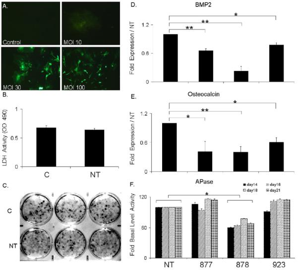
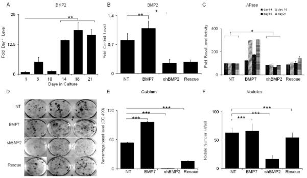
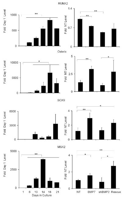
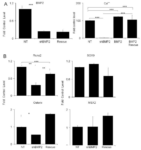
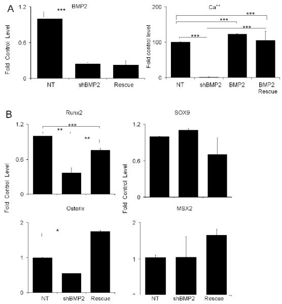
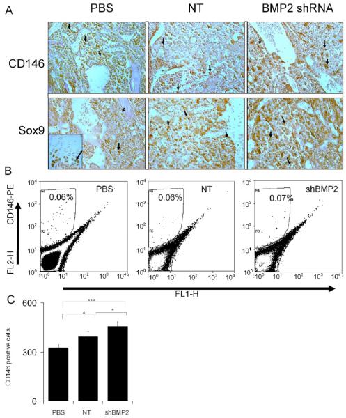
The effects of BMP2 on bone marrow stromal cell differentiation and bone formation after bone marrow ablation were determined using C57 BL/6J (B6) mice. Inhibition of BMP2 expression with lentiviral BMP2 shRNA prevented both mineralized nodule formation in vitro and bone formation in vivo, and blocked the expression of Runx2 and osterix, transcriptional determinants of terminal osteogenic differentiation. No effect was observed on the expression of Sox9, a transcription factor, which is the one of the first transcriptional determinant to be expressed in committed chondroprogenitor and osteoprogenitor cells. In vitro studies showed that exogenously added BMP7 rescued the expression of osterix and enhanced the expression of Sox9, but had no effect on the expression of Runx2, while it only partially recovered the development of mineral deposition in the cultures. On the other hand, the exogenous addition of BMP2 rescued both Runx2 and osterix expression, did not enhance the expression of Sox9, but fully recovered the inhibition of mineral deposition in the cultures. Using antibodies against CD146 and Sox9, immunohistological examination of the cell populations found in the medullary space three days after bone marrow ablation, showed qualitatively equal numbers of cells expressing these skeletal progenitor and stem cell markers in control and BMP2 shRNA treated animals. Fluorescence Activated Cell Sorting (FACS) analysis of the cells found with the marrow cavities at three days after marrow ablation using CD146 antibody showed near equal numbers of immunopositive cells in both control and shRNA treated animals. In summary, the differences observed in vitro for BMP2 and BMP7 on osteogenic gene expression and mineralization suggest that they have differing effects on bone cell differentiation. These results further demonstrate that in vivo BMP2 is a central morphogenetic regulator of post natal osteoprogenitor differentiation, but does not affect recruitment of progenitors to the osteoblastic lineage.


Figures

Fig. 1
Fig. 1. Characterization of Marrow Stromal Culture Transduction with Lentiviral BMP2 shRNA Expression Constructs
C= control no virus transduction and NT= transduced with non target virus. A) Assessment of the optimal multiplicity of infection (MOI) for lentiviral transduction by green fluorescent protein expression. Viral titers are as indicated in the figure. Representative phase contrast microscopy images taken under UV light excitation are depicted (100X magnification). Percentage optimal cell transduction was validated by FACs analysis. B) Effect of lentiviral transduction on cellar viability after 21days of culture growth. Viability was measured by LDH enzyme release as quantified by OD405nm showed no significant difference. . C) Effect of lentiviral transduction on formation of alizarin red stained osteogenic nodules. Representative images are of mineralized nodule formation in triplicate 33mm culture wells after 21 days of growth in mineralization media. D) Identification of the optimal BMP2 shRNA for BMP2 RNA silencing in MSC cultures. Cultures were separately transduced three different lentiviral constructs each containing a unique shRNA to BMP2. The expression of BMP2 mRNAs are presented as the relative value to NT transduced cultures. Values were determined at 21 days in culture. Numeric notation of the separate lentivirus constructs are as provided by Sigma Aldrich Corporation from The MISSION shRNA library of The RNAi Consortium. E) Functional effect of the various BMP2 shRNAs on osteocalcin mRNA expression relative to NT transduced cultures was determined at 21 days. F) Functional effect of effect of various BMP-2 shRNA on alkaline phospatase (Apase) activity. Activities were assessed at multiple time points as indicated in the figure. Lentivirus BMP2 shRNA clone 878 showed significant down regulation of BMP2 mRNA, osteocalcin mRNA and APase activity. Error bars represent standard deviation from replicates measurements from three experiments. P-value reflects the comparison of different treatment groups calculated by t-test and denoted as * p<0.05, ** p<0.01.
Fig. 2
Fig. 2. Rescue of Osteogenic Differentiation with Exogenous Addition of BMP7 in BMP2 shRNA Treated MSCs
NT= cultures transduced with non targeted; BMP7= transduced with NT lentivirus and treated with 200ng/ml BMP7; shBMP2= cultures transduced with lentivirus containing BMP2 shRNA; and Rescued= cultures transduced with lentivirus containing BMP2 shRNA and treated with 200ng/ml BMP7. All RNA measurements made from three separate preparations of cells and error bars are the SD of the triplicate measurements from 3 separate cell preparations. A) Steady state BMP2 mRNA expression levels over the time course of in vitro osteogenic differentiation. RNA measurements are presented as a relative fold of expression to day 1. B) Steady state BMP2 mRNA expression levels after 21 days growth in cultures under various experimental conditions. RNA measurements are presented as a relative value to the NT control. C) APase activities were assessed at 21 days time points. Error bars are the SD from three separate cell culture preparations. D) Representative images of mineralized nodule formation in triplicate 33mm culture wells after 21 days of growth in mineralization media. E) Quantification of effect of lentiviral transduction on Ca++ accumulation. Calcium bound alizarin dye was released as described in the materials and absorbance was analyzed. F) Effect of lentiviral transduction on formation of Alizarin red stained nodules. Error bars represent standard deviation from replicates measurements from three experiments. P-value reflects the comparison of different treatment groups calculated by t-test and denoted as * p<0.05, ** p<0.01, *** p<0.001.
Fig. 3
Fig. 3. Effect of Exogenous Addition of BMP7 in BMP2 shRNA Treated MSCs on the Expression of Various Transcription Factors that Control Skeletal Stem Cell Lineage Progression
The nature of each mRNA that was assayed is indicated in the figure. Left panels in the figure present the steady state mRNA expression levels of the various transcription factors over the time course of in vitro osteogenic differentiation. RNA measurements are presented as a relative fold of expression to day 1. Right panels show the transcription factor mRNA expression levels after 21 days growth in cultures under various experimental conditions. RNA measurements are presented as a relative value to the NT control. Mean values are measurements made from three separate preparations of cells. Error bars represent standard deviation from replicates measurements from three experiments. P-value reflects the comparison of different treatment groups calculated by t-test and denoted as * p<0.05, ** p<0.01, *** p<0.001.
Fig. 4
Fig. 4. Affect of Exogenous Addition of BMP2 on BMP2 shRNA Treated Culture
All data is presented as the fold of the control untreated MSC cultures at 21 days. A) The exogenous addition of BMP2 protein rescue of nodule and mineral deposition in BMP2 shRNA inhibited MSCs cultures. Left panel shows the fold change in BMP2 mRNA expression in response to both BMP2 shRNA treated and BMP2 shRNA in the presence of exogenous addition of BMP2. Right panel shows the total mineral accumulation as measured by alizarin dye uptake by BMP2 rescue in comparison of NT shRNA and NT shRNA with BMP2 protein. B) Affect of Exogenous Addition of BMP2 in BMP2 shRNA Treated MSCs on the Expression of Various Transcription Factors that Control Skeletal Stem Cell Lineage Progression. Error bars represent standard deviation from replicate measurements from three separate cell preparations. P-value reflects the comparison of different treatment groups calculated by t-test and denoted as * p<0.05, ** p<0.01, *** p<0.001.
Fig. 5
Fig. 5. In vivo Effects of BMP2 shRNA Inhibition on Bone Formation after Marrow Ablation
A) Gross staining of tibiae to detect β-galactosidase activity. Individual tibia from mice injected with either PBS control or lentiviral particles that expression β-galactosidase. Representative images of three bones that had been cut in half and reacted with X-gal staining are shown. Staining is detected as dark green to green brown areas and is denoted with arrows. B) Representative in vivo measurements of bioluminescent intensities from mice injected with lentivirus overexpressing luciferase (lenti-Luc) compared to PBS injected controls. The data are reported as the photon flux (p/s) from a defined region of interest in tibia of lentivirus expressing luciferase compared to noninjected control mice. C) MicroCT assessment of endosteal bone formation in response to surgical marrow ablation. All images are orientated with the proximal end of the bone at the top and are from 7 days after the time of surgery. Representative μCT 3D renderings of bone formation from control mice and mice injected with NT and BMP2 shRNA lentiviral particles are shown in the top panels. Graphical measurements of percent bone tissue (BV/TV) as determined from the μCT analysis are presented in the below the microCT images. The Comparisons for all pairs using one way ANOVA showed statistically significant difference in mean in shBMP2 group (mean=0.66) compared to PBS (Mean =0.14) and NT (mean= 0.10) and p value<.0001 denoted as ***showing significant differences in each group. Analysis by t-test showed also showed significance for the comparison of shBMP2 with p <.05 whereas with PBS p<.0001. D) Histological Analysis of bone formation in response to surgical marrow ablation. All images are orientated with the proximal end of the bone at the right side of the micrographs. Sections were cut in longitudinal orientations and sections were selected from the central regions such that the surrounding cortices are seen. Sections were stained with H&E. In shRNA injected groups, there is observable loss of trabecular structure and bone loss indicated by arrow.
Fig. 6
Fig. 6. Effect of BMP2 shRNA Lentiviral Treatment on the Expression of Various Transcription Factors that Control Skeletal Stem Cell Lineage Progression within Bone Tissues Formed After Surgical Ablation
Individual samples are as denoted in the figure and the legend. Levels of expression are relative to unoperated control tibia bones. Error bars represent standard deviation from replicates measurements from three experiments. P-value reflects the comparison of different treatment groups calculated by t-test and denoted as * p<0.05, ** p<0.01, *** p<0.001.
Fig. 7
Fig. 7. Effect of BMP2 shRNA Lentiviral Treatment on the Development of Immunoreactive Cells Showing Stem Cell and Osteogenic Progenitor Marker Expression
A) Immunohistology of CD146 and Sox9 reactive cells. Tibiae were collected three days post bone marrow ablation. Tissues were cut in longitudinal orientations and sections were selected from the central regions. Sections were reacted with antibodies directed against mouse specific CD146 or Sox9. All photomicrographs were taken at 200X magnification. Immunopositive cells stain reddish brown. The experimental group and antibody used for the immunostaining are indicated in the figure. The inset in left lower (PBS/Sox9) image depicts positive immunostaining for Sox 9 in the growth plate from the same specimen. B) Quantification of CD146 positive osteoprogenitor stems cells in different treatment groups. Representative FACs analysis of CD146 positive cells observed in the total population of cells flushed from the marrow space three days post surgery and plated overnight before FACs. The gated population was optimized for nonspecific stating by isotype control antibody. Left Panel Shows 0.06% CD146 positive cells in gate R4 from tibia of mice injected with PBS as a control. Middle panel shows this cell population in nontarget shRNA treated group (006%). Right panel shows that the BMP2 shRNA treatment produced a slight increase in CD146 cells (0.07%). The total number of CD146 cells sorted cells from three separate experiments plotted, showed statistical significant difference in shRNA treated groups compared to PBS (p= 0.00154) and NT (0.028139). Error bars are standard deviation from replicates measurements from three experiments. P-value reflects the comparison of different treatment groups calculated by t-test and denoted as * p<0.05, ** p<0.01, *** p<0.001.

References

    1. Hollinger JO, Schmitt JM, Buck DC, Shannon R, Joh SP, Zegzula HD, Wozney J. Recombinant human bone morphogenetic protein-2 and collagen for bone regeneration. J Biomed Mater Res. 1998;43:356–64. - PubMed
    1. Tsiridis E, Morgan EF, Bancroft JM, Song M, Kain M, Gerstenfeld L, Einhorn TA, Bouxsein ML, Tornetta P. Effects of OP-1 and PTH in a new experimental model for the study of metaphyseal bone healing. J Orthop Res. 2007;25:1193–2033. - PubMed
    1. Welch RD, Jones AL, Bucholz RW, Reinert CM, Tjia JS, Pierce WA, Wozney JM, Li XJ. Effect of recombinant human bone morphogenetic protein-2 on fracture healing in a goat tibial fracture model. J Bone Miner Res. 1998;13:1483–90. - PubMed
    1. Hogan BL. Bmps: multifunctional regulators of mammalian embryonic development. Harvey Lect. 1996;92:83–98. - PubMed
    1. Solloway MJ, Dudley AT, Bikoff EK, Lyons KM, Hogan BL, Robertson EJ. Mice lacking Bmp6 function. Dev Genet. 1998;22:321–39. - PubMed

Publication types

MeSH terms

Substances