. 2009 Jun;15(6):1078-89.
doi: 10.1261/rna.1363109. Epub 2009 Apr 27.

Mammalian GW182 contains multiple Argonaute-binding sites and functions in microRNA-mediated translational repression

Affiliations

Mammalian GW182 contains multiple Argonaute-binding sites and functions in microRNA-mediated translational repression

Koji Takimoto et al. RNA. 2009 Jun.

Abstract

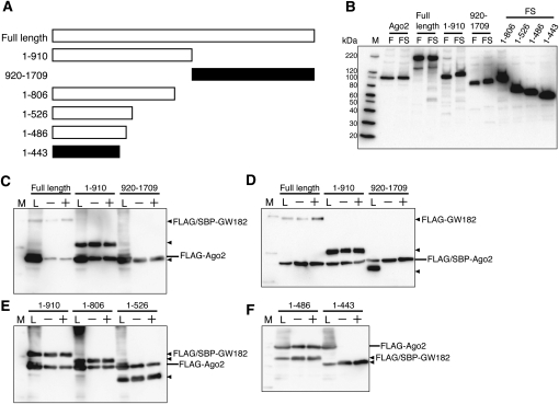
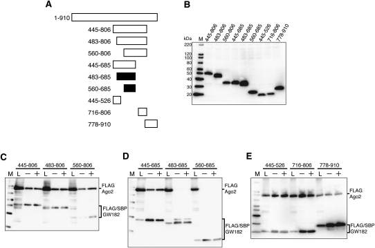
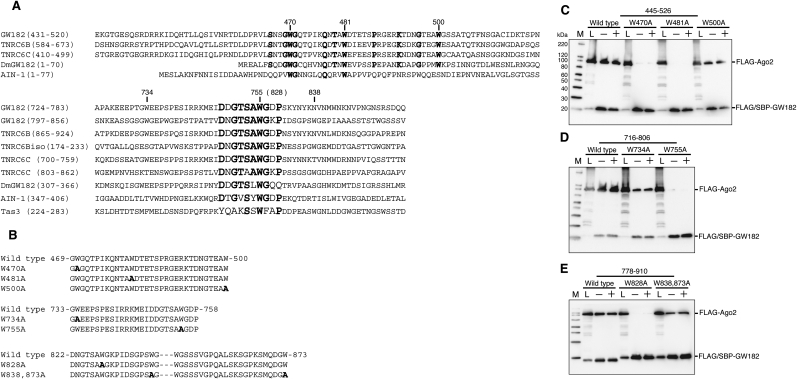
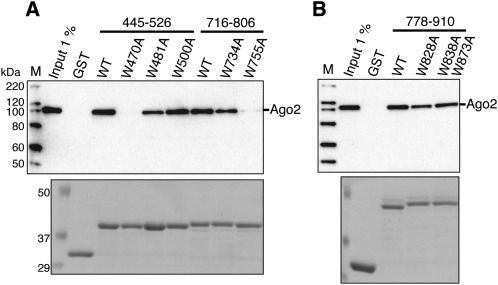
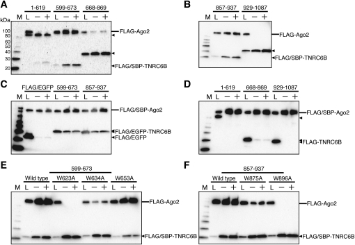
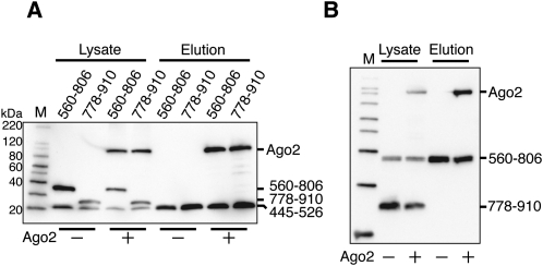
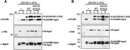
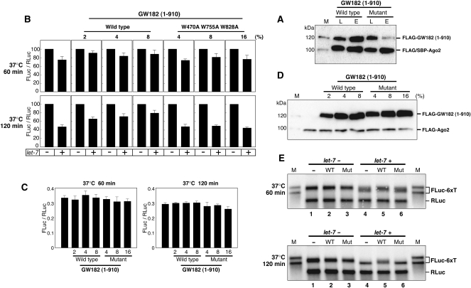
In mammalian cells, microRNAs (miRNAs) are incorporated into miRNA-induced silencing complexes (miRISCs), which regulate protein expression post-transcriptionally through binding to 3'-untranslated regions of target mRNAs. Argonaute2 (Ago2), a key component of the miRISC, recruits GW182, a component of the processing body (GW/P-body), to the target mRNAs. To elucidate the function of GW182 in an miRNA-mediated translational repression, we analyzed Argonaute-binding sites in GW182. We found that human GW182 contains three binding sites for Ago2, within the amino-terminal glycine tryptophan (GW/WG)-repeated region that is characteristic of the GW182 family proteins. We also found that the first and second Ago2-binding site is conserved within the amino-terminal half of TNRC6B, which is a paralog of GW182. Each of the Ago-binding sites is alone sufficient to bind Ago2. Furthermore, we demonstrated that multiple Argonaute proteins were connected via the GW182 protein. A GW182 fragment containing the Ago2-binding region partially relieved let-7-mediated repression of protein synthesis in a mammalian cell-free system. Coincidentally, let-7-directed target mRNA deadenylation was delayed. Together, these results strongly suggested that the interactions of GW182 with Argonautes may induce the formation of large complexes containing miRNA target mRNAs, and may be critical for miRNA-mediated translational repression.


Figures

FIGURE 1.
FIGURE 1.
The amino-terminal half of GW182, containing GW/WG repeats, binds Ago2. (A) Schematic representation of the GW182 fragments used in the SBP pull-down assay. The polypeptides that bound Ago2 are represented by the white bars, while the black bars represent fragments that did not bind Ago2. (B) Expression of Ago2 and the GW182 fragments in HEK293F cells. The expressed proteins were detected by Western blotting with an anti-FLAG M2 antibody. The sizes of the molecular markers are indicated on the left (lane M). (Lanes F,FS) FLAG- and FLAG/SBP-tagged proteins, respectively. (C–F) SBP pull-down assay of Ago2 and GW182. HEK293F cells were cotransfected with FLAG/SBP or FLAG-tagged Ago2 and the GW182 expression vectors. After 48 h, the cells were harvested and the prepared cell lysates were subjected to SBP pull-down assays with (+) or without (−) RNaseA. The precipitates were analyzed by Western blotting using an anti-FLAG M2 antibody. The positions of the GW182 fragments are indicated with triangles on the right. (Lane L) The cell lysate sample.
FIGURE 2.
FIGURE 2.
Multiple Ago2-binding sites are present in the amino-terminal half of GW182. (A) Schematic representation of the GW182 fragments used in the SBP pull-down assay. The polypeptides that can bind Ago2 are represented by the white bars, while the black bars represent those that cannot bind Ago2. (B) Expression of Ago2 and each GW182 fragment in HEK293F cells. The expressed proteins were detected by Western blotting with an anti-FLAG M2 antibody. (C–E) SBP pull-down assays of Ago2 and GW182. HEK293F cells were cotransfected with FLAG-Ago2 and FLAG/SBP-GW182 expression vectors. SBP-pull down assays and Western blotting were performed as shown in Figure 1C.
FIGURE 3.
FIGURE 3.
A conserved tryptophan residue is important for Ago2 binding. (A) Alignment of the highly conserved regions of human GW182 (AY035864), TNRC6B (AB029016), TNRC6C (NM_018996), TNRC6Bisoform2 (NM_001024843), Drosophila melanogaster (Dm) GW182 (NP_726596), Caenorhabditis elegans AIN-1 (NM_078286), and Schizosaccharomyces pombe Tas3 (O94687). Highly conserved amino acids are indicated in bold. (B) The partial amino acid sequences with the point mutations in GW182 445-526 (top), 716-806 (middle), and 778-910 (bottom). Mutated amino acid residues are indicated in bold. (C–E) SBP pull-down assays of Ago2 and GW182 mutants. HEK293F cells were cotransfected with FLAG-Ago2 and FLAG/SBP-GW182 mutant expression vectors. SBP pull-down assays and Western blotting were performed as shown in Figure 1C.
FIGURE 4.
FIGURE 4.
GST pull down assay of Ago2 and GST-fused GW182 mutants. (A) The wild-type and mutated GST-GW182 (445–526 and 716–806) fragments were each expressed in E. coli. (B) The wild-type and mutated GST-GW182 (778–910) fragments were each expressed in E. coli. HEK293F cell extracts overexpressing FLAG-Ago2 were incubated with each MagneGST particle-immobilized GST-GW182 fragment. The resulting complexes were resolved by SDS-PAGE, followed by Coomassie Brilliant Blue (CBB) staining (bottom), and were analyzed by Western blotting using an anti-FLAG M2 antibody (top).
FIGURE 5.
FIGURE 5.
Two Ago2-binding sites exist in the amino-terminal half of TNRC6B. (A,B,D) SBP pull-down assays of Ago2 and TNRC6B. HEK293F cells were cotransfected with FLAG/SBP or FLAG-tagged Ago2 and the TNRC6B expression vectors. (C) HEK293F cells were cotransfected with FLAG-Ago2 and FLAG-EGFP-TNRC6B or FLAG-EGFP expression vectors as a negative control. (E,F) HEK293F cells were cotransfected with FLAG-Ago2 and wild-type or mutant FLAG/SBP-TNRC6B expression vectors. SBP-pull down assays and Western blotting were performed as shown in Figure 1C.
FIGURE 6.
FIGURE 6.
Each Ago2-binding domain of GW182 binds to an overlapping region of Ago2. (A) HEK293F cells were cotransfected with the FLAG/SBP-GW182 445–526, FLAG-GW182 560–806, or 778–910 expression vector, with or without the FLAG–Ago2 expression vector. (B) HEK293F cells were cotransfected with the FLAG/SBP-GW182 560–806 and FLAG-GW182 778–910 expression vectors, with or without the FLAG-Ago2 expression vector. SBP pull-down assays and Western blotting were performed as described in Figure 1C, in the presence of RNaseA.
FIGURE 7.
FIGURE 7.
GW182 can associate with multiple Ago2 proteins. FLAG/SBP-Ago2 and HA-Ago2 were coexpressed with or without FLAG-GW182 1–910 or FLAG-GW182 1–910 (W470A and W755A) in HEK293F cells. (A) SBP pull-down assays were performed, and the precipitates were analyzed by Western blotting using an anti-FLAG M2 antibody (top), an anti-HA antibody (middle), and an anti-hAgo2 antibody (bottom). (Lane L) Cell lysate (1.25 μL equivalent); (lane E) fraction eluted with biotin (300 μL of cell lysate was subjected to the assay and 14% of the eluted fraction was loaded). (B) HA-Ago2 protein was precipitated with anti-HA agarose beads, and the precipitates were analyzed by an anti-FLAG M2 (top), an anti-HA (middle), and an anti-hAgo2 (bottom) Western blotting. (Lane L) Cell lysate (1.25 μL equivalent); (lane E) fraction eluted with 100 μg/mL of HA peptide solution. (300 μL of cell lysate was subjected to the assay and 14% of the eluted fraction was loaded).
FIGURE 8.
FIGURE 8.
Let-7-mediated translational repression is inhibited by a GW182 fragment containing an Ago2-binding site. (A) FLAG/SBP-Ago2 was coexpressed with FLAG-GW182 (1–910) or FLAG-GW182 (1–910, W470A, W755A, W828A) in HEK293F cells. SBP pull-down assays were performed, as described in the Figure 7A legend, and the precipitates were analyzed by Western blotting using an anti-FLAG antibody. Lane L, cell lysate; lane E, fraction eluted with biotin. (B) In vitro translation experiments were performed with HEK293F extracts (containing 10% [v/v] Ago2 extract) along with 2%, 4%, and 8% (v/v) of the wild-type, or 4%, 8%, and 16% (v/v) of the mutant FLAG-GW182 (1–910) extract. Capped FLuc-6xT with a poly(A) tail mRNA was translated in the absence (−) or presence (+) of let-7. The FLuc and RLuc activities were measured, and the FLuc-to-RLuc activity ratio in the reaction without let-7 was set at 100. The data shown constitute an average of three independent experiments, with standard deviations. (C) Comparison of the translation efficiencies of the extracts with different ratios of FLAG-GW182 (1–910) extract in the absence of let-7. The average number of light units produced by the translation of FLuc mRNAs in the absence of FLAG-GW182 (1–910) lysate was ∼2,200,000. (D) Expression of FLAG-Ago2 and wild-type or mutant FLAG-GW182 (1–910) in the cell extract mixture using the translation experiment. The expressed proteins were detected by Western blotting using an anti-FLAG M2 antibody. (E) Analysis of capped and polyadenylated FLuc-6xT and RLuc mRNAs by Northern blotting with an FLuc probe and an RLuc probe. Translation reactions of mRNAs were performed with the cell extract mixture containing 8% (v/v) of the wild-type GW182 (1–910) and 16% (v/v) of the mutant GW182 (1–910). As molecular markers, nonadenylated (shorter) and polyadenylated (longer) transcripts were loaded in lane M. After 60 min (upper panel) and 120 min (lower panel) of translation, the RNAs were extracted from the reaction mixture, and similar amounts were loaded in each lane.

Similar articles

Cited by

References

    1. Ambros V. The functions of animal microRNAs. Nature. 2004;431:350–355. - PubMed
    1. Bartel D.P. MicroRNAs: Genomics, biogenesis, mechanism, and function. Cell. 2004;116:281–297. - PubMed
    1. Behm-Ansmant I., Rehwinkel J., Doerks T., Stark A., Bork P., Izaurralde E. mRNA degradation by miRNAs and GW182 requires both CCR4:NOT deadenylase and DCP1:DCP2 decapping complexes. Genes & Dev. 2006;20:1885–1898. - PMC - PubMed
    1. Chendrimada T.P., Finn K.J., Ji X., Baillat D., Gregory R.I., Liebhaber S.A., Pasquinelli A.E., Shiekhattar R. MicroRNA silencing through RISC recruitment of eIF6. Nature. 2007;447:823–828. - PubMed
    1. Ding L., Han M. GW182 family proteins are crucial for microRNA-mediated gene silencing. Trends Cell Biol. 2007;17:411–416. - PubMed

Publication types

LinkOut - more resources