. 2009 Jul;77(7):2908-18.
doi: 10.1128/IAI.01068-08. Epub 2009 Apr 27.

Nonhematopoietic cells control the outcome of infection with Listeria monocytogenes in a nucleotide oligomerization domain 1-dependent manner

Affiliations

Nonhematopoietic cells control the outcome of infection with Listeria monocytogenes in a nucleotide oligomerization domain 1-dependent manner

Ahmed Mosa et al. Infect Immun. 2009 Jul.

Abstract

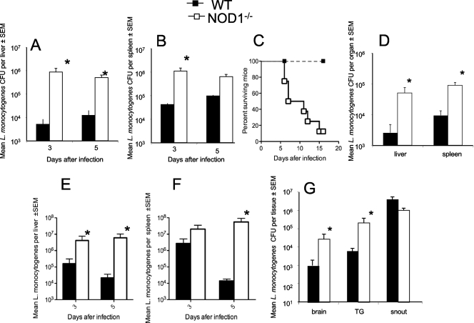
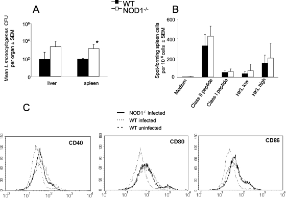
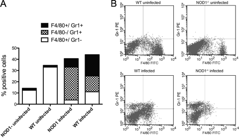
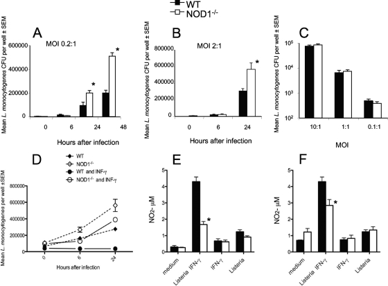
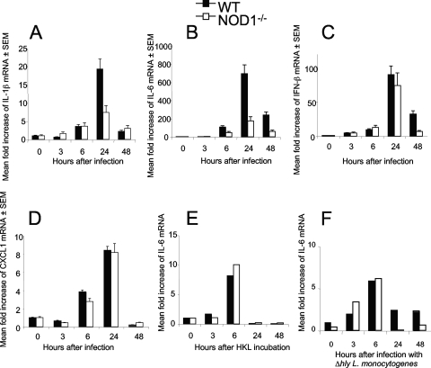
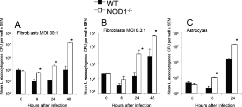
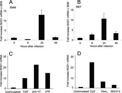
We analyzed the defensive role of the cytosolic innate recognition receptor nucleotide oligomerization domain 1 (NOD1) during infection with Listeria monocytogenes. Mice lacking NOD1 showed increased susceptibility to systemic intraperitoneal and intravenous infection with high or low doses of L. monocytogenes, as measured by the bacterial load and survival. NOD1 also controlled dissemination of L. monocytogenes into the brain. The increased susceptibility to reinfection of NOD1(-/-) mice was not associated with impaired triggering of listeria-specific T cells, and similar levels of costimulatory molecules or activation of dendritic cells was observed. Higher numbers of F480(+) Gr1(+) inflammatory monocytes and lower numbers of F480(-) Gr1(+) neutrophils were recruited into the peritoneum of infected WT mice than into the peritoneum of infected NOD1(-/-) mice. We determined that nonhematopoietic cells accounted for NOD1-mediated resistance to L. monocytogenes in bone marrow radiation chimeras. The levels of NOD1 mRNA in fibroblasts and bone marrow-derived macrophages (BMM) were upregulated after infection with L. monocytogenes or stimulation with different Toll-like receptor ligands. NOD1(-/-) BMM, astrocytes, and fibroblasts all showed enhanced intracellular growth of L monocytogenes compared to WT controls. Gamma interferon-mediated nitric oxide production and inhibition of L. monocytogenes growth were hampered in NOD1(-/-) BMM. Thus, NOD1 confers nonhematopoietic cell-mediated resistance to infection with L. monocytogenes and controls intracellular bacterial growth in different cell populations in vitro.


Figures

FIG. 1.
FIG. 1.
NOD1 is required for resistance to infection with L. monocytogenes in vivo. The course of infection with L. monocytogenes in NOD1−/− and WT mice was analyzed. Mice were infected i.p. (A to D) or i.v. (E and F) with 105 (A to C), 2 × 10⁴ (E and F), or 2 × 10³ (D) L. monocytogenes CFU and sacrificed at indicated time points (A, B, E, and F) or 4 days after infection (D). The mean L. monocytogenes CFU titers in livers and spleens of six mice per group are shown. The error bars indicate standard errors of the means (SEM). *, P < 0.05 for a comparison with WT mice (Student's t test). (C) Cumulative mortality of NOD1−/− and WT mice (eight mice per group) after i.p. infection with 105 L. monocytogenes CFU. (G) Numbers of L. monocytogenes CFU in the snout, trigeminal nerve (TG), and brain of six NOD1−/− mice and six WT mice 4 days after infection via the snout. The bacterial loads are expressed in CFU per g of tissue (mean ± standard error of the mean). The differences between the NOD1−/− and WT groups are significant for all tissues except the snout (P < 0.05, Student's t test; indicated by an asterisk).
FIG. 2.
FIG. 2.
NOD1 is redundant for induction of adaptive immune responses. Mice infected with 2 × 10³ L. monocytogenes CFU were reinfected i.p. with a 50-fold-larger bacterial inoculum 20 days after the primary infection. (A) Mean numbers of L. monocytogenes CFU in livers and spleens from mice sacrificed 4 days after reinfection. The error bars indicate standard errors of the means (SEM). *, P < 0.05 for a comparison with WT mice (Student's t test), indicating a significant difference. (B) Spleen cells from uninfected WT or NOD1−/− mice or from mice 12 days after reinfection with 105 L. monocytogenes CFU (six mice per group) were cultured overnight with or without 2 μM LLO 296-304, 2 μM LLO 190-201, or 0.2 (low) or 0.8 (high) HKL per cell. The data are the numbers of IFN-γ-secreting cells as determined by an ELISPOT assay. All mice showed concanavalin A responses well above 100 SFC per 105 cells, confirming the viability of the splenocyte cultures (data not shown). Uninfected mice showed no SFC following addition of L. monocytogenes whole-cell lysates or following peptide-specific stimulation (data not shown). (C) Spleen CD11c+ DC from NOD1−/− and WT mice at 0 or 4 days after infection with L. monocytogenes were examined by flow cytometry for expression of CD40, CD80, and CD86.
FIG. 3.
FIG. 3.
NOD1 affects L. monocytogenes-induced recruitment of granulocytes and resident and inflammatory monocytes to the inflammatory site. NOD1−/− and WT peritoneal exudate cells were harvested from mice before (five WT mice and four NOD1−/− mice) or 2 days after (6 WT mice and six NOD1−/− mice) i.p. infection with 105 L. monocytogenes CFU. The mean percentages of cells that were stained with anti-Gr1 and/or anti-F4/80 to differentiate between polymorphonuclear leukocytes and inflammatory monocytes and resting macrophages and monocytes in the peritoneal exudates from individual mice are shown. Differences between the total numbers of harvested peritoneal cells from L. monocytogenes-infected and uninfected NOD1−/− and WT animals are not significant. Differences between the percentages of F4/80+ Gr1 cells in infected and uninfected peritoneal exudates from WT or NOD1−/− mice are significant (P < 0.001, Student's t test). Differences between the percentages of F4/80+ Gr1 cells in peritoneal exudates from infected or uninfected WT and NOD1−/− mice are significant (P < 0.001, Student's t test). Differences between the percentages of Gr1+ F4/80 cells from infected WT and NOD1−/− mice are significant (P < 0.001, Student's t test). Differences between the percentages of Gr1+ F4/80+ peritoneal exudate cells from WT and NOD1−/− mice are significant (P < 0.02, Student's t test). The results of one of two independent experiments are shown. (B) Levels of expression of F4/80+ and Gr1+ cells in the peritoneal exudates of representative mice before or after infection with L. monocytogenes. FITC, fluorescein isothiocyanate; PE, phycoerythrin.
FIG. 4.
FIG. 4.
NOD1-mediated resistance to infection with L. monocytogenes is due to the activity of nonhematopoietic cells. NOD1−/− or WT bone marrow cells were inoculated i.v. into irradiated NOD1−/− or WT mice. Six weeks after inoculation of the cells, mice were infected i.p. with 105 L. monocytogenes CFU. Mice were sacrificed 5 days after infection. The numbers of CFU in lungs from mice treated as described above were determined. The data are the means ± standard errors of the means (SEM) and are the numbers of CFU per liver (A) or spleen (B) for six mice per group. *, P < 0.05 for a comparison of chimeric mice and WT→WT sham chimera mice (Student's t test), indicating a significant difference.
FIG. 5.
FIG. 5.
NOD1 controls the growth of L. monocytogenes in BMM. (A and B) NOD1−/− and WT BMM in triplicate wells were incubated with L. monocytogenes at a multiplicity of infection (MOI) of 0.2 (A) or 2 (B) and lysed at the indicated time points after infection, and the numbers of L. monocytogenes CFU in the BMM lysates were determined. Two independent experiments were performed, and the results of one of them are shown. (C) NOD1−/− and WT BMM were infected with L. monocytogenes at the indicated multiplicities of infection. After 1 h, cells were extensively washed and lysed, and the numbers of CFU were determined. (D) NOD1−/− and WT BMM were incubated with 100 U recombinant IFN-γ (BD, Pharmingen) 18 h before infection with L. monocytogenes at a multiplicity of infection of 0.2. One hour after infection cells were extensively washed, and IFN-γ was replenished. Cells were lysed at the indicated time points after infection, and the numbers of L. monocytogenes CFU in the BMM lysates were determined. *, P < 0.05 for a comparison with WT mice (Student's t test), indicating a significant difference. (E and F) NO2 levels in culture supernatants of BMM incubated with 100 U IFN-γ 18 h before incubation with HKL (multiplicity of infection, 10:1), of BMM incubated with IFN-γ, or of BMM incubated with HKL alone 24 h (E) and 48 h (F) after addition of the bacterial lysate.
FIG. 6.
FIG. 6.
NOD1 modulates IL-6 and IL-1β responses after incubation with L. monocytogenes but not after incubation with heat-killed bacterial lysates or with bacteria unable to escape from the phagosome into the cytosol. Total RNA from BMM was extracted before and 6 h after infection with L. monocytogenes at a multiplicity of infection of 0.2 (A to D) or with Δhly L. monocytogenes at a multiplicity of infection of 10 (F) or after incubation with HKL (E). The titers of IL-1β, IL-6, IFN-β, CXCL1 (A to F) and HPRT mRNA were determined by real-time RT-PCR. The mean increases in cytokine or HPRT levels for triplicate (A to D) or duplicate (E and F) wells are shown. Two independent experiments were performed, and the results of one of them are shown.
FIG. 7.
FIG. 7.
NOD1 controls growth of L. monocytogenes in murine embryonic fibroblasts and astrocytes. NOD1−/− and WT fibroblasts (A and B) and astrocytes (C) in triplicate wells were incubated with L. monocytogenes at multiplicities of infection (MOI) of 30 (A), 0.3 (B), and 10 (C) and lysed at the indicated time points after infection. The numbers of L. monocytogenes CFU in the cell lysates were determined. *, P < 0.05 for a comparison with WT mice (Student's t test), indicating a significant difference. SEM, standard error of the mean.
FIG. 8.
FIG. 8.
NOD1 mRNA levels are increased in fibroblasts and macrophages after stimulation with L. monocytogenes or incubation with TLR ligands. The levels of NOD1 mRNA transcripts in WT BMM (A, C, and D) or fibroblasts (B) infected with L. monocytogenes (A and B) or stimulated with CpG (C and D), poly(I-C) (C), LPS (C), Pam3 (D), or MALP-2 (D) were determined by real-time RT-PCR. The mean increases in the level of NOD1 mRNA for duplicate wells for each condition compared to the level for unstimulated wells are shown. SEM, standard error of the mean.

Similar articles

Cited by

References

    1. Beckerman, K. P., H. W. Rogers, J. A. Corbett, R. D. Schreiber, M. L. McDaniel, and E. R. Unanue. 1993. Release of nitric oxide during the T cell-independent pathway of macrophage activation. Its role in resistance to Listeria monocytogenes. J. Immunol. 150888-895. - PubMed
    1. Carneiro, L. A., L. H. Travassos, and D. J. Philpott. 2004. Innate immune recognition of microbes through Nod1 and Nod2: implications for disease. Microbes Infect. 6609-616. - PubMed
    1. Chamaillard, M., S. E. Girardin, J. Viala, and D. J. Philpott. 2003. Nods, Nalps and Naip: intracellular regulators of bacterial-induced inflammation. Cell. Microbiol. 5581-592. - PubMed
    1. Chamaillard, M., M. Hashimoto, Y. Horie, J. Masumoto, S. Qiu, L. Saab, Y. Ogura, A. Kawasaki, K. Fukase, S. Kusumoto, M. A. Valvano, S. J. Foster, T. W. Mak, G. Nunez, and N. Inohara. 2003. An essential role for NOD1 in host recognition of bacterial peptidoglycan containing diaminopimelic acid. Nat. Immunol. 4702-707. - PubMed
    1. Chin, A. I., P. W. Dempsey, K. Bruhn, J. F. Miller, Y. Xu, and G. Cheng. 2002. Involvement of receptor-interacting protein 2 in innate and adaptive immune responses. Nature 416190-194. - PubMed

Publication types

MeSH terms

Substances