. 2009 Jul;77(7):2624-34.
doi: 10.1128/IAI.01531-08. Epub 2009 Apr 27.

Subcellular localization of the Staphylococcus aureus heme iron transport components IsdA and IsdB

Affiliations

Subcellular localization of the Staphylococcus aureus heme iron transport components IsdA and IsdB

Gleb Pishchany et al. Infect Immun. 2009 Jul.

Abstract

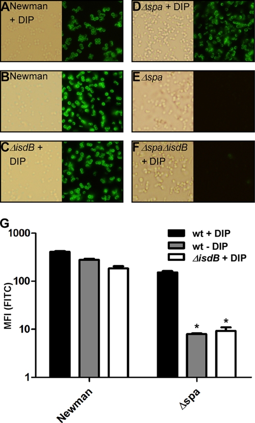
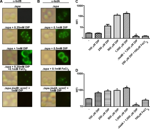
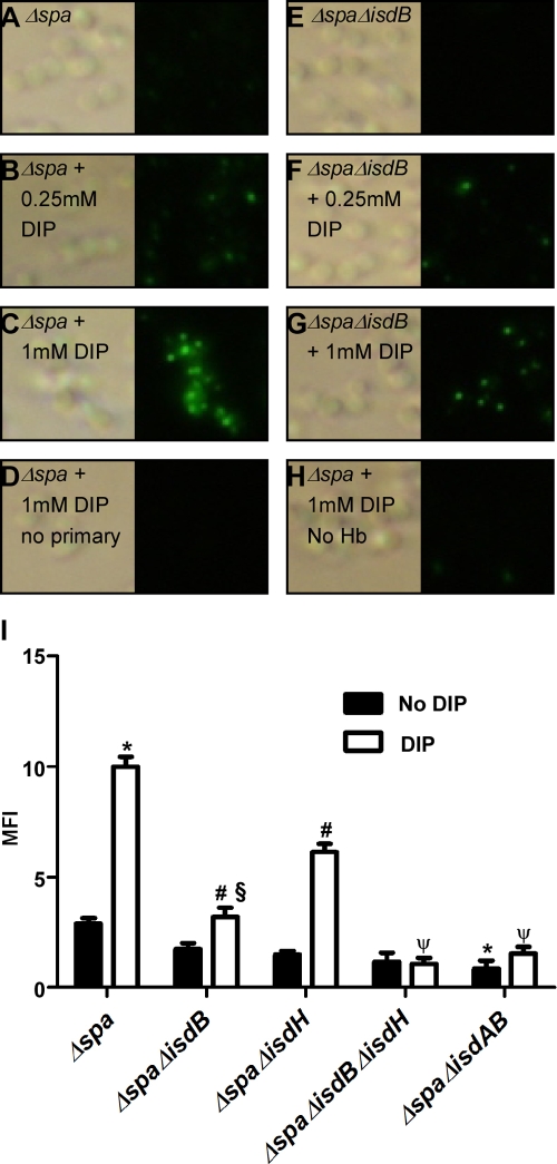
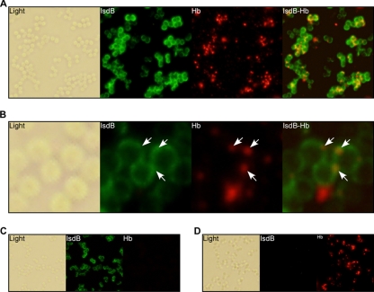
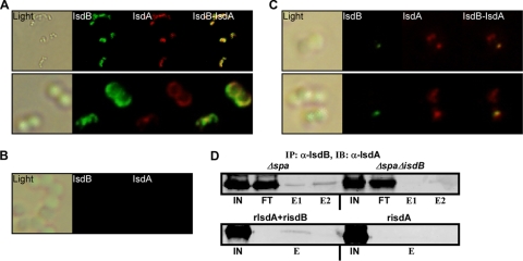
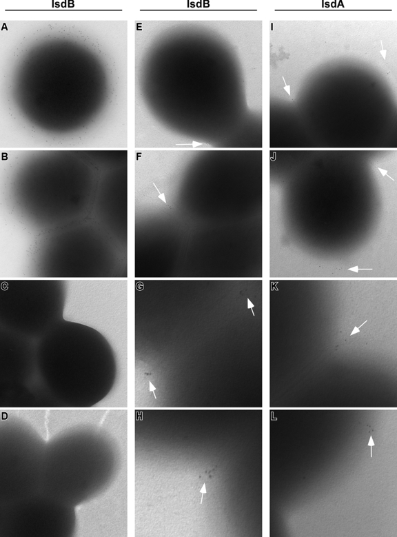
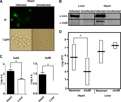
Staphylococcus aureus is a human pathogen that represents a tremendous threat to global public health. An important aspect of S. aureus pathogenicity is the ability to acquire iron from its host during infection. In vertebrates, iron is sequestered predominantly within heme, the majority of which is bound by hemoglobin. To acquire iron, S. aureus binds hemoglobin, removes heme, and transports it into the cytoplasm, where heme is degraded. This process is carried out by the iron-regulated surface determinant system (Isd); however, the mechanism by which hemoglobin recognition occurs is not completely understood. Here we report that the surface receptor components of the Isd system, IsdA and IsdB, physically interact with each other and are anchored to a discrete location within the cell wall. This organized localization pattern is dependent upon the iron status of the bacterium. Furthermore, we have found that hemoglobin colocalizes with IsdB at discrete sites within the cell wall. Virulence studies revealed that IsdB is required for the efficient colonization of the heart and that IsdB is differentially expressed within infected organs, suggesting that S. aureus experiences various degrees of iron starvation depending on the site of infection. These findings significantly expand our understanding of hemoglobin iron acquisition and demonstrate an orchestrated pattern of regulation and localization for the S. aureus heme iron acquisition system.


Figures

FIG. 1.
FIG. 1.
Creation of a protein A-deficient strain for specific labeling of S. aureus surface-exposed proteins. (A to F) S. aureus strains were grown overnight in TSB with 1 mM of the iron-chelating agent DIP where indicated. Bacteria were subsequently labeled with rabbit anti-IsdB antibody and Alexa Fluor 488 goat anti-rabbit IgG(H+L). (G) Bacteria grown under the same conditions and labeled similarly to A to F were subjected to FACS analysis to determine mean fluorescence intensity (MFI). Error bars represent standard errors. Asterisks indicate statistically significant differences in relation to Δspa cells with DIP as determined by a Student's t test (P < 0.05). FITC, fluorescein isothiocyanate; wt, wild type.
FIG. 2.
FIG. 2.
Iron availability influences the expression and localization of IsdB and IsdA on the staphylococcal surface. Δspa cells were grown overnight in medium supplemented with the indicated concentrations of DIP. (A and B) Bacteria were subsequently labeled with rabbit anti-IsdB (α-IsdB) (A) or rabbit anti-IsdA (B) antibodies, followed by Alexa Fluor 488 goat anti-rabbit IgG(H+L). (C and D) Quantification of relative amounts of IsdB (C) and IsdA (D) expressed on the surface of Δspa cells was determined by FACS analysis. Asterisks indicate statistically significant differences in relation to Δspa cells grown in plain TSB (−) as determined by a Student's t test (P < 0.05). In fluorescent images, green was increased by 100% in A and by 25% in B. MFI, mean fluorescence intensity.
FIG. 3.
FIG. 3.
Iron- and Isd-dependent hemoglobin binding to the surface of S. aureus. (A to G) Δspa (A to D) or Δspa ΔisdB (E to G) cells were grown overnight in TSB supplemented with the indicated concentrations of DIP. Bacteria were then incubated in PBS with human hemoglobin (Hb) and washed with PBS. Cells were subsequently labeled with mouse anti-hemoglobin IgG, followed by Alexa Fluor 488 goat anti-mouse IgG(H+L). (I) Quantification of the relative amounts of human hemoglobin bound to the surface of the indicated strains, grown in the presence of 1 mM DIP where indicated, determined by FACS. Error bars represent standard errors. The symbols indicate statistically significant differences as determined by a Student's t test (P < 0.05) in relation to the Δspa strain with no DIP (*), the Δspa strain with DIP (#), the Δspa ΔisdB strain with no DIP (§), and the Δspa ΔisdB strain with DIP (ψ).
FIG. 4.
FIG. 4.
Hemoglobin colocalizes with IsdB on the surface of S. aureus. (A) Δspa cells were grown overnight in TSB supplemented with 1 mM DIP. Bacteria were then incubated with human hemoglobin (Hb) and washed with PBS. Cells were then simultaneously labeled with rabbit anti-IsdB and mouse anti-hemoglobin antibodies, followed by Alexa Fluor 488 goat anti-rabbit and Alexa Fluor 555 goat anti-mouse IgG(H+L). (B) Close-up of A. The arrows point to locations on the cell surface where hemoglobin is bound. To control for antibody cross-reactivity, either mouse anti-hemoglobin (C) or rabbit anti-IsdB (D) antibodies were omitted. In fluorescent images, green was increased by 25% and red was increased by 75%.
FIG. 5.
FIG. 5.
IsdA and IsdB colocalize and interact on the cell wall of S. aureus. (A) Δspa cells were grown overnight in TSB supplemented with 1 mM DIP. Bacteria were then sequentially labeled with rabbit anti-IsdB (α-IsdB), Alexa Fluor 488 goat anti-rabbit IgG(H+L), biotinylated rabbit anti-IsdA, and streptavidin Alexa Fluor 555 conjugate. (B and C) Cells from the same culture were subjected to trypsin digestion, washed, resuspended in TSB plus 1 mM DIP, and incubated. Aliquots were taken after 0 min (B) or 15 min (C) and labeled as described above (A). (D, top) Δspa or Δspa ΔisdB cells were grown overnight in TSB plus 1 mM DIP. The cell wall proteins were solubilized by lysostaphin. IsdB was pulled down with a Seize X protein A immunoprecipitation kit and rabbit anti-IsdB antibody. Input (IN), nonprecipitated flowthrough (FT), and eluted proteins (E1 and E2) were subjected to SDS-PAGE, transferred onto nitrocellulose, and immunoblotted (IB) with anti-IsdA antibody. (Bottom) rIsdA and rIsdB were combined and incubated at 37°C for 30 min. Immunoprecipitation (IP) was performed as described above (top), and input (IN) and elution (E) are shown.
FIG. 6.
FIG. 6.
IsdA and IsdB are anchored at sites of nascent cell wall formation. Δspa cells were grown overnight in TSB supplemented with 1 mM DIP. Cells were treated with trypsin, washed, resuspended in TSB plus 1 mM DIP, and incubated. Aliquots were taken at different time points, washed, attached to nickel Formvar grids, and sequentially labeled with indicated primary antibodies and secondary 6-nm colloidal gold-Affinipure goat anti-rabbit IgG(H+L). (A and B) Nontrypsinized Δspa cells labeled for IsdB. (C) Nontrypsinized Δspa ΔisdB cells labeled for IsdB. (D) Trypsinized Δspa cells labeled for IsdB. (E to L) Δspa cells upon trypsin treatment and 5 min of recovery in TSB, labeled for IsdB (E to H) or IsdA (I to L).
FIG. 7.
FIG. 7.
IsdB is expressed within the hearts of infected animals and contributes to cardiac colonization. (A) C57BL/6J mice were retro-orbitally infected with 107 CFU of the Δspa strain in 100 ml PBS and sacrificed 96 h postinfection. Hearts and livers were removed and homogenized in 1 ml sterile PBS. Bacteria were then partially separated from the mammalian cells by centrifugation, washed, and immunofluorescently labeled with anti-IsdB. IF, immunofluorescence. (B) Wild-type S. aureus cells recovered from the organs of C57BL/6J mice were separated from the mammalian cells as described above (A), normalized to 1 × 10⁵ CFU, lysed with lysostaphin to release cell wall proteins that were separated on SDS-PAGE gels, and transferred onto a nitrocellulose membrane. The membrane was immunoblotted with anti-IsdA (α-IsdA) and anti-IsdB antibodies. (C) Relative amounts of IsdA and IsdB in the infected organs were quantified based on immunoblot intensity. Error bars represent standard errors. Asterisks indicate statistically significant differences as determined by a Student's t test (P < 0.05). a.u., arbitrary units. (D) Organ colonization was estimated based on CFU quantification by serial dilution and plating onto tryptic soy agar. The horizontal bars represent the means, and boxes represent standard deviations. Asterisks indicate statistically significant differences as determined by a Student's t test (P < 0.05). Each group included at least nine mice.

References

    1. Bae, T., and O. Schneewind. 2006. Allelic replacement in Staphylococcus aureus with inducible counter-selection. Plasmid 5558-63. - PubMed
    1. Bullen, J. J., and E. Griffiths. 1999. Iron and infection: molecular, physiological and clinical aspects. John Wiley & Sons, New York, NY.
    1. Carlsson, F., M. Stalhammar-Carlemalm, K. Flardh, C. Sandin, E. Carlemalm, and G. Lindahl. 2006. Signal sequence directs localized secretion of bacterial surface proteins. Nature 442943-946. - PubMed
    1. Christou, L., G. Pappas, and M. E. Falagas. 2007. Bacterial infection-related morbidity and mortality in cirrhosis. Am. J. Gastroenterol. 1021510-1517. - PubMed
    1. Clarke, S. R., and S. J. Foster. 2008. IsdA protects Staphylococcus aureus against the bactericidal protease activity of apolactoferrin. Infect. Immun. 761518-1526. - PMC - PubMed

Publication types

LinkOut - more resources