. 2009 Jun 19;284(25):17340-17351.
doi: 10.1074/jbc.M109.010108. Epub 2009 Apr 27.

Autoprocessing of the Escherichia coli AIDA-I autotransporter: a new mechanism involving acidic residues in the junction region

Affiliations

Autoprocessing of the Escherichia coli AIDA-I autotransporter: a new mechanism involving acidic residues in the junction region

Marie-Ève Charbonneau et al. J Biol Chem. .

Abstract

The cleavage of the autotransporter adhesin involved in diffuse adherence (AIDA-I) of Escherichia coli yields a membrane-embedded fragment, AIDAc, and an extracellular fragment, the mature AIDA-I adhesin. The latter remains noncovalently associated with AIDAc but can be released by heat treatment. In this study we determined the mechanism of AIDA-I cleavage. We showed that AIDA-I processing is an autocatalytic event by monitoring the in vitro cleavage of an uncleaved mutant protein isolated from inclusion bodies. Furthermore, by following changes in circular dichroism spectra and protease resistance of the renaturated protein, we showed that the cleavage of the protein is correlated with folding. With site-directed deletions, we showed that the catalytic activity of the protein lies in a region encompassing amino acids between Ala-667 and Thr-953, which includes the conserved junction domain of some autotransporters. With site-directed point mutations, we also found that Asp-878 and Glu-897 are involved in the processing of AIDA-I and that a mutation preserving the acidic side chain of Asp-878 was tolerated, giving evidence that this carboxylic acid group is directly involved in catalysis. Last, we confirmed that cleavage of AIDA-I is intramolecular. Our results unveil a new mechanism of auto-processing in the autotransporter family.


Figures

FIGURE 1.
FIGURE 1.
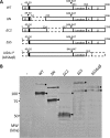
Cleavage of AIDA-I* in vitro. A, schematic diagram of the AIDA-I* construct, showing plasmid pAngH (WT) and pAngHΔSSΔαβ (ΔSSΔαβ or AIDA-I*). The filled box represents the His tag at the N terminus of mature AIDA-I. B, inclusion bodies containing the AIDA-I* protein were collected, solubilized in 0.1 m Tris-HCl, pH 8, 6 m guanidinium hydrochloride, and purified by immobilized metal affinity chromatography. Renaturation of the AIDA-I protein was performed by first exchanging the buffer on a 1-ml His trap HP column for 2 h with renaturation buffer (TBS, pH 8, 0.1% Triton X-100). The protein was then eluted and left at 4 °C. At different times after buffer exchange, aliquots were taken and boiled in SDS-PAGE loading buffer. The proteins were then resolved by SDS-PAGE and the gel-stained with Coomassie Blue. The uncleaved (proprotein, circles) and cleaved (mature AIDA-I*, arrowheads) forms of the protein are indicated. The intensities of the bands were quantified and normalized to the maximal intensity. C, the WT and the uncleaved (D878N*) proteins 145 h after buffer exchange were separated by SDS-PAGE on a 16% Tris-Tricine gel. The star indicates the C-terminal cleaved peptide. D, far-UV CD spectra of the AIDA-I* protein at different times after buffer exchange. The ellipticities were recorded between 205 and 260 nm. E, the ellipticities at 218 nm were recorded at different times after the initial renaturation. MRE, mean residual ellipticity.
FIGURE 2.
FIGURE 2.
Protease sensitivity of AIDA-I* during refolding. Limited proteolysis of pure AIDA-I* was conducted for 30 min with 37 μg·ml−1 (final concentration) of trypsin at different times after buffer exchange. The proteins were resolved by SDS-PAGE and the gel stained with Coomassie Blue.
FIGURE 3.
FIGURE 3.
Effect of pH on the in vitro folding of AIDA-I*. Renaturation of the AIDA-I* protein was performed by first exchanging the buffer on a 1-ml His trap HP column for 2 h with renaturation buffer at pH 8. After elution, an equal amount of protein was diluted in buffer with different pH values (50 mm sodium citrate, pH 4 or 6; 50 mm Tris, pH 8; 50 mm sodium carbonate, pH 10, all supplemented with 150 mm NaCl and 0.1% Triton X-100) and left at 4 °C. A, far-UV CD spectra of the AIDA-I* protein at different times after buffer exchange for pH4 (top) pH 6 (middle) or pH 10 (bottom). The ellipticities were recorded between 205 and 260 nm. MRE, mean residual ellipticity. B, at different times after the buffer exchange aliquots were taken and boiled in SDS-PAGE loading buffer. C, limited proteolysis at 145 h after buffer exchange was performed as described in Fig. 2. For pH 4, the proteolysis was performed after a pH shift at pH 8. The proteins were resolved by SDS-PAGE and the gel-stained with Coomassie Blue. The uncleaved (proprotein, circles) and cleaved (mature AIDA-I*, arrowheads) forms of the protein are indicated.
FIGURE 4.
FIGURE 4.
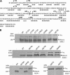
Mapping of the region involved in AIDA-I cleavage. A, schematic diagram of the deletion constructs showing plasmid pAngH (WT), pAngHΔN (ΔN), pAngHΔC2 (ΔC2), pAngHΔSS (ΔSS), and pAngHΔSSΔαβ (ΔSSΔαβ or AIDA-I*). The filled box represents the His tag at the N terminus of mature AIDA-I. B, whole-cell lysates were obtained from overnight cultures of C600 harboring an empty vector (−), expressing wild-type AIDA-I (WT), or expressing the ΔN, ΔC2, ΔSS or ΔSSΔαβ mutants were obtained and probed with anti-His antibodies, which allowed detection of the proprotein (circles) and mature AIDA-I (arrowheads). All plasmids were transformed in bacteria expressing the Aah glycosyltransferase.
FIGURE 5.
FIGURE 5.
Mutational analysis of the region involved in AIDA-I cleavage. A, predicted secondary structure of the region of AIDA-I encompassing the amino acids between Asn-708 and Ser-953; E, β-strand; x, no prediction. The positions corresponding to modified amino acids are indicated. The dashed line indicates the cleavage site. The numbering of amino acids corresponds to the position in full-length pre-proprotein. B, whole-cell lysates were obtained from overnight cultures of C600 harboring an empty vector (−), plasmid pAgH (WT), or one of the plasmids bearing a point mutation in pAgH. Proteins separated by SDS-PAGE were probed by immunoblotting with antibodies against the His tag, which allowed the detection of the proprotein (Pro., circles) and the mature AIDA-I (arrowheads).
FIGURE 6.
FIGURE 6.
Characterization of the D878N protein. A, far-UV CD spectra of the purified AIDA-I wild-type protein (gray circles) and the uncleaved D878N mutant (black squares). The ellipticities were recorded between 205 and 260 nm. B, the ellipticities at 218 nm of the WT protein (gray circles) or the D878N mutant (black squares) were recorded with the temperature varying between 25 and 80 °C at a rate of 5 °C/min. MRE, mean residual ellipticity. C, purified uncleaved (D878N) and cleaved (WT) proteins were incubated in the presence of different concentrations of trypsin. The proteins were resolved by SDS-PAGE, and the gel was stained with Coomassie Blue.
FIGURE 7.
FIGURE 7.
Renaturation and folding kinetics of D878N* mutant. The D878N* protein was purified as described in Fig. 1. A, at different times after buffer exchange, aliquots were taken and boiled in SDS-PAGE loading buffer. The proteins were then resolved by SDS-PAGE, and the gel was stained with Coomassie Blue. The uncleaved (proprotein D878N*, circles) form of the protein is indicated. B, far-UV CD spectra of the D878N* protein. The ellipticities were recorded between 205 and 260 nm. The CD spectrum of the WT AIDA-I* after 145 h of renaturation is indicated. C, the ellipticities at 218 nm were recorded at different times after the initial renaturation. MRE, mean residual ellipticity. D, limited proteolysis was performed as described in Fig. 2. The proteins were resolved by SDS-PAGE, and the gel was stained with Coomassie Blue.
FIGURE 8.
FIGURE 8.
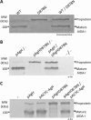
Intramolecular cleavage of AIDA-I. A, wild-type AIDA-I (WT) or the uncleaved (D878N) proteins were incubated separately or together for 30 min at room temperature and resolved by SDS-PAGE, and the gel was stained with Coomassie Blue. B, whole-cell lysates were obtained from overnight cultures of E. coli C600 harboring plasmids pTRC99a (−), pAgH (WT), or pAgHD878N (D878N) coincubated for 30 min at 30 °C. The samples were resolved by SDS-PAGE and probed with anti-His tag antibodies, which allowed the detection of the proprotein (circle) and the mature AIDA-I (arrowheads). C, whole-cell lysates from overnight cultures of E. coli C600 harboring plasmids pTRC99a and/or PACYC184 (−), pAgH, pACYC-AgH, pAgHD878N, or pACYC-AgH and pAgHD878N were resolved by SDS-PAGE and probed with anti-His tag antibodies.
FIGURE 9.
FIGURE 9.
Structural model and alignment of the cleavage site of AIDA-I. Left panel, a three-dimensional structural model of the junction region and the cleavage site of AIDA-I (amino acids Ile-807—Leu-956). The amino acids constituting the cleavage site, Ser-846 and Ala-847, are highlighted in cyan, and the amino acids Asp-878 and Glu-897 are highlighted in red and depicted with their side chains. The bottom panel shows a close-up view of the cleavage site and the Asp-878 and Glu-897 residues using the same color scheme. Right panel, alignment of the junction region of several autotransporters. The secondary structure features, either modeled (for AIDA-I) or experimentally determined (for Hbp (48) and Prn (39)), are indicated. The positions of Ser-846, Ala-847, Asp-878, and Glu-897 are highlighted in cyan and red, as in A. The accession numbers for the sequence data and the amino acids range used for the alignment are: ABS20376, amino acids 807–956 (E. coli AIDA-I); Q9XD84, amino acids 470–629 (E. coli TibA); P39180, amino acids 540–704 (E. coli Ag43); O88093, amino acids 919–1073 (E. coli Hbp); Q7BS42, amino acids 915–1058 (E. coli Pic); O68900, amino acids 836–991 (E. coli Pet); AAC44731, amino acids 851–1022 (E. coli EspC); Q7BSW5, amino acids 840–996 (E. coli EspP); CAA88252, amino acids 911–1062 (S. flexneri SepA); CAC05837, amino acids 572–739 (S. flexneri IcsA); Q03035, amino acids 416–569 (B. pertussis Prn); AAA51646, amino acids 551–706 (B. pertussis BrkA); CAA45708, amino acids 828–984 (H. influenzae IgA protease); and P45387, amino acids 800–977 (H. influenzae Hap).

Similar articles

Cited by

References

    1. Henderson I. R., Navarro-Garcia F., Desvaux M., Fernandez R. C., Ala'Aldeen D. ( 2004) Microbiol. Mol. Biol. Rev. 68, 692– 744 - PMC - PubMed
    1. Henderson I. R., Nataro J. P. ( 2001) Infect. Immun. 69, 1231– 1243 - PMC - PubMed
    1. Girard V., Mourez M. ( 2006) Res. Microbiol. 157, 407– 416 - PubMed
    1. Peterson J. H., Szabady R. L., Bernstein H. D. ( 2006) J. Biol. Chem. 281, 9038– 9048 - PubMed
    1. Oomen C. J., van Ulsen P., van Gelder P., Feijen M., Tommassen J., Gros P. ( 2004) EMBO J. 23, 1257– 1266 - PMC - PubMed

Publication types

MeSH terms

LinkOut - more resources