. 2009 Jul;29(13):3710-21.
doi: 10.1128/MCB.01869-08. Epub 2009 Apr 27.

Targeted knock-in mice expressing mutations of CD28 reveal an essential pathway for costimulation

Affiliations

Targeted knock-in mice expressing mutations of CD28 reveal an essential pathway for costimulation

Lindzy F Dodson et al. Mol Cell Biol. 2009 Jul.

Abstract

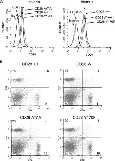
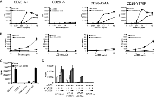
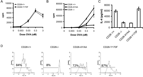
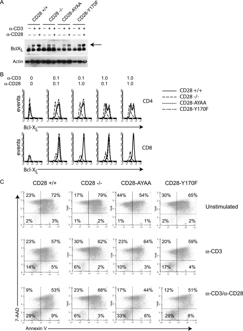
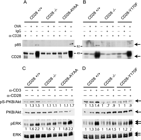
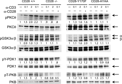
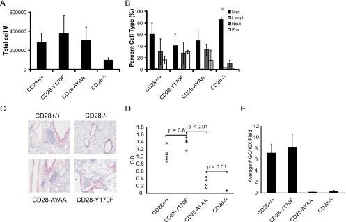
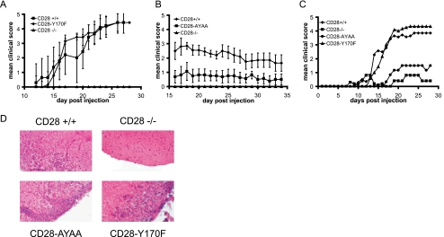
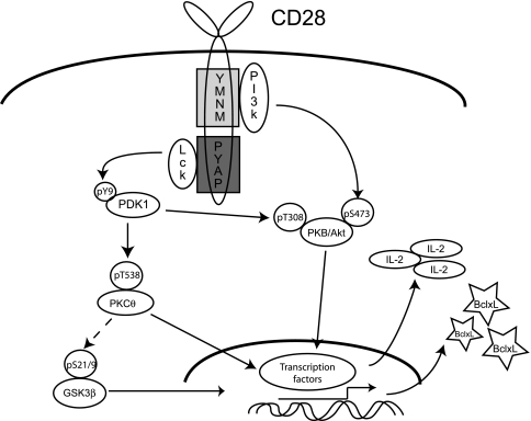
Despite extensive study, the role of phosphatidylinositol 3-kinase (PI3-kinase) activation in CD28 function has been highly contentious. To definitively address this question, we generated knock-in mice expressing mutations in two critical domains of the cytoplasmic tail of CD28. Mutation of the proximal tyrosine motif interrupted PI3-kinase binding and prevented CD28-dependent phosphorylation of protein kinase B (PKB)/Akt; however, there was no detectable effect on interleukin-2 (IL-2) secretion, expression of Bcl-X(L), or on T-cell function in vivo. Furthermore, we demonstrate that signaling initiated by the C-terminal proline motif is directly responsible for tyrosine phosphorylation of phosphoinosotide-dependent kinase 1, protein kinase C theta, and glycogen synthase kinase 3beta, as well as contributing to threonine phosphorylation of PKB. T cells mutated in this domain were profoundly impaired in IL-2 secretion, and the mice had marked impairment of humoral responses as well as less severe disease manifestations in experimental allergic encephalomyelitis. These data demonstrate that the distal proline motif initiates a critical nonredundant signaling pathway, whereas direct activation of PI3-kinase by the proximal tyrosine motif of CD28 is not required for normal T-cell function.


Figures

FIG. 1.
FIG. 1.
(A) Expression of CD28 on CD4+ splenocytes and thymocytes from the wild-type and mutant knock-in mice. (B) CD4 and CD8 expression on splenocytes isolated from wild-type and CD28 mutant knock-in mice.
FIG. 2.
FIG. 2.
Proliferation and IL-2 secretion depend on the distal proline motif and not the proximal tyrosine motif. Splenocytes were isolated from mice of each genotype and stimulated with graded doses of anti-CD3 alone or in combination with anti-CD28 antibody (1 μg/ml). (A) Proliferation was determined by tritiated thymidine incorporation. (B) IL-2 was measured in culture supernatants by ELISA. (C) Cells were stimulated with PMA (5 ng/ml) alone or in combination with anti-CD28 (1 μg/ml), and proliferation was determined by tritiated thymidine incorporation. (D) Splenocytes were isolated from mice heterozygous for wild-type or mutant CD28 on the CD28−/− background, therefore expressing only one CD28 allele, and stimulated with anti-CD3 (0.1 μg/ml) alone, in combination with CTLA4 Ig (10 μg/ml), or with both CTLA4 Ig and increasing doses of anti-CD28 (0.03, 0.3, or 1.0 μg/ml). Proliferation was determined by tritiated thymidine incorporation. All experiments were repeated a minimum of three times and representative data are presented. Data are the means ± standard deviations of quadruplicate wells.
FIG. 3.
FIG. 3.
Proliferation and IL-4 secretion in response to antigen is impaired in the CD28-AYAA knock-in mice but not CD28-Y170F mice. Splenocytes were isolated from mice of each genotype in the DO11.10 background. (A and B) Cells were stimulated with OVA(323-339) peptide and proliferation was measured after 72 h by tritiated thymidine incorporation. (C) Cells were cultured with OVA(323-339) peptide plus recombinant IL-4 and anti-IL-12 for 7 days. The cells were then washed, rested overnight, and restimulated with peptide for 48 h. The culture supernatant was collected and IL-4 content was assayed by ELISA. (D) Splenocytes were isolated from mice of each genotype and labeled with CFSE. A total of 5 × 10⁶ cells were then injected i.v. into naïve BALB/c mice. The following day, the recipient mice were injected with OVA-alum (50 μg) i.p. Mesenteric lymph nodes were collected 72 h later, stained for CD4 and KJ1-26, and analyzed by flow cytometry. The CFSE profile of the KJ1-26-positive cells is presented. Representative data of three independent experiments are presented.
FIG. 4.
FIG. 4.
Neither the proximal nor distal motif is absolutely required for T-cell survival or Bcl-XL expression. (A) Splenocytes were cultured in medium or stimulated with anti-CD3 alone (1.0 mg/ml) or with anti-CD28 (1.0 mg/ml). After 48 h, the cells were lysed and Bcl-XL expression levels were determined by Western blotting. The arrow points to the specific band. (B) Splenocytes from mice of each genotype were isolated and cultured either with medium alone or stimulated with anti-CD3 (0.1 or 1.0 μg/ml) and anti-CD28 (0.1 or 1.0 μg/ml) in the presence of CTLA4 Ig (10 μg/ml) for 48 h. The cells were surface stained with anti-CD4 or anti-CD8 followed by an intracellular stain for Bcl-XL. Shown is the Bcl-XL expression of CD4+ (top row) and CD8+ (bottom row) cells. (C) Purified T cells were stimulated as indicated for 18 h and then washed, and equal numbers of cells were exposed to γ-irradiation (3 Gy). After 24 h, cell viability was determined by staining with CD4-PE, annexin V-APC, and 7-amino-actinomycin D (7-AAD) and analyzed by flow cytometry. Representative data of three independent experiments are presented.
FIG. 5.
FIG. 5.
p85 binding and serine phosphorylation of PKB/Akt are impaired in the CD28-Y170F mutant. (A and B) Splenocytes from mice of each genotype in the DO11.10 background were isolated and cultured with OVA(323-339) peptide for 72 h. Equal numbers of cells were isolated and lysed in RIPA buffer, and CD28 was immunoprecipitated. The immunoprecipitated protein was separated by SDS-polyacrylamide gel electrophoresis, transferred to polyvinylidene difluoride, and blotted for p85 or CD28. Arrows indicate the specific bands. (C and D) Purified T cells were isolated from mice of each genotype and cultured with either medium alone or stimulated for 5 min with anti-CD3 (0.1 μg/ml) alone or in combination with anti-CD28 (8 μg/ml) with cross-linking antibodies as described in Materials and Methods. Following stimulation, Western blot analysis was performed for each protein as indicated. Densitometry was performed as described in Materials and Methods, and the ratio with the unstimulated control is shown.
FIG. 6.
FIG. 6.
Phosphorylation of PKCθ, GSK3β, and PDK1 depends on the distal proline motif but not the proximal tyrosine motif of CD28, whereas threonine phosphorylation of PKB requires both motifs. Purified T cells were isolated from mice of each genotype and cultured with either medium alone or stimulated for 5 min with the indicated doses of anti-CD3 alone or in combination with anti-CD28 (8 μg/ml) with cross-linking antibodies as described in Materials and Methods. Following stimulation, Western blot analysis was performed for each protein as indicated. Arrows indicate the specific bands. The lane order for the CD28-AYAA and CD28-Y170F pT-PKB and total PKB samples was modified from the original blot by using Adobe Photoshop software to be consistent with the other samples presented in this figure. In the original gel, the samples were loaded with CD28-AYAA in lanes 1 to 4 and CD28-Y170F in lanes 5 to 8. Densitometry was performed as described in Materials and Methods, and the ratio with the unstimulated control is shown.
FIG. 7.
FIG. 7.
Allergic airway inflammation in the CD28 knock-in mice. Mice of each genotype were sensitized and given an inhaled challenged of OVA. (A and B) Total cell number (A) and differential analysis of cells (B) recovered in the bronchoalveolar lavage fluid. Mac, macrophages; Lymph, lymphocytes; Neut, neutrophils; Eos, eosinophils. (C) Histologic sections of lung tissue stained with H&E. (D) OVA-specific IgG1 titers in the serum of mice of each genotype following systemic sensitization. (E) Germinal center number as determined by staining sections of spleens with peanut agglutinin and anti-IgD.
FIG. 8.
FIG. 8.
Disease severity in EAE is reduced in CD28-AYAA mice. Mice of each genotype were immunized with myelin oligodendrocyte protein as described in Materials and Methods. The clinical score was determined in a blinded fashion. For each experiment 8 to 10 mice of each genotype were included. Independent experiments were performed comparing CD28+/+ and CD28−/− mice with either CD28-Y170F (A) or CD28-AYAA (B) or all genotypes together (C). For clarity, error bars are not shown in this panel. There were no statistically significant differences in disease severity for the CD28+/+ and CD28-Y170F mice at any point. At later time points, there was a significant difference between the CD28-AYAA and CD28−/− mice. (D) Representative sections of brain stained with H&E.
FIG. 9.
FIG. 9.
Model of CD28 signaling pathways initiated by the distal and proximal motifs. Pathways mediated by the proximal tyrosine motif are initiated by the binding of the p85 subunit of PI3-kinase, leading to S473 phosphorylation of PKB/AKT, which can activate gene transcription through NF-κB. Additionally, the distal proline motif activates Lck, which in turn results in tyrosine phosphorylation of PDK1. PDK1 can then phosphorylate PKB on T308 as well as PKCθ on T538. Phosphorylation of GSK3β is also dependent on the distal proline motif, perhaps through PKCθ. Both PKCθ phosphorylation and GSKβ phosphorylation lead to increased gene transcription through NF-κB, NFAT, and AP-1. The critical, nonredundant signaling pathway is primarily provided by the distal proline motif, in that mutation of the proximal tyrosine motif does not functionally impair T-cell responses, whereas mutation of the proline severely impairs CD28 function.

References

    1. Alessi, D. R., S. R. James, C. P. Downes, A. B. Holmes, P. R. Gaffney, C. B. Reese, and P. Cohen. 1997. Characterization of a 3-phosphoinositide-dependent protein kinase which phosphorylates and activates protein kinase Bα. Curr. Biol. 7261-269. - PubMed
    1. Ali, A., K. P. Hoeflich, and J. R. Woodgett. 2001. Glycogen synthase kinase-3: properties, functions, and regulation. Chem. Rev. 1012527-2540. - PubMed
    1. Andres, P. G., K. C. Howland, A. Nirula, L. P. Kane, L. Barron, D. Dresnek, A. Sadra, J. Imboden, A. Weiss, and A. K. Abbas. 2004. Distinct regions in the CD28 cytoplasmic domain are required for T helper type 2 differentiation. Nat. Immunol. 5435-442. - PubMed
    1. Appleman, L. J., A. A. van Puijenbroek, K. M. Shu, L. M. Nadler, and V. A. Boussiotis. 2002. CD28 costimulation mediates down-regulation of p27kip1 and cell cycle progression by activation of the PI3K/PKB signaling pathway in primary human T cells. J. Immunol. 1682729-2736. - PubMed
    1. Ballou, L. M., P.-Y. Tian, H.-Y. Lin, Y.-P. Jiang, and R. Z. Lin. 2001. Dual regulation of glycogen synthase kinase-3β by the α1A-adrenergic receptor. J. Biol. Chem. 27640910-40916. - PubMed

Publication types

MeSH terms