. 2009 Aug;57(8):775-85.
doi: 10.1369/jhc.2009.953810. Epub 2009 Apr 27.

Epstein-Barr virus-associated gastric carcinoma: a distinct carcinoma of gastric phenotype by claudin expression profiling

Affiliations

Epstein-Barr virus-associated gastric carcinoma: a distinct carcinoma of gastric phenotype by claudin expression profiling

Aya Shinozaki et al. J Histochem Cytochem. 2009 Aug.

Abstract

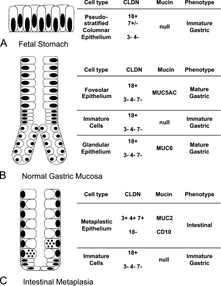
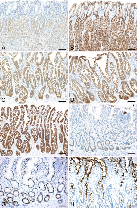
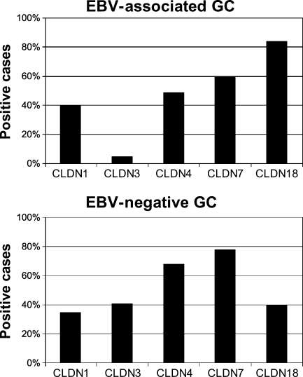
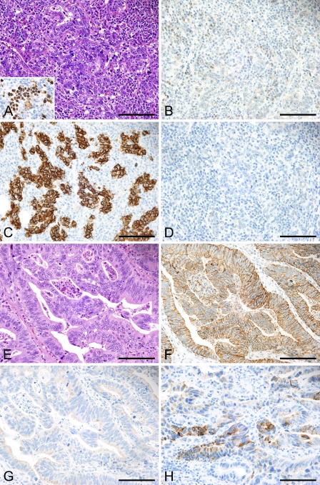
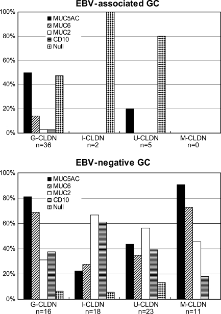
Epstein-Barr virus (EBV)-associated gastric carcinoma (GC) is a distinct subtype with characteristic clinicopathological features. To better characterize its cellular characteristics, 43 cases of EBV-associated GC, 68 cases of EBV-negative GC, and non-neoplastic gastric mucosa in adults and fetuses were examined immunohistochemically. We quantified the expression of the major tight-junction protein claudin (CLDN) -1, -3, -4, -7, and -18 together with gastric mucins (MUC5AC and MUC6), intestinal mucin (MUC2), and CD10. EBV-associated GC showed a high frequency of CLDN18 expression (84%) and a low frequency of CLDN3 expression (5%). This expression profile corresponded to that of normal gastric epithelium in adults and fetuses. Almost half of the EBV-associated GC cases demonstrated gastric mucin expression, whereas the other half lacked mucin or CD10 expression. In contrast, as demonstrated by the expression profiles of CLDN3 and CLDN18, EBV-negative GC comprised a heterogeneous group of four different CLDN phenotypes: gastric, intestinal, mixed, and an undifferentiated type with variable expression patterns of mucins. These results indicate that EBV-associated GC is considerably homogenous with regard to cellular differentiation and that it preserves well the nature of the cells of origin. EBV-associated GC may undergo distinct carcinogenic processes, which differ from those of EBV-negative GC.


Figures

Figure 1
Figure 1
Claudin (CLDN) and mucin expression profiles in non-neoplastic gastric mucosa. Schematic illustration of normal gastric mucosa in the fetus (A), adult (B), and gastric mucosa with intestinal metaplasia (C). The expression patterns of each CLDN and mucin and their respective phenotypes are shown by different cell types. In the fetal stomach, CLDN7 expression was observed at the 9th week of gestational age, but diminished by the 23rd week.
Figure 2
Figure 2
Immunoreactivity for CLDN3 and CLDN18 in non-neoplastic gastric mucosa. In normal gastric mucosa, the expression of CLDN3 (A) is negative, whereas that of CLDN18 (B) is positive in both the foveolar and glandular epithelia. Gastric mucosa with intestinal metaplasia is positive for CLDN3 (C), -4 (D), and -7 (E), and the expression of CLDN18 (F) is diminished, with only scattered positive cells remaining at the base. MUC2 expression (G) and CD10 expression (H) are also observed. The fetal stomach at the 9th week of gestational age is lined by pseudostratified columnar epithelia (I), which are positive for CLDN18 (J) and CLDN7 (K). (L–P) Fetal stomach at the 23rd week of gestational age. CLDN18 expression remains (M), whereas CLDN7 expression is diminished (N). Surface epithelium is positive for MUC5AC (O), and proper gland epithelium is positive for MUC6 (P). Bar = 100 μm.
Figure 2
Figure 2
Immunoreactivity for CLDN3 and CLDN18 in non-neoplastic gastric mucosa. In normal gastric mucosa, the expression of CLDN3 (A) is negative, whereas that of CLDN18 (B) is positive in both the foveolar and glandular epithelia. Gastric mucosa with intestinal metaplasia is positive for CLDN3 (C), -4 (D), and -7 (E), and the expression of CLDN18 (F) is diminished, with only scattered positive cells remaining at the base. MUC2 expression (G) and CD10 expression (H) are also observed. The fetal stomach at the 9th week of gestational age is lined by pseudostratified columnar epithelia (I), which are positive for CLDN18 (J) and CLDN7 (K). (L–P) Fetal stomach at the 23rd week of gestational age. CLDN18 expression remains (M), whereas CLDN7 expression is diminished (N). Surface epithelium is positive for MUC5AC (O), and proper gland epithelium is positive for MUC6 (P). Bar = 100 μm.
Figure 3
Figure 3
CLDN expression in Epstein-Barr virus (EBV)-associated gastric carcinoma (GC) and EBV-negative GC. The percentage of positive cases for each CLDN in EBV-associated GC and EBV-negative GC is shown. The expression of CLDN18 is significantly higher in EBV-associated GC compared with EBV-negative GC (p<0.0001, Fisher's exact test), whereas CLDN3 expression is significantly higher in EBV-negative GC compared with EBV-associated GC (p<0.0001, Fisher's exact test).
Figure 4
Figure 4
Immunoreactivity for CLDN and mucin in gastric carcinoma EBV-associated GC (A–D). Dense lymphocytic infiltrate within the tumor represents lymphoepithelioma-like histology (A, inset: EBER1 in situ hybridization). This case is negative for CLDN3 (B). Strong membranous staining for CLDN18 highlights tumor cells (C). The tumor is negative for gastric mucin (MUC6) (D). (E–H) EBV-negative GC. This case shows moderately differentiated tubular structures (E), which are positive for CLDN3 (F), and negative for CLDN18 (G). An intestinal mucin marker, MUC2 (H) is positive in tumor cells. Bar = 100 μm.
Figure 5
Figure 5
Associations between CLDN phenotype and mucin expression. Percentages of positive cases for mucin markers and CD10 in each CLDN phenotype are shown. Cases without expression of any of these markers were defined as the null type; the percentage of each CLDN phenotype is given. G-CLDN, gastric CLDN; I-CLDN, intestinal CLDN; U-CLDN, undifferentiated CLDN; M-CLDN, mixed CLDN.

References

    1. Barua RR, Uozaki H, Chong JM, Ushiku T, Hino R, Chang MS, Nagai H, et al. (2006) Phenotype analysis by MUC2, MUC5AC, MUC6, and CD10 expression in Epstein-Barr virus-associated gastric carcinoma. J Gastroenterol 41:733–739 - PubMed
    1. Chang MS, Uozaki H, Chong JM, Ushiku T, Sakuma K, Ishikawa S, Hino R, et al. (2006) CpG island methylation status in gastric carcinoma with and without infection of Epstein-Barr virus. Clin Cancer Res 12:2995–3002 - PubMed
    1. Chong JM, Sakuma K, Sudo M, Ushiku T, Uozaki H, Shibahara J, Nagai H, et al. (2003) Global and non-random CpG-island methylation in gastric carcinoma associated with Epstein-Barr virus. Cancer Sci 94:76–80 - PMC - PubMed
    1. Fukayama M, Hayashi Y, Iwasaki Y, Chong J, Ooba T, Takizawa T, Koike M, et al. (1994) Epstein-Barr virus-associated gastric carcinoma and Epstein-Barr virus infection of the stomach. Lab Invest 71:73–81 - PubMed
    1. Fukayama M, Hino R, Uozaki H (2008) Epstein-Barr virus and gastric carcinoma: virus-host interactions leading to carcinoma. Cancer Sci 99:1726–1733 - PMC - PubMed

Publication types

MeSH terms