. 2009 May 4;185(3):397-407.
doi: 10.1083/jcb.200903088. Epub 2009 Apr 27.

Active establishment of centromeric CENP-A chromatin by RSF complex

Affiliations

Active establishment of centromeric CENP-A chromatin by RSF complex

Marinela Perpelescu et al. J Cell Biol. .

Abstract

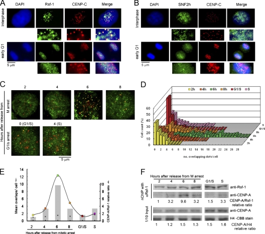
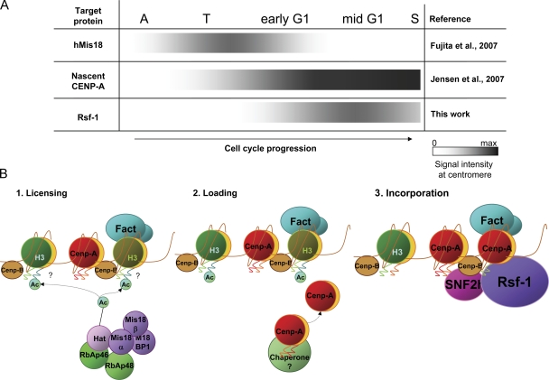
Centromeres are chromosomal structures required for equal DNA segregation to daughter cells, comprising specialized nucleosomes containing centromere protein A (CENP-A) histone, which provide the basis for centromeric chromatin assembly. Discovery of centromere protein components is progressing, but knowledge related to their establishment and maintenance remains limited. Previously, using anti-CENP-A native chromatin immunoprecipitation, we isolated the interphase-centromere complex (ICEN). Among ICEN components, subunits of the remodeling and spacing factor (RSF) complex, Rsf-1 and SNF2h proteins, were found. This paper describes the relationship of the RSF complex to centromere structure and function, demonstrating its requirement for maintenance of CENP-A at the centromeric core chromatin in HeLa cells. The RSF complex interacted with CENP-A chromatin in mid-G1. Rsf-1 depletion induced loss of centromeric CENP-A, and purified RSF complex reconstituted and spaced CENP-A nucleosomes in vitro. From these data, we propose the RSF complex as a new factor actively supporting the assembly of CENP-A chromatin.


Figures

Figure 1.
Figure 1.
RSF complex interacts with CENP-A chromatin at the mononucleosome level. (A) Ethidium bromide–stained gel showing DNA nucleosomal ladder of bulk chromatin that had undergone mild (200 U/ml × min, lane 1) and extensive (12,000 U/ml × min, lane 2) digestion with micrococcal nuclease in the presence of 0.3 M NaCl. Percentages of mono-, di-, and tri-nucleosomes for lane 2 are shown at the right. (B) The bulk chromatin of asynchronous HeLa cells was mildly digested with MNase as in A, immunoprecipitated using anti-CENP-A (lane 2), anti-CENP-H (lane 3), and anti-SNF2h antibodies (lane 4), and nonimmune IgG (lane 5). Input bulk chromatin, one-tenth of the other samples, was applied in lane 1. Samples were run on a 5–20% gel and immunostained with ACA serum on the same membrane. The apparent molecular weight of CENP-A is calculated as ∼17 kD. The bottom panel shows a Coomassie blue–stained duplicate SDS-PAGE gel with histones as loading controls. The ratio of CENP-A to histone H4 of lanes 2 and 4 relative to the input sample (lane 1) was calculated and is depicted at the bottom. (C) Identification of proteins recovered by nChIP with anti-SNF2h antibodies of bulk chromatin after mild digestion with MNase (A, lane 1). The proteins eluted using the antigen peptide p4c were separated on 7.5% SDS-PAGE, transferred to a PVDF membrane, and identified with Coomassie Brilliant blue staining (lane 1) or with anti-Rsf-1 (lane 2) and anti-SNF2h (lane 3) antibodies. Fig. S1 shows mass spectrometry analysis of an identical sample. The apparent molecular weight of Rsf-1 or SNF2h is calculated from the molecular weight marker as ∼250 kD or ∼135 kD, respectively. (D) The anti-CENP-A nChIP samples of mild (lane 1) and extensive (lane 2) MNase digestion were separated in a 5–20% gel and immunostained using ACA serum, and anti-Rsf-1 and -SNF2h antibodies. (E) Western blot analysis of the nChIP samples using antibodies against SNF2h (lanes 1 and 2), Rsf-1 (lanes 3 and 4), and control IgG (lanes 5 and 6) after mild (lanes 1, 3, and 5) and extensive (2, 4, and 6) MNase digestion of bulk chromatin.
Figure 2.
Figure 2.
Rsf-1/RSF transits centromeres in middle G1. (A and B) Immunolocalization of Rsf-1 (A, green) and SNF2h (B, green) regarding centromeres recognized by anti-CENP-C antibodies (red). DAPI (blue) was used for DNA staining. The enlarged insets show clear overlapping and nonoverlapping. (C) HeLa cells synchronized at G1 at 2, 4, 6, and 8 h after release from TN16-induced mitotic arrest; G1/S border at 0 h and S phase at 4 h after release from thymidine block were coimmunofluorescent stained using mouse monoclonal antibody against anti-Rsf-1 (green) and rat monoclonal antibody against CENP-A (red). (D) Quantification of Rsf-1 and CENP-A overlapping signals in each of the 100 cells evaluated per interphasic time-point. (E) Interaction of Rsf-1 with CENP-A is maximal at 6 h after release from mitotic arrest. The colored boxes show results from D as the mean number of Rsf-1 and CENP-A overlapping signals per cell for each interphasic stage (n = 1). Shaded bars show the relative ratio of CENP-A to Rsf-1 recovered after nChIP with anti-Rsf-1 antibody from F, of the cells synchronized as in C (n = 1). Asterisks show relative ratio of CENP-A to histone H4 of input samples in F (n = 1). (F) Cell cycle dependency of association of the Rsf-1/RSF complex with CENP-A chromatin. Bulk chromatin of approximately 3 × 10⁸ HeLa cells synchronized as in C was extensively digested with MNase (4,000 U/ml x min) and subjected to nChIP using anti-Rsf-1 antibody. The relative amounts of CENP-A to Rsf-1 were determined on immunoblotted membrane by densitometer tracing of the bands and drawn in E as shaded columns. Input bulk chromatin fractions (one-tenth of nChIP sample) were separately run on two duplicate gels, one transferred to a membrane and probed for CENP-A, and one stained with Coomassie Brilliant blue to visualize histone H4. The amounts of CENP-A relative to histone H4 in input samples were measured and depicted as asterisks in E. Relative molecular mass of Rsf-1 or CENP-A is ∼250 kD or ∼17 kD, respectively.
Figure 3.
Figure 3.
siRNA-mediated depletion of Rsf-1 and SNF2h of RSF delays cell cycle progression and causes kinetochore misalignment. (A) Immunostaining of HeLa cells with anti-Rsf-1 (top panels) and anti-SNF2h antibodies (bottom panels) at 48 h after mock and specific siRNA transfection. (B) Western blot analysis of Rsf-1 and SNF2h levels in cells siRNA-depleted of Rsf-1 (right) or SNF2h (left) at 0, 24, 48, and 72 h post-transfection. WB, Western blot; CBB, Coomassie Brilliant blue staining of a gel-duplicate. Rsf-1 = ∼250 kD; SNF2h = ∼135 kD. (C) Results of microscopic observation of cell stage distribution in G2 (blue), prometaphase (green), metaphase (red), and anaphase/telophase (yellow) at 2–6 d after siRNA-transfection of Rsf-1, SNF2h, and co-depletion (RSF) (n = 200–400 cells/depletion type/day post-transfection). Discrimination of each cell stage was based on the shape and condensation degree of chromosomes, distribution pattern of centromeres, and/or presence or absence of mitotic microtubules. Differences between Pm and M are shown in E as an example. (D) Prometaphase to metaphase cell ratio at 4 d post-transfection for each depletion type in C, and siCENP-A–transfected cells (see also Fig. S3). (E) Co-immunofluorescent staining of HeLa prometaphase and metaphase cells in control and siRNA-depleted samples using anti-β-tubulin (green) and anti-CENP-C (red) antibodies. DNA is shown in blue. (F) Percentage of abnormal prometaphase and metaphase cells at 4 d post-transfection, from C (n = 1).
Figure 4.
Figure 4.
Rsf-1 depletion impairs deposition of CENP-A to the centromeric core chromatin. (A) Immunoblot of Rsf-1 and CENP-A in CENP-A– (a) and Rsf-1–depleted (b–d) cells. Analysis was performed in whole-cell lysate (a and b), nuclei after low salt (c), and high salt (0.6 M NaCl) wash (d). “Core chromatin” represents the high salt insoluble nuclear fraction and “nuclear extract” represents the high salt soluble nuclear fraction, prepared as schematically depicted in e. Depletion was induced by two rounds of specific siRNA transfection at d 0 and d 2 before sample preparation at d 5 for Rsf-1, and d 4 for CENP-A. The intensity of each CENP-A band of mock and siRNA-treated sample was quantified, and the intensity relative to mock sample is shown at the bottom of each lane. Histone H4 was visualized on a duplicate gel stained with Coomassie Brilliant blue. Rsf-1 = ∼250 kD; SNF2h = ∼135 kD; CENP-A = ∼17 kD. (B) The effect of high salt wash on centromere stability of CENP-A immunofluorescent signals, in siCENP-A– and siRsf-1–depleted cells. Mock-, CENP-A–, and Rsf-1–depleted cells were fixed and washed in low salt (0.15 M NaCl; top) or high salt (0.5 M NaCl; bottom). Depletion of each protein was induced by two rounds of siRNA transfection as in A. Nuclei were visualized by DAPI staining. Images showing CENP-A (green) and CENP-C (red) were merged. (C) Quantification of the CENP-A fluorescent signals on photographed cells prepared in B. The intensity of the fluorescent signal was measured for each of 300–1,000 cells per depletion type, and the mean intensity value per cell was normalized with mock sample in each washing condition. Error bars represent ± SEM.
Figure 5.
Figure 5.
RSF complex can reconstitute and regularly space CENP-A nucleosomes using CENP-A core histones and naked DNA template. (A) Partially purified Flag-tagged (2 µl, left) and native (35 µl, right) RSF fractions were resolved on 7.5% SDS-PAGE and visualized with Coomassie Brilliant blue. Concentration of Rsf-1 was measured by densitometer tracing as 50 ng/µl for recombinant and 1 ng/µl for native fraction using BSA as control protein. (B) 15% SDS-PAGE stained with Coomassie Brilliant blue showing an equimolar ratio of reconstituted H3 (left) and CENP-A (right) core histones. CENP-A = ∼17 kD. (C) RSF-mediated H3 and CENP-A nucleosomes assembly assay. The ability of the RSF complex to reconstitute and space H3 and CENP-A nucleosomes was evaluated by the presence of periodic nucleosomal ladders following partial micrococcal nuclease digestion at two different dilutions (lanes 1–14) or a single dilution (15–18). Reactions were performed without (lanes 1–2), or with 10 µl each of recombinant (lanes 3–8) or native RSF fraction (9–18). The native fraction's activity was inhibited by immunodepletion with anti-Rsf-1 antibody (lane 17) and restored by the readdition of the same RSF sample (lane 18). Immunodepletion reduced Rsf-1 to ∼15% as detected from the analysis of the immunoblot shown under lanes 16 and 17. The nucleosomal ladder was resolved on a 1% agarose gel and stained with ethidium bromide.
Figure 6.
Figure 6.
Sequential establishment in two steps of centromeric CENP-A chromatin. (A) Timing cascade of centromere localization of hMis18, nascent CENP-A, and Rsf-1. Dark shade gradients represent gradual accumulation or loss of the indicated factor at centromeres throughout the cell cycle progression from anaphase to the synthetic phase. (B) Putative two-step model for CENP-A nucleosomes establishment at centromeres. Establishment of CENP-A nucleosomes at the centromere region occurs after a licensing process engaged by the Mis18 complex (1) in two sequences: first, loading of CENP-A occurs at or around centromere regions (2), followed by incorporation/assembly into the centromeric nucleosomes and spacing by RSF remodeling function (3).

References

    1. Ando S., Yang H., Nozaki N., Okazaki T., Yoda K. 2002. CENP-A, -B, and -C chromatin complex that contains the I-type alpha-satellite array constitutes the prekinetochore in HeLa cells.Mol. Cell. Biol. 22:2229–2241 - PMC - PubMed
    1. Belotserkovskaya R., Oh S., Bonarenko V.A., Orphanides B., Studitsky V.M., Reinberg D. 2003. FACT facilitates transcription-dependent nucleosome alteration.Science. 301:1090–1093 - PubMed
    1. Black B.E., Bassett E.A. 2008. The histone variant CENP-A and centromere specification.Curr. Opin. Cell Biol. 20:91–100 - PubMed
    1. Black B.E., Foltz D.R., Chakravarthy S., Luger K., Woods V.L., Jr., Cleveland D.W. 2004. Structure determinants for generating centromeric chromatin.Nature. 430:578–582 - PubMed
    1. Buchwitz B.J., Ahmad K., Moore L.L., Roth M.B., Henikoff S. 1999. A histone-H3-like protein in C. elegans.Nature. 401:547–548 - PubMed

Publication types

MeSH terms