Review
. 2009 Aug;66(15):2427-43.
doi: 10.1007/s00018-009-0030-6. Epub 2009 Apr 28.

Intracellular protein degradation in mammalian cells: recent developments

Affiliations
Review

Intracellular protein degradation in mammalian cells: recent developments

Erwin Knecht et al. Cell Mol Life Sci. 2009 Aug.

Abstract

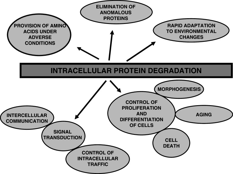
In higher organisms, dietary proteins are broken down into amino acids within the digestive tract but outside the cells, which incorporate the resulting amino acids into their metabolism. However, under certain conditions, an organism loses more nitrogen than is assimilated in the diet. This additional loss was found in the past century to come from intracellular proteins and started an intensive research that produced an enormous expansion of the field and a dispersed literature. Therefore, our purpose is to provide an updated summary of the current knowledge on the proteolytic machinery involved in intracellular protein degradation and its physiological and pathological relevance, especially addressed to newcomers in the field who may find further details in more specialized reviews. However, even providing a general overview, this is an extremely wide field and, therefore, we mainly focus on mammalian cells, while other cells will be mentioned only for comparison purposes.


Figures

Fig. 1
Fig. 1
Intracellular protein degradation fulfills many functions in mammalian cells. This process, in addition to survey and degrade invasive microorganisms, brings several advantages for the cells and organisms as indicated, including: (1) the provision, under adverse conditions such as starvation, of amino acids to be used as an energy source and/or for the synthesis of proteins essential for survival; (2) the rapid degradation of defective proteins; (3) a better and quick adaptation of cell metabolism to the environment, by modifying the levels of proteins; (4) the control of many important processes such as cell proliferation (division and growth) and differentiation, morphogenesis and regression of retired tissues, cell aging and death (by necrosis and apoptosis), etc.; and (5) signal transduction, including the regulation of intercellular (for example, in the generation of antigenic peptides) and intracellular communication, and the control of protein traffic. All this, taken together, justifies that intracellular protein degradation is universal
Fig. 2
Fig. 2
Different regulatory particles can bind to two ends or to one end of the 20S proteasome. The best known of these complexes is formed when 20S proteasomes bind, in an ATP-dependent process, a 19S regulatory particle (RP), composed of a base and a lid, with the indicated subunits, to produce the 26S proteasome that degrades polyubiquitinated proteins. Upon substrate degradation and ATP hydrolysis, the 26S proteasome separates into four parts, 20S proteasome, lid, base and subunit Rpn10, suggesting a cycle of proteasome assembly and dissociation during proteolysis. Also, the 20S proteasomes can form the PA28-proteasomes when PA28α/β heteroheptamers, whose expression is intensified in the presence of γ-interferon, are bound. These proteasomes and the immunoproteasomes, which are formed from 20S proteasomes when three interferon-inducible subunits (β1i, β2i and β5i) substitute the corresponding three constitutive subunits in the nascent proteasomes, facilitate the generation of antigenic peptides. The functions of other proteasome complexes with PA200, PA28γ, PI31 or PR39 are still poorly known. In addition, hybrid proteasomes are formed when 20S proteasomes bind two different regulatory particles at each end, but this has been firmly established only for proteasomes with the 19S and the PA28α/β RPs
Fig. 3
Fig. 3
The ubiquitin-proteasome system is the major non-lysosomal pathway of intracellular proteolysis. The process has two parts: (1) substrate polyubiquitination (on the left) and degradation of the tagged protein by the downstream 26S/30S proteasome (on the right). Canonical ubiquitination involves three steps: (1) activation of ubiquitin by E1 enzyme in an ATP-dependent manner, (2) transfer of ubiquitin from E1 to an E2 enzyme, and (3) direct or indirect transfer of ubiquitin to a specific protein substrate recognized by an E3 enzyme. Further incorporation of other activated ubiquitin molecules generates a polyubiquitin chain. Although polyubiquitination is a reversible process, most proteins with polyubiquitin chains of four or more ubiquitins (Ub) attached to an inner Lys of the substrate and each other by their Lys48 are recognized by a subunit (Ub-R) on the 19S regulatory particle of the 26S/30S proteasome, deubiquitinated to generate free ubiquitin, and degraded in the 20S catalytic core to peptides (P) by processes that also consume ATP
Fig. 4
Fig. 4
Ubiquitination of proteins controls many cellular processes by either proteolytic or non-proteolytic means. The figure illustrates some of the possibilities of ubiquitination (Ubq) of a protein that are important to decide its final fate. a Polyubiquitination at Lys48 in the ubiquitin molecule is the canonical signal for the 26S/30S proteasomal-dependent degradation of cellular proteins. b In contrast, mono or oligoubiquitination (<4 ubiquitins) targets plasma membrane proteins, via endocytosis, to early endosomes, multivesicular bodies and, finally, lysosomes, where they are degraded. In both proteasomal and lysosomal degradation, ubiquitin is first released for further use. Monoubiquitination can also target proteins to different cell compartments or have other roles as mentioned in the text. c Polyubiquitination at Lys63 in the ubiquitin molecule can serve as a non-degradative signal functioning in several cell processes as indicated
Fig. 5
Fig. 5
Proteins can be incorporated into lysosomes for degradation by different mechanisms. Macroautophagy (upper part) is the main lysosomal degradative route and involves the sequestration by a segregating structure of large areas of cytoplasm, typically including whole organelles, to make up autophagosomes. These pre-lysosomes fuse with endosomes and lysosomes to form autolysosomes that degrade its cytoplasmic content. In addition to macroautophagy, other mechanisms (lower part) have been described whereby lysosomes could also participate in intracellular protein degradation, including endocytosis, crinophagy, microautophagy and chaperone-mediated autophagy (for details, see text)
Fig. 6
Fig. 6
Covalent binding of LC3 to phosphatidylethanolamine (PE) is essential for autophagosome formation. In mammalian cells, cytosolic LC3-I (Atg8 in yeast) is synthesized as a precursor (Pro-LC3-I). Immediately after its synthesis, a C-terminal fragment is cleaved by Atg4 to produce LC3-I with an exposed glycine residue that binds covalently to PE on the pre-autophagosome membrane to form LC3-II. In this process, the mammalian homologues of Atg7 and Atg3 work, respectively, as the E1- and E2-like enzymes of the ubiquitination system, and the Atg16L complex probably functions as an E3-like enzyme. Once the autophagosome is formed, LC3-II localizes both at the cytosolic and luminal faces of its double membrane. After fusion of the autophagosome with endosomes/lysosomes to form an autolysosome, the luminal LC3-II is degraded by lysosomal cathepsins, while Atg4 recycles LC3-I and PE from LC3-II on the cytosolic face of the autolysosome membrane
Fig. 7
Fig. 7
Macroautophagy is mainly regulated by the TOR signalling pathway. The kinase mammalian TOR (mTOR) is the principal negative regulator of macroautophagy. mTOR exists in two distinct complexes: one which is sensitive to the drug rapamycin (C1) and a second, which is not (C2). Regulatory amino acids activate mTORC1 and, thus, inhibit macroautophagy by a still unknown pathway. Insulin and some growth factors inhibit macroautophagy through the insulin receptors (IRS) and class I PI3K (PI3K–I) that produces phosphatidylinositol 3,4,5-trisphosphate (PIP3). The well-known tumor suppressor phosphatase and tensin homologue (PTEN) antagonizes the activity of PI3K–I by dephosphorylating PIP3 to.phosphatidylinositol 4,5-bisphosphate (PIP2). PIP3 recruits the oncoprotein Akt (also called proteinase kinase B or PKB) to the cytosolic face of the plasma membrane where it is phosphorylated at different residues (T308 and S473) by the 3-phosphoinositide-dependent kinase 1 (PDK1) and the mTORC2 complex, respectively. Akt phosphorylates one member (TSC2) of the tuberous sclerosis complex TSC1 (hamartin), TSC2 (tuberin), which is inhibited. TSC1,2 is a negative regulator with GAP activity of the small GTPase Rheb, which in its GTP-bound form activates mTOR-Raptor, inhibiting macroautophagy. On the other hand, a high AMP/ATP ratio or an increase in cytosolic Ca++ produces, through the serine/threonine protein kinase LKB1 or the Ca++/calmodulin-dependent protein kinase kinase beta (CaMKK-β), respectively, the phosphorylation and the activation of the AMP-activated protein kinase (AMPK), which increases macroautophagy, via TSC1,2. mTORC1 phosphorylates its substrates, including 4EBP1 and S6K1, and rapamycin is a strong and quite specific inhibitor of mTORC1. To prevent the overactivation of the mTOR signalling pathway, S6K1 can phosphorylate the insulin receptor substrate 1 (IRS1), inhibiting in this way the proximal part of the insulin signalling pathway. Finally, other signalling pathways are also known to regulate macroautophagy, including those involving the MAP kinases ERK1,2 (Ras-Raf-MEK-ERK pathway) and the p38MAPK, which activate and inhibit, respectively, macroautophagy. ERK1,2, like Akt, also inhibits TSC1,2, while how p38MAPK inhibits macroautophagy is less known

References

    1. Ciechanover A. Intracellular protein degradation: from a vague idea through the lysosome and the ubiquitin-proteasome system, and onto human diseases and drug targeting. Cell Death Differ. 2005;12:1178–1190. doi: 10.1038/sj.cdd.4401692. - DOI - PubMed
    1. Hershko A. The ubiquitin system for protein degradation and some of its roles in the control of the cell division cycle. Cell Death Differ. 2005;12:1191–1197. doi: 10.1038/sj.cdd.4401702. - DOI - PubMed
    1. Varshavsky A. Discovery of cellular regulation by protein degradation. J Biol Chem. 2008;283:34469–34489. doi: 10.1074/jbc.X800009200. - DOI - PMC - PubMed
    1. Yewdell JW, Nicchitta CV. The DRiP hypothesis decennial: support, controversy, refinement and extension. Trends Immunol. 2006;27:368–373. doi: 10.1016/j.it.2006.06.008. - DOI - PubMed
    1. Grisolía S, Hernández-Yago J, Knecht E. Regulation of mitochondrial protein concentration: a plausible model which may permit assessing protein turnover. Curr Topics Cell Regul. 1985;27:387–396. - PubMed

Publication types

LinkOut - more resources