

Varicella zoster virus is not a disease-relevant antigen in multiple sclerosis
- PMID: 19399839
- PMCID: PMC2844106
- DOI: 10.1002/ana.21605
Varicella zoster virus is not a disease-relevant antigen in multiple sclerosis
Abstract
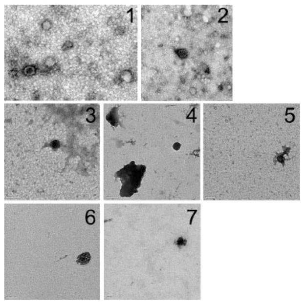
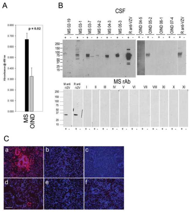
Herpesvirions and varicella zoster virus (VZV) DNA were recently reported in all 15 cerebrospinal fluid (CSF) samples from patients with relapsing-remitting multiple sclerosis (MS) obtained within 1 week of exacerbation. Using identical electron microscopic and polymerase chain reaction techniques, including additional primer sets representing different regions of the VZV genome, we found no herpesvirions or VZV DNA in MS CSF or acute MS plaques. Although enzyme-linked immunosorbent assay analysis demonstrated a higher titer of VZV antibody in MS CSF than in inflammatory control samples, recombinant antibodies prepared from clonally expanded MS CSF plasma cells did not bind to VZV. VZV is not a disease-relevant antigen in MS.
Conflict of interest statement
Potential conflict of interest: Nothing to report.
Figures
Similar articles
-
Varicella-zoster virus in cerebrospinal fluid at relapses of multiple sclerosis.Ann Neurol. 2008 Mar;63(3):303-11. doi: 10.1002/ana.21316. Ann Neurol. 2008. PMID: 18306233
-
Varicella-Zoster Virus in Cerebrospinal Fluid at Relapses of Multiple Sclerosis is Infective in Vitro.Arch Med Res. 2018 Jul;49(5):350-355. doi: 10.1016/j.arcmed.2018.10.001. Epub 2018 Oct 17. Arch Med Res. 2018. PMID: 30342846
-
The participation of varicella zoster virus in relapses of multiple sclerosis.Clin Neurol Neurosurg. 2014 Apr;119:44-8. doi: 10.1016/j.clineuro.2013.12.020. Epub 2014 Jan 10. Clin Neurol Neurosurg. 2014. PMID: 24635924
-
Brief presence of varicella-zoster vral DNA in mononuclear cells during relapses of multiple sclerosis.Arch Neurol. 2004 Apr;61(4):529-32. doi: 10.1001/archneur.61.4.529. Arch Neurol. 2004. PMID: 15096401
-
On the viral hypothesis of multiple sclerosis: participation of varicella-zoster virus.J Neurol Sci. 2007 Nov 15;262(1-2):113-6. doi: 10.1016/j.jns.2007.07.001. Epub 2007 Jul 30. J Neurol Sci. 2007. PMID: 17663004 Review.
Cited by
-
Viral Proteins with PxxP and PY Motifs May Play a Role in Multiple Sclerosis.Viruses. 2022 Jan 28;14(2):281. doi: 10.3390/v14020281. Viruses. 2022. PMID: 35215874 Free PMC article. Review.
-
Antigen microarrays identify CNS-produced autoantibodies in RRMS.Neurology. 2012 Feb 21;78(8):532-9. doi: 10.1212/WNL.0b013e318247f9f3. Epub 2012 Jan 18. Neurology. 2012. PMID: 22262743 Free PMC article.
-
Antibodies produced by clonally expanded plasma cells in multiple sclerosis cerebrospinal fluid.Ann Neurol. 2009 Jun;65(6):639-49. doi: 10.1002/ana.21641. Ann Neurol. 2009. PMID: 19557869 Free PMC article.
-
Varicella zoster virus and relapsing remitting multiple sclerosis.Mult Scler Int. 2011;2011:214763. doi: 10.1155/2011/214763. Epub 2011 Mar 30. Mult Scler Int. 2011. PMID: 22096629 Free PMC article.
-
Subclinical reactivation of varicella zoster virus in all stages of HIV infection.J Neurol Sci. 2011 May 15;304(1-2):22-4. doi: 10.1016/j.jns.2011.02.030. Epub 2011 Mar 17. J Neurol Sci. 2011. PMID: 21419427 Free PMC article.
References
-
- Bennett JL, Yu X, Gilden DH, et al. Infectious agents and multiple sclerosis. In: Raine CS, McFarland HF, Hohlfeld R, editors. Multiple sclerosis: a comprehensive text. New York: Saunders Elsevier; 2008. pp. 226–236.
-
- Sotelo J, Martinez-Palomo A, Ordonez G, et al. Varicella-zoster virus in cerebrospinal fluid at relapses of multiple sclerosis. Ann Neurol. 2008;63:303–311. - PubMed
-
- Yu X, Gilden DH, Ritchie AM, et al. Specificity of recombinant antibodies generated from multiple sclerosis cerebrospinal fluid probed with a random peptide library. J Neuroimmunol. 2006;172:121–131. - PubMed
Publication types
MeSH terms
Substances
Grants and funding
LinkOut - more resources
Full Text Sources
