. 2009 Jul 1;515(1):15-30.
doi: 10.1002/cne.22037.

Effect of levodopa priming on dopamine neuron transplant efficacy and induction of abnormal involuntary movements in parkinsonian rats

Affiliations

Effect of levodopa priming on dopamine neuron transplant efficacy and induction of abnormal involuntary movements in parkinsonian rats

Kathy Steece-Collier et al. J Comp Neurol. .

Abstract

Clinical trials of neural grafting for Parkinson's disease (PD) have produced variable, but overall disappointing, results. One particular disappointment has been the development of aberrant motor complications following dopamine (DA) neuron grafting. Despite a lack of consistent benefit, the utility of dopamine neuron replacement remains supported by clinical and basic data. In a continued effort to elucidate factors that might improve this therapy, we used a parkinsonian rat model to examine whether pregraft chronic levodopa affected graft efficacy and/or graft-induced dyskinesia (GID) induction. Indeed, all grafted PD patients to date have had a pregraft history of long-term levodopa. It is well established that long-term levodopa results in a plethora of long-lasting neurochemical alterations and genomic changes indicative of altered structural and synaptic plasticity. Thus, therapeutic dopamine terminal replacement in a striatal environment complicated by such changes could be expected to lead to abnormal or inappropriate connections between graft and host brain and to contribute to suboptimal efficacy and/or postgraft GID behaviors. To investigate the effect of pregraft levodopa, one group of parkinsonian rats received levodopa for 4 weeks prior to grafting. A second levodopa-naïve group was grafted, and the grafts were allowed to mature for 9 weeks prior to introducing chronic levodopa. We report here that, in parkinsonian rats, preexposure to chronic levodopa significantly reduces behavioral and neurochemical efficacy of embryonic dopamine grafts. Furthermore, dopamine terminal replacement prior to introduction of chronic levodopa is highly effective at preventing development of levodopa-induced dyskinesias, and GID-like behaviors occur regardless of pregraft levodopa status.


Figures

Figure 1
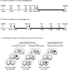
Figure 1. Experimental timeline
Animals were given a unilateral 6-OHDA-induced lesion of the nigrostriatal dopamine system, and lesion success confirmed with amphetamine rotations. Parkinsonian rats were grafted with either a localized single-site graft of 200,000 total VM cells, or widespread graft consisting of either 200,000 total VM cells deposited into 6 sites, or 500,000 total VM cells grafted into 2 sites. Sham grafted rats received cell-free media using all three implantation paradigms. (A) Grafts in the non-primed groups were allowed to mature prior to introduction of levodopa. Graft maturation and efficacy were tested in these animals with amphetamine rotations performed at 4, 6 and 8 weeks post-grafting. Nine weeks after grafting, this group began chronic levodopa (*12.5 mg/kg levodopa: 12.5 mg/kg benserazide, i.p.). (B) Primed groups received levodopa (*) beginning two weeks after 6-OHDA, and for four weeks prior to grafting. All animals were euthanized approximately 10 weeks after evaluation of graft + levodopa interaction. Abbreviations: LD= levodopa; Amph(et)= amphetamine; 200K= 200,000; 500K= 500,000. Stippled bar above timeline indicates levodopa administration.
Figure 2
Figure 2. Levodopa-induced Dyskinesia in Levodopa Primed (A) and Non-primed (B) rats
(A) In rats primed with levodopa there was a significant groups effect (F(3,185)= p<0.05) with all dopamine grafted groups showing a significant decrease in dyskinesia severity from sham grafted rats by week 4 post-grafting (*p<0.05). It is notable that parkinsonian rats grafted with the largest VM cell number (500,000) showed a more rapid decrease, with significant difference from sham-grafted rats by 2 weeks post-grafting (**p<0.05). There was, however, no significant difference in any of the dopamine graft recipients, regardless of the graft cell number or distribution, after 4 weeks post-grafting (p>0.05). (B) In non-primed rats that began chronic levodopa 9 weeks post-grafting, again, all dopamine grafted groups showed significantly less LIDs compared with sham-grafted rats (weeks 12, 16, 20 post-grafting, ##p<0.05). However, in contrast to primed rat, non-primed rats receiving 500,000 cells showed an additional significantly lower level of LIDs than rats receiving 200,000 cell grafts (weeks 12, # p<0.05; and 20 (# p<0.05) post-grafting).
Figure 3
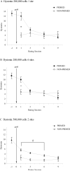
Figure 3. Total Body Dystonia in Levodopa Primed versus Non-Primed Grafted Rats
Rats receiving 200,000 VM cell grafts to 1 site (A) or 200,000 cells to 6 sites (B) both showed a significant difference between primed and non-primed rats only at rating session 1 (the first rating session following grafting; *p<0.001, +p=0.03). For rats receiving 500,000 VM cell grafts (C) non-primed rats showed significantly less dystonia on rating sessions 1,3 and 6 (#p<0.05) compared to their levodopa primed counterpart.
Figure 4
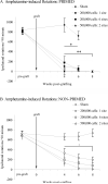
Figure 4. Amphetamine-induced Rotations in Levodopa Primed (A) and Non-primed (B) Rats
(A) Primed rats showed a significant decrease in amphetamine-induced rotational behavior weeks 4 through 8 post-grafting (* p<0.05, all 200,000 VM grafts versus sham; **p>0.05 500,000 VM grafts: 4 wks versus 8 weeks). (B) Non-primed rats showed a significant decrease in amphetamine-induced rotations at 4, 6 and 8 weeks post-grafting (+p<0.05, all VM grafts versus sham), with complete reversal in all three dopamine graft groups at week 8 post-grafting. Comparative analyses between primed and non-primed groups showed no significant differences at any of the time-points analyzed (p<0.05).
Figure 5
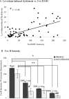
Figure 5. Dorsolateral FosB/ΔFosB Staining Intensity: Correlation with Levodopa-induced Dyskinesias
(A) The percent increase in FosB/ΔFosB staining intensity in the dopamine-depleted dorsolateral striatum over that of the intact hemisphere correlated significantly with the severity of levodopa-induced dyskinesia in all rats (p<0.001). (B) The implantation of dopamine cells to the dopamine-depleted striatum ameliorated the increase in FosB/ΔFosB staining intensity in the dorsolateral striatum in all grafted groups in both primed (+ p<0.001) and non-primed (++ p<0.001) rats. In rats receiving 200,000 cells to 1 site (# p=0.03) or 500,000 cells to 6 sites (## p=0.05), non-primed rats showed a significantly greater decrease in FosB/ΔFosB staining intensity compared to their levodopa primed counterpart.
Figure 6
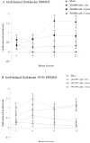
Figure 6. Graft-induced Dyskinesia Profiles for Levodopa Primed (A) and Non-Primed (B) Grafted Rats
Rats receiving 200,000 VM cell grafts to 1 site or 200,000 VM cells to 6 sites showed no significant difference between primed and non-primed rats at all rating sessions (p≥ 0.105). (A) Primed rats grafted with 200,000 VM cells into a single site, but not grafted into 6 sites, had significantly elevated level of GIDs at rating sessions 8 and 12 compared to sham grafted rats (p<0.025). Primed rats grafted with 500,000 VM cell showed significantly less graft-induced dyskinesia on rating session 3 (p=0.02). (B) Non-primed rats grafted with 200,000 VM cells, either into 1 or 6 sites, had significantly elevated level of GIDs at rating sessions 4, 8 and 12 compared to sham grafted rats (p<0.018). Rating Sessions: (A) For the primed cohort, the rating sessions corresponded to: ‘1’= -3 weeks (pre-graft), ‘2’= 2 weeks post-graft, ‘3’= 6 weeks post-graft, ‘4’= 10 weeks post-graft. (B) For the non-primed cohort, the rating sessions corresponded to: ‘1’= 9 wks post-graft (1 wk post-levodopa); ‘2’= 12 weeks post-graft, ‘3’= 16 weeks post-graft, ‘4’= 20 weeks post-graft.
Figure 7
Figure 7. TH- positive Graft Cell Number
The percentage of surviving TH-positive grafted cells (total TH-positive cells counted/total VM cells injected) did not differ significantly between any dopamine grafted primed and non-primed groups. (Raw cell counts: 200,000 cells to 1 site: primed= 2887±471, non-primed= 2429±437; 200,000 cells to 6 sites: primed= 2495±447, non-primed=2873±790, 500,000 cells to 2 sites: primed= 9680±2181, non-primed= 7377±1000).
Figure 8
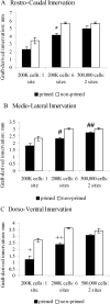
Figure 8. Area of TH-positive Innervation in Levodopa Primed and Non-primed dopamine Grafted Rats for the Rostro-caudal Axis (A), Medio-lateral Axis (B) and Dorso-ventral Axis (C)
(A) TH-positive fiber outgrowth in the rostral-caudal axis, revealed a significant increase for non-primed rats receiving 200,000 cells delivered to 6 sites compared to their primed counterpart group (*p<0.001). There was no significant difference between primed and non-primed rats in graft-derived reinnervation for rats receiving 200,000 cells to 1 injections site (p=0.10) or 500,000 cells (p>0.05). (B) In the medio-lateral axis, there was a significant increase in TH-positive outgrowth in the non-primed rats receiving 200,000 cells grafts to 6 sites (#p<0.001), and 500,000 cells (p<0.01). No significant effect of priming was noted in this dimension in rats receiving 200,000 cell grafts delivered to a single site (p=0.052). (C) In the dorso-ventral axis, there was a significant increase in TH-positive outgrowth in non-primed compared to primed groups for rats receiving 200,000 cell grafts to 1 site (+p<0.001) and 6 sites (++p<0.001), but not in rats receiving 500,000 cells (p>0.05).

References

    1. Andersson M, Hilbertson A, Cenci MA. Striatal fosB expression is causally linked with l-DOPA-induced abnormal involuntary movements and the associated upregulation of striatal prodynorphin mRNA in a rat model of Parkinson's disease. Neurobiol Dis. 1999;6:461–474. - PubMed
    1. Andersson M, Westin JE, Cenci MA. Time course of striatal DeltaFosB-like immunoreactivity and prodynorphin mRNA levels after discontinuation of chronic dopaminomimetic treatment. Eur J Neurosci. 2003;17:661–666. - PubMed
    1. Bolam JP, Izzo PN. The postsynaptic targets of substance P-immunoreactive terminals in the rat neostriatum with particular reference to identified spiny striatonigral neurons. Exp Brain Res. 1988;70:361–77. - PubMed
    1. Brown P, Williams D. Basal ganglia local field potential activity: character and functional significance in the human. Clin Neurophysiol. 2005;116(11):2510–9. Review. - PubMed
    1. Caligiuri MP, Lohr JB. Worsening of postural tremor in patients with levodopa-induced dyskinesia: a quantitative analysis. Clin Neuropharmacol. 1993;16:244–250. - PubMed

Publication types

MeSH terms