

In vitro and in vivo suppression of hepatocellular carcinoma growth by midkine-antisense oligonucleotide-loaded nanoparticles
- PMID: 19399928
- PMCID: PMC2675086
- DOI: 10.3748/wjg.15.1966
In vitro and in vivo suppression of hepatocellular carcinoma growth by midkine-antisense oligonucleotide-loaded nanoparticles
Abstract
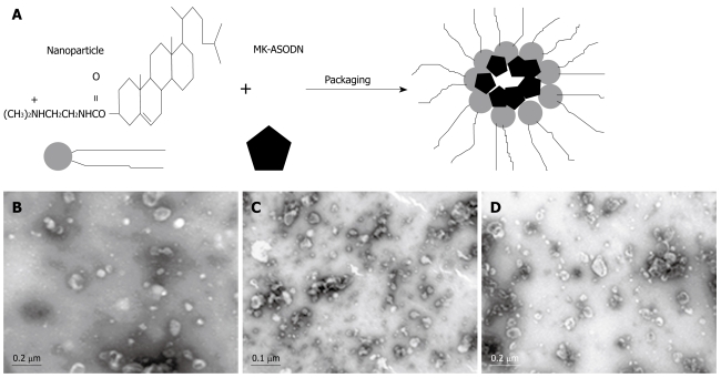
Aim: To synthesize antisense oligonucleotides (ASODNs) of midkine (MK), package the ASODNs with nanoparticles, and to inhibit hepatocellular carcinoma (HCC) growth using these nanoparticles.
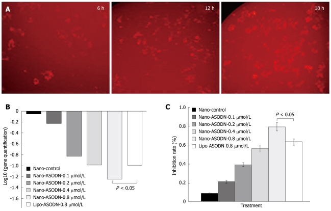
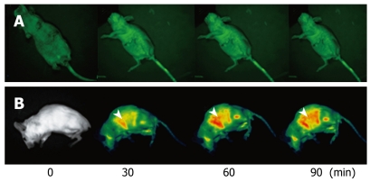
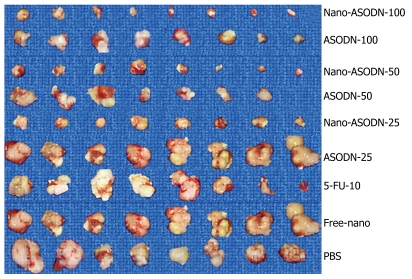
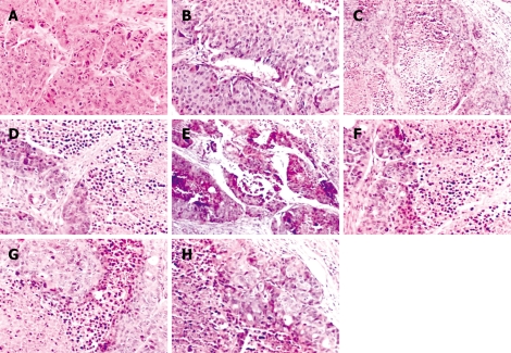
Methods: HepG2 cell proliferation was analyzed in vitro using the 3-(4,5-dimethythiazol-2-yl)-5-(3-carboxymethoxyphenyl)-2-(4-sulfophenyl)-2Htetrazolium, inner salt assay. The in vivo activity of nanoparticles delivering the MK-ASODNs was analyzed by histopathological and immunohistochemical staining and quantitative real time polymerase chain reaction (PCR).
Results: The in vitro proliferation of HepG2 cells was significantly inhibited by the nanoparticles packaged with MK-ASODNs (NANO-ASODNs). Furthermore, the NANO-ASODNs significantly inhibited the growth of HCC in the mouse model.
Conclusion: NANO-ASODNs can significantly suppress the growth of HCC in vitro and in vivo.
Figures
References
-
- Yang L, Parkin DM, Ferlay J, Li L, Chen Y. Estimates of cancer incidence in China for 2000 and projections for 2005. Cancer Epidemiol Biomarkers Prev. 2005;14:243–250. - PubMed
-
- Parkin DM, Bray F, Ferlay J, Pisani P. Global cancer statistics, 2002. CA Cancer J Clin. 2005;55:74–108. - PubMed
-
- Michikawa M, Xu RY, Muramatsu H, Muramatsu T, Kim SU. Midkine is a mediator of retinoic acid induced neuronal differentiation of embryonal carcinoma cells. Biochem Biophys Res Commun. 1993;192:1312–1318. - PubMed
-
- Garver RI Jr, Radford DM, Donis-Keller H, Wick MR, Milner PG. Midkine and pleiotrophin expression in normal and malignant breast tissue. Cancer. 1994;74:1584–1590. - PubMed
-
- Take M, Tsutsui J, Obama H, Ozawa M, Nakayama T, Maruyama I, Arima T, Muramatsu T. Identification of nucleolin as a binding protein for midkine (MK) and heparin-binding growth associated molecule (HB-GAM) J Biochem. 1994;116:1063–1068. - PubMed
Publication types
MeSH terms
Substances
LinkOut - more resources
Full Text Sources
