Review
. 2009:4:517-50.
doi: 10.1146/annurev.pathol.4.110807.092322.

The brainstem and serotonin in the sudden infant death syndrome

Affiliations
Review

The brainstem and serotonin in the sudden infant death syndrome

Hannah C Kinney et al. Annu Rev Pathol. 2009.

Abstract

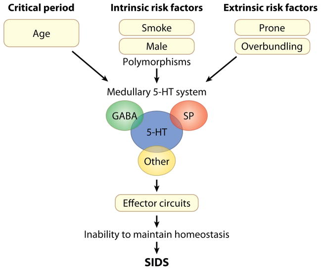
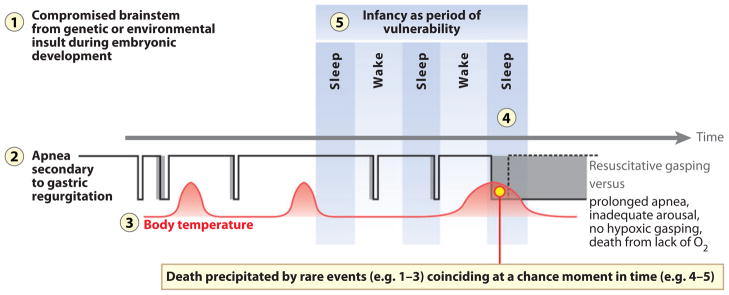
The sudden infant death syndrome (SIDS) is the sudden death of an infant under one year of age that is typically associated with sleep and that remains unexplained after a complete autopsy and death scene investigation. A leading hypothesis about its pathogenesis is that many cases result from defects in brainstem-mediated protective responses to homeostatic stressors occurring during sleep in a critical developmental period. Here we review the evidence for the brainstem hypothesis in SIDS with a focus upon abnormalities related to the neurotransmitter serotonin in the medulla oblongata, as these are the most robust pathologic findings to date. In this context, we synthesize the human autopsy data with genetic, whole-animal, and cellular data concerning the function and development of the medullary serotonergic system. These emerging data suggest an important underlying mechanism in SIDS that may help lead to identification of infants at risk and specific interventions to prevent death.


Figures

Figure 1
Figure 1
Schematic representation of our concept of sudden infant death syndrome (SIDS) as a disorder of homeostasis due to abnormalities in the medullary serotonin (5-HT) system. This disorder involves multiple neurotransmitters and neuromodulators that interact with the defective medullary 5-HT system; multiple intrinsic and extrinsic (exogenous) stressors acting simultaneously; and the critical developmental period, i.e., the first six months of postnatal life when 90% of SIDS cases occur. Abbreviations: GABA, γ-aminobutyric acid; SP, substance P.
Figure 2
Figure 2
(a) Sagittal view of whole human brain upon which the site of the 5-hydroxytryptamine (5-HT) neuronal cell bodies (red circles) and their projections (red lines) are superimposed. Serotonergic neurons are located in the brainstem in either the rostral or the caudal domain, each of which has different projections and functions. In the rostral domain, 5-HT neurons are present in the rostral pons and midbrain and project rostrally to the cerebral cortex, thalamus, hypothalamus, hippocampus, and basal ganglia to help modulate cognition and mood, among other functions. In the caudal domain, 5-HT neurons are located primarily in the medulla and project caudally to other brainstem regions, cerebellum, and spinal cord; this domain, known as the medullary 5-HT system, is postulated to integrate and modulate homeostatic function relative to the individual’s state. Interconnections exist between 5-HT neurons in the rostral and caudal domains but are poorly characterized. (b) Cross-sectional diagrams of the medullary 5-HT system at representative rostral-and midlevels. Panel b based upon the human brainstem atlas of Olszewski & Baxter (188). The nuclei shown in red are the site of 5-HT cell bodies, positioned in the midline raphé, extra-raphé, and ventral surface. The nucleus of the solitary tract (NTS) and the hypoglossal nucleus (HG) (blue), along with the principal inferior olive (PIO) (tan), are a major target site of 5-HT projections within the medulla. Abbreviations: ARC, arcuate nucleus; IRZ, intermediate reticular zone; nGC, nucleus gigantocellularis; nPGCL, nucleus paragigantocellularis; ROB, raphé obscurus; RPA, raphé pallidus.
Figure 3
Figure 3
Schematic diagram of the inputs and outputs of the caudal raphé in the medulla relevant to multiple homeostatic functions. The 5-hydroxytryptamine (5-HT) neurons (red dots) innervate the specific effector systems (right). Inputs into the caudal raphé (left) include known transmitter and receptor phenotypes that are specifically on 5-HT neurons (circles) and unknown transmitter/modulator phenotypes (triangles and question marks). The caudal raphé innervates the major effector nuclei of respiration, chemosensitivity, upper airway control, and autonomic regulation, including temperature. It receives inputs from the limbic system, the hypothalamus, other brainstem regions, and the spinal cord. Abbreviations: BAT, brown adipose tissue; GABA, γ-aminobutyric acid; HG, hypoglossal nucleus; IML, intermediolateral column of the spinal cord; LHA, lateral hypothalamic area; NE, norepinephrine; NTS, nucleus of the solitary tract; PAG, periaqueductal gray; PGCL, paragigantocellularis lateralis; PreBötC, pre-Bötzinger complex; RTN, retrotrapezoid nucleus; RVLM, rostral ventrolateral medulla; VLPO, ventrolateral preoptic area.
Figure 4
Figure 4
Multiple abnormalities in 5-hydroxytryptamine (5-HT) markers in sudden infant death syndrome (SIDS) infants compared to controls adjusted for postconceptional age in cases from the San Diego Medical Examiner’s Office, 1996–2005 (modified from Reference 25). (a) Binding to 5-HT1A receptors is significantly reduced in nuclei that contain 5-HT neurons and comprise the source neurons of the medullary 5-HT system. (b) The number of 5-HT neurons is increased in the same regions in SIDS cases compared to controls in the same data set. (c) The ratio of granular (immature) 5-HT neurons to total 5-HT neurons is significantly increased in the SIDS cases compared to controls, whereas the ratio of multipolar (mature or well-differentiated) 5-HT neurons to total 5-HT neurons is decreased, suggesting a developmental failure in 5-HT neuronal differentiation in the SIDS cases. (d ) The relative binding of the 5-HT transporter (SERT) to total number of 5-HT neurons is proportionately reduced in SIDS cases compared to controls, suggesting a relative failure in 5-HT transporter regulation relative to 5-HT cell number in SIDS. Abbreviations: ARC, arcuate nucleus; GC, ganglion cells; IRN, intermediate reticular nucleus; PGCL, paragigantocellularis lateralis; ROB, raphé obscurus.
Figure 5
Figure 5
(a) Serotonergic neuronal firing in response to changes in carbon dioxide (CO2) and pH. Serotonergic neurons respond to CO2/acidosis with an increase in firing rate. Shown is the membrane potential of a 5-hydroxytryptamine (5-HT) neuron from a rat in primary cell culture recorded using a patch clamp electrode. Hypercapnic acidosis (high CO2 plus low pH) causes an increase in firing rate. This response is due to intrinsic chemosensitivity. Panel a modified from Reference with permission. (b) Pharmacological “rescue” of abnormal CO2 chemosensitivity by intraventricular infusion of 5-HT in Lmx1b f/f/p mice. Central CO2 chemosensitivity is severely depressed in the absence of 5-HT neurons in these mice but can be rescued by therapeutic replacement of 5-HT. Ventilation in response to an increase in inhaled CO2 from 0% to 7% is shown for wild-type mice (blue curve), mice in which development of all 5-HT neurons is prevented by selective deletion of the gene for Lmx1b in all 5-HT neuron precursor cells (Lmx1b f/f/p) (red curve), and Lmx1b f/f/p mice (orange curve), whose lateral cerebral ventricles are infused with 5-HT via a catheter. Baseline ventilation is normal in the Lmx 1b mice, but the hypercapnic ventilatory response is decreased by 50%. Exogenous 5-HT stimulates baseline ventilation, and also restores the CO2 response to normal. Panel b adapted from Reference with permission.
Figure 6
Figure 6
The laryngeal chemoreflex (LCR) induced by intralaryngeal water in a 14-day-old male rat anesthetized with chloralose and urethane. The tracings represent (a) baseline, (b) hyperthermic, and (c) recovery conditions. Apnea is prolonged during hyperthermia. Modified from Reference . Abbreviation: EMG, electromyography.
Figure 7
Figure 7
A typical experiment showing the effects of 8-hydroxy-2-di-n-propylaminotetralin (8-OH-DPAT, a 5-HT1A receptor agonist) dialyzed into the paragigantocellularis lateralis (PGCL) of the piglet (165). Data include electroencephalography (EEG), electrooculogram (EOG), neck electromyography (EMG), delta power (DELTA), the ratio of theta to delta power (TD ratio), and a hypnogram showing near–rapid eye movement (NREM) sleep (red bars), REM sleep ( yellow bars), and wakefulness (WAKE) (orange bars). Periods of NREM sleep are characterized by increases in EEG amplitude and DELTA power, no rapid eye movements on the EOG, and relatively increased neck EMG activity. REM is indicated by low EEG amplitude and DELTA power, an increased TD ratio, rapid eye movements, and low neck EMG activity. (a) A recording during a control period where artificial cerebrospinal fluid (aCSF) is dialyzed into the PGCL. (b) A recording after 30 min of dialyzing DPAT into the PGCL. After 5-HT1A receptor activation, there is sleep fragmentation characterized by alternating periods of NREM and WAKE and a complete absence of REM sleep.
Figure 8
Figure 8
The organization of 5-hydroxytryptamine (5-HT) neurons as defined by embryonic origin and developmental gene expression profile. This organization differs from that based historically on anatomical architecture. (ac) Schematics of mouse embryo at 10–12 days postcoitum (d.p.c.) illustrate the sagittal section (with the brain shown in white) (a), the transverse section (b), and the dorsal view of the hindbrain (c). The dashed lines in panels a and c represent the level of section in panel b. The 5-HT primordium is situated on either side of the floor plate and spans almost the entire length of the hindbrain. (d ) Sagittal schematic of adult brainstem compressed along the mediolateral axis. Blue represents the 5-HT progenitor cells in r1 (c) or r1-derived mature 5-HT neurons (d ); orange, r2 5-HT progenitors (c) or descendants (d ); magenta, r3; pink, presumed r5; gray, r6–r7. B1–B9 refer to the names given to nuclei defined anatomically. The dashed lines represent coronal sections (eh) presented rostral to caudal, left to right. r1-derived 5-HT neurons (blue) populate the B7, B6, and B4 groups in their entirety, as well as aspects of the B9, B8, and B5 nuclei. r2-derived 5-HT neurons (orange) populate the B9, B8, and B5 nuclei intermingled with both the r1-derived (blue) and the r3-derived (magenta) 5-HT neurons. Presumed r5-derived 5-HT neurons ( pink) populate the B3 nucleus intermingled with more caudal (r6–r7) 5-HT neurons ( gray). Figure modified from Reference .
Figure 9
Figure 9
Schematic diagram of the concept of sudden infant death syndrome (SIDS) as the biologic version of the perfect storm, in which the chance combination of multiple events results in sleep-related sudden death during a critical developmental period and in which the combination of events is far more powerful than each individual event alone. Depicted here is one possible lethal scenario, in which the vulnerable infant with an underlying abnormality in the medullary 5-HT system (influenced by adverse prenatal exposures and/or genetic susceptibilities) (1) passes through the critical postnatal period in homeostatic development (5) and experiences regurgitation of gastric contents and triggering of the laryngeal chemoreflex (LCR), i.e., a life-threatening challenge (2). The infant may be slightly febrile due to an otherwise trivial upper respiratory tract infection (3); as a consequence, the apnea component of the LCR is inordinately prolonged by mild hyperthermia (4). Further, if the infant’s ventilatory response to the progressive hypoxia and hypercapnia during the apnea is depressed, and if the hypoxic gasping and/or arousal mechanism is abnormal, oxygen lack from uninterrupted apnea results (red circle). Ultimately death occurs within minutes to hours.

References

    1. Krous HF, Beckwith JB, Byard RW, Rognum TO, Bajanowski T, et al. Sudden infant death syndrome and unclassified sudden infant deaths: a definitional and diagnostic approach. Pediatrics. 2004;114:234–38. - PubMed
    1. Hoyert DL, Mathews TJ, Menacker F, Strobino DM, Guyer B. Annual summary of vital statistics: 2004. Pediatrics. 2006;117:168–83. - PubMed
    1. Mathews TJ, MacDorman MF. Infant mortality statistics from the 2003 period linked birth/infant death data sets. Natl Vital Stat Rep. 2006;54:1–29. - PubMed
    1. Willinger M, Hoffman HJ, Hartford RB. Infant sleep position and risk for sudden infant death syndrome: report of meeting held January 13 and 14, 1994, National Institutes of Health, Bethesda, MD. Pediatrics. 1994;93:814–19. - PubMed
    1. Corwin MJ, Lesko SM, Heeren T, Vezina RM, Hunt CE, et al. Secular changes in sleep position during infancy: 1995–1998. Pediatrics. 2003;111:52–60. - PubMed

Publication types