Review
. 2009:11:109-34.
doi: 10.1146/annurev.bioeng.10.061807.160521.

Patient-specific modeling of cardiovascular mechanics

Affiliations
Review

Patient-specific modeling of cardiovascular mechanics

C A Taylor et al. Annu Rev Biomed Eng. 2009.

Abstract

Advances in numerical methods and three-dimensional imaging techniques have enabled the quantification of cardiovascular mechanics in subject-specific anatomic and physiologic models. Patient-specific models are being used to guide cell culture and animal experiments and test hypotheses related to the role of biomechanical factors in vascular diseases. Furthermore, biomechanical models based on noninvasive medical imaging could provide invaluable data on the in vivo service environment where cardiovascular devices are employed and on the effect of the devices on physiologic function. Finally, patient-specific modeling has enabled an entirely new application of cardiovascular mechanics, namely predicting outcomes of alternate therapeutic interventions for individual patients. We review methods to create anatomic and physiologic models, obtain properties, assign boundary conditions, and solve the equations governing blood flow and vessel wall dynamics. Applications of patient-specific models of cardiovascular mechanics are presented, followed by a discussion of the challenges and opportunities that lie ahead.


Figures

FIGURE 1
FIGURE 1
Schematic of solid model construction from imaging data. (a) Volume-rendered image of a contrast-enhanced magnetic resonance angiogram illustrating abdominal aortic anatomy. (b) Centerline paths were created along the vessels of interest. (c) Two-dimensional segmentations of vessel lumen were taken perpendicularly to the vessel path. Segmentations were found using a level set method. (d) Two-dimensional segmentations were lofted to form solid models for each vessel which were then joined to form a complete three-dimensional solid model of the aorta and its branches. (e) The solid model was discretized into a finite element mesh (gold) and is shown with the original volume-rendered magnetic resonance angiogram (7, 104).
FIGURE 2
FIGURE 2
Anisotropic, adapted boundary layer mesh for a healthy human abdominal aorta. A boundary layer mesh is observed in the top right panel and anisotropic elements are highlighted in the bottom left panel (figure courtesy of Dr. Kenneth Jansen).
FIGURE 3
FIGURE 3
Simulation of blood flow in the pulmonary arteries of a 16-year-old male with repaired tetralogy of Fallot and left pulmonary arterial stenosis. Main, right, and left pulmonary arterial flow and pressure are shown for simulations performed with and without left pulmonary artery stenosis. Main pulmonary arterial flow and morphometry-based impedance outlet boundary conditions were prescribed. The predicted blood flow to the left lung is less than that to the right lung, and unaffected by removal of the stenosis (47).
FIGURE 4
FIGURE 4
Configurations of the blood and vascular structure domains.
FIGURE 5
FIGURE 5
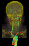
Volume rendering of velocity magnitude for patient-specific model of blood flow from aorta to cerebral arteries for a patient with a right middle cerebral aneurysm. Simulation results are visualized together with CT image data to provide anatomic context.
FIGURE 6
FIGURE 6
Velocity magnitude and pressure fields at peak systole and mid-diastole for a patient-specific simulation of blood flow and vessel dynamics in the aorta of a 10 year old patient with aortic coarctation.
FIGURE 7
FIGURE 7
Overview of simulation-based medical planning approach as applied to designing bypass surgery for patient with occlusive cardiovascular disease in aorta and iliac arteries. Shown from left are magnetic resonance angiography data, preoperative geometric solid model (red), operative plan (proposed aortofemoral bypass graft shown in yellow), and computed blood flow velocity in aorta and proximal end of bypass (103).
FIGURE 8
FIGURE 8
Iterative loop and information transferred in the coupling between the FSI and G&R parts of a FSG framework (122).

References

    1. Moore JA, Rutt BK, Karlik SJ, Yin K, Ethier CR. Computational Blood Flow Modeling Based on In Vivo Measurements. Annals of Biomedical Engineering. 1999;27:627–40. - PubMed
    1. Taylor CA, Draney MT, Ku JP, Parker D, Steele BN, et al. Predictive medicine: computational techniques in therapeutic decision-making. Comput Aided Surg. 1999;4:231–47. - PubMed
    1. Taylor CA, Hughes TJR, Zarins CK. Finite Element Modeling of Blood Flow in Arteries. Computer Methods in Applied Mechanics and Engineering. 1998;158:155–96.
    1. Long Q, Xu XY, Ariff B, Thom SA, Hughes AD, et al. Reconstruction of blood flow patterns in a human carotid bifurcation: a combined CFD and MRI study. J Magn Reson Imaging. 2000;11:299–311. Stant. - PubMed
    1. Steinman DA. Image-based computational fluid dynamics modeling in realistic arterial geometries. Ann Biomed Eng. 2002;30:483–97. - PubMed

Publication types

LinkOut - more resources