Review
. 2009:32:383-412.
doi: 10.1146/annurev.neuro.051508.135614.

Axon growth and guidance: receptor regulation and signal transduction

Affiliations
Review

Axon growth and guidance: receptor regulation and signal transduction

Michael O'Donnell et al. Annu Rev Neurosci. 2009.

Abstract

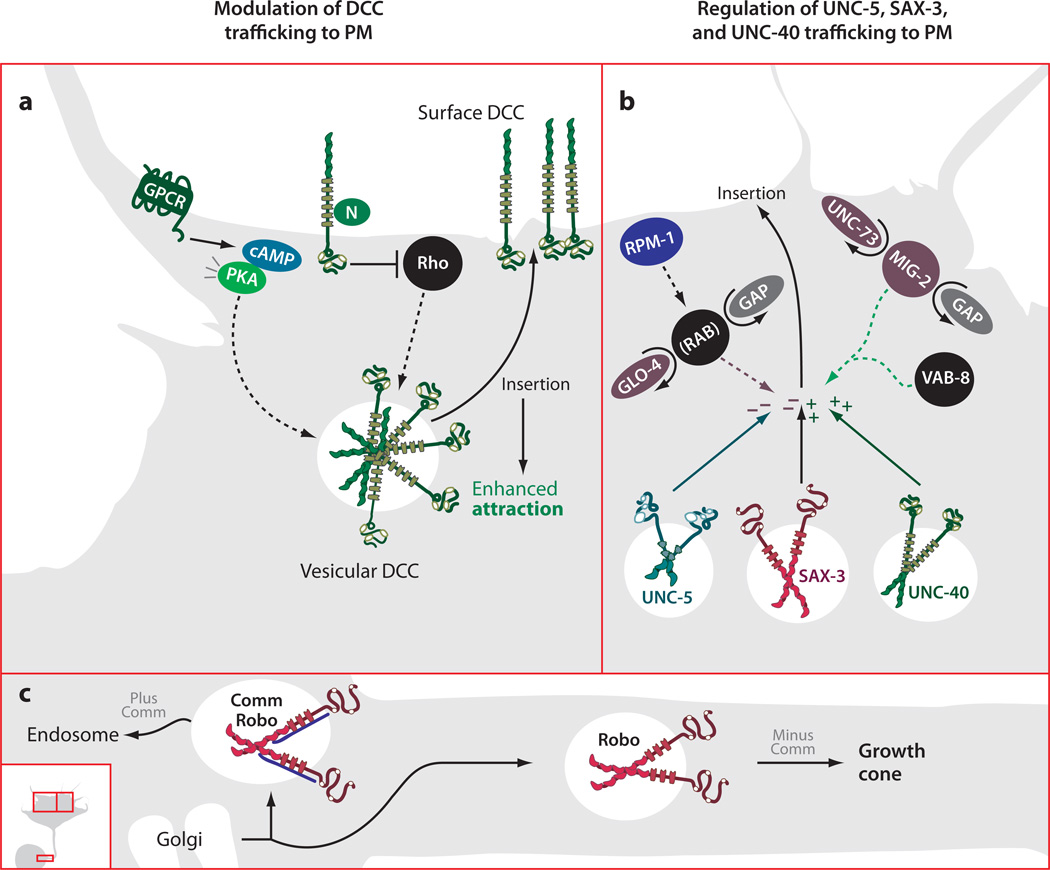
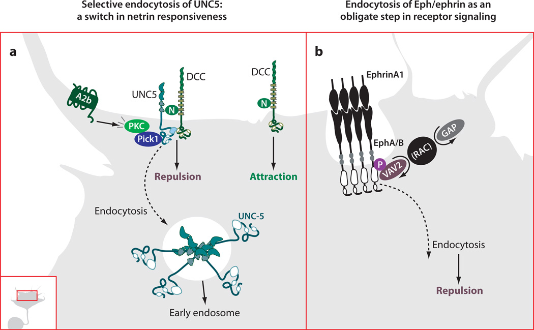
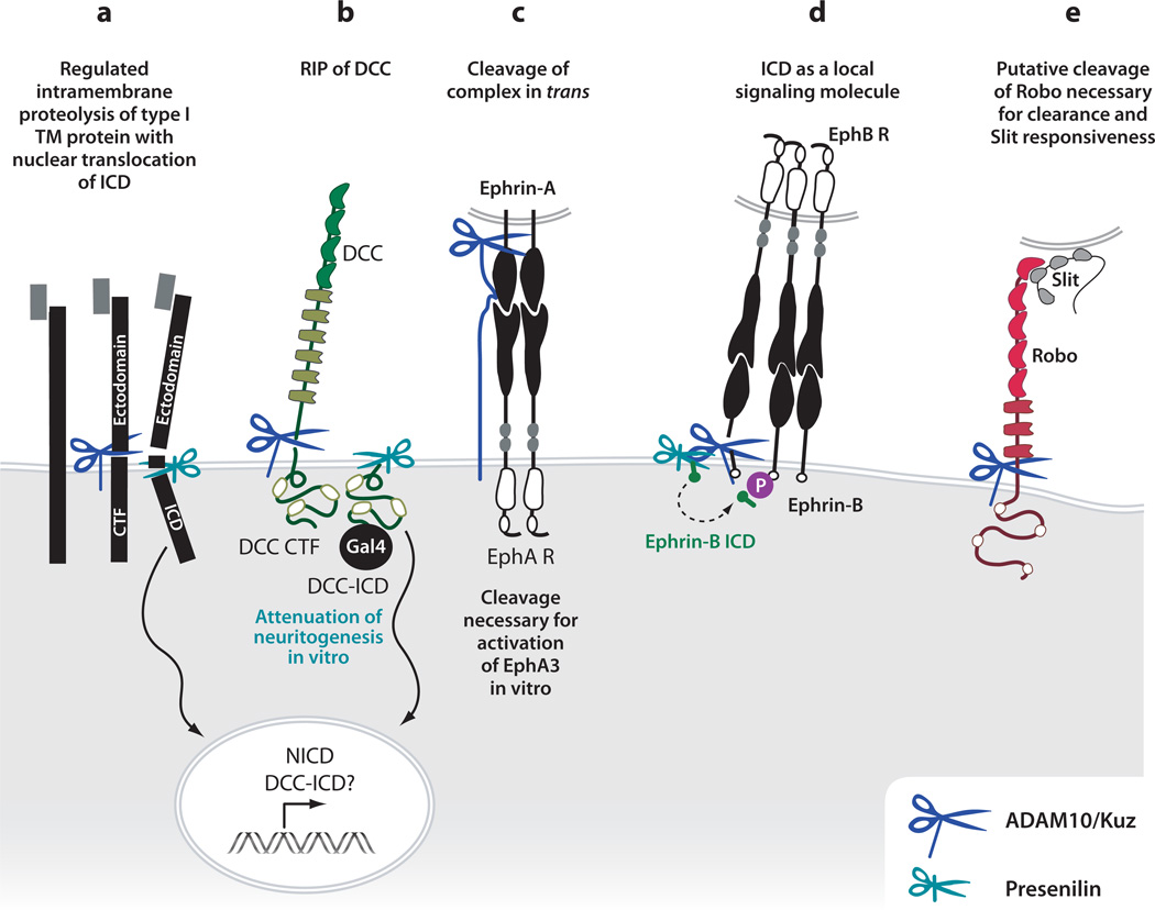
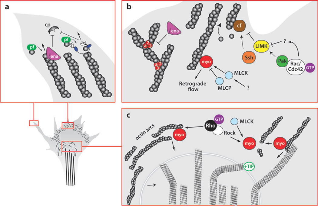
The development of precise connectivity patterns during the establishment of the nervous system depends on the regulated action of diverse, conserved families of guidance cues and their neuronal receptors. Determining how these signaling pathways function to regulate axon growth and guidance is fundamentally important to understanding wiring specificity in the nervous system and will undoubtedly shed light on many neural developmental disorders. Considerable progress has been made in defining the mechanisms that regulate the correct spatial and temporal distribution of guidance receptors and how these receptors in turn signal to the growth cone cytoskeleton to control steering decisions. This review focuses on recent advances in our understanding of the mechanisms mediating growth cone guidance with a particular emphasis on the control of guidance receptor regulation and signaling.


Figures

Figure 1
Figure 1
Trafficking guidance receptors to the growth cone plasma membrane (PM). (a) Both PKA activation and Rho inhibition positively regulate mobilization of an intracellular, vesicular pool of DCC. Increases in surface DCC lead to increases in Netrin responsiveness. (b) Positive and negative regulation of membrane expression of UNC-5, SAX-3, and UNC-40. RPM-1 activates GLO-4, a RAB GEF, which negatively regulates surface levels of UNC-5 and SAX-3. Activation of UNC-73, a MIG-2 (Rac) GEF, as well as activation of VAB-8, positively regulates surface levels of UNC-40 and SAX-3. Vesicles leaving the trans-Golgi network containing Robo are subjected to differential trafficking depending on the presence or absence of Comm. Vesicles containing Comm along with Robo are sorted to the endosome, whereas those containing Robo alone are trafficked down the axon toward the growth cone.
Figure 2
Figure 2
Regulated endocytosis in axon guidance. (a) Adenosine2b receptor (A2b) activity leads to PKC-dependent endocytosis of UNC5, which requires a physical interaction between PKC, Pick1, and the cytoplasmic domain of UNC5. This change in receptor composition at the plasma membrane leads to a switch in responsiveness to netrin from repulsion mediated by UNC5 alone, or by an UNC5/DCC complex, to attraction mediated by DCC. N, netrin. (b) The Vav family of Rac GEFs is required for endocytosis of ephrin ligand/Eph receptor complexes in retinal ganglion cell growth cones. Vav2 is recruited to the ephrin-stimulated juxtamembrane phosphorylated tyrosine of EphA and EphB receptors and then stimulates endocytosis. This endocytotic event is an obligate step in the forward signaling leading to growth cone retraction or repulsion.
Figure 3
Figure 3
Regulated proteolysis regulates guidance receptor function. (a) Processive proteolysis of a prototypical type I transmembrane (TM) protein, such as Notch or APP. Upon ligand binding, cleavage by an ADAM10 in the juxta-membrane region causes release of an N-terminal fragment into the extracellular space (ectodomain) and generates a C-terminal fragment (CTF) with a small extracellular stub. A second, constitutive cleavage by the gamma-secretase complex within the plane of the plasma membrane releases the intracellular domain (ICD). In the case of Notch, the ICD translocates to the nucleus, where it regulates transcription. (b) Regulated proteolysis of DCC occurs by ADAM10-mediated creation of a CTF, followed by gamma-secretase-mediated intramembraneous cleavage releasing DCC ICD. This ICD is competent to translocate to the nucleus when fused to Gal4. The cleavage event by ADAM10 leads to attenuation of neuritogenesis in vitro. (c) Following ligand-receptor complex formation, ADAM10 cleaves the ephrin-A5 ligand. This regulated proteolytic event leads to release from the initial cell-cell adhesion, allowing for growth cone retraction, and is necessary for the transduction of the EphA3 forward signal. (d) Processive cleavage in the ephrinB/ephB system indicates that the released ephrinB ICD may activate SRC-family kinases to contribute to reverse signaling. On the other hand, cleavage of the EphB2 receptor, in this case by matrix metalloproteases, is required for activation in vitro. (e) Kuzbanian appears to act positively in the Slit-Robo signaling pathway. On the basis of genetic observations and the abnormal presence of Robo protein on the commissural portions of axons in kuz mutants, we speculate that Kuz may cleave Robo to regulate receptor activity.
Figure 4
Figure 4
The growth cone cytoskeleton: structural components and regulatory proteins. (a) Filopodial actin dynamics. Guidance cues (netrin and Slit) may regulate the activity of Ena/VASP (ena) proteins, which in turn promote filament elongation either by antagonizing capping protein (CP) or by barbed-end G-actin addition (gray circles, bound to profilin, pf). Diaphanous-related formins (F) act as actin nucleators and promote barbed-end addition of G-actin, in addition to inhibiting CP. (b) Actin dynamics in the lamellipodium. Ena/VASP antagonizes Arp2/3-dependent filament branching, promoting filopodia formation. Cofilin severs actin filament pointed ends, providing a fresh pool of actin monomers. Rac/Cdc42 inhibit cofilin function through Pak/LIMK, whereas Ssh activates cofilin. Myosin-II (myo)-dependent retrograde actin flow toward the central domain is regulated by myosin light chain kinase (MLCK) and myosin light chain phosphatase (MLCP). (c) Actin/microtubule dynamics in the growth cone central domain. Rho/ROCK regulate myosin-dependent actin arc contractility. Myosin also promotes actin filament severing, as well as microtubule bundling. Microtubule advancement is regulated by retrograde actin flow in addition to microtubule-associated proteins (MAPs), such as the +TIP protein, orbit/CLASP.

References

    1. Aizawa H, Wakatsuki S, Ishii A, Moriyama K, Sasaki Y, et al. Phosphorylation of cofilin by LIM-kinase is necessary for semaphorin 3A-induced growth cone collapse. Nat. Neurosci. 2001;4:367–373. - PubMed
    1. Ang LH, Kim J, Stepensky V, Hing H. Dock and Pak regulate olfactory axon pathfinding in Drosophila. Development. 2003;130:1307–1316. - PubMed
    1. Arakawa Y, Bito H, Furuyashiki T, Tsuji T, Takemoto-Kimura S, et al. Control of axon elongation via an SDF-1α/Rho/mDia pathway in cultured cerebellar granule neurons. J. Cell Biol. 2003;161:381–391. - PMC - PubMed
    1. Arber S, Barbayannis FA, Hanser H, Schneider C, Stanyon CA, et al. Regulation of actin dynamics through phosphorylation of cofilin by LIM-kinase. Nature. 1998;393:805–809. - PubMed
    1. Arimura N, Menager C, Kawano Y, Yoshimura T, Kawabata S, et al. Phosphorylation by Rho kinase regulates CRMP-2 activity in growth cones. Mol. Cell Biol. 2005;25:9973–9984. - PMC - PubMed

Substances

LinkOut - more resources