Review
. 2009:32:347-81.
doi: 10.1146/annurev.neuro.31.060407.125536.

Establishment of axon-dendrite polarity in developing neurons

Affiliations
Review

Establishment of axon-dendrite polarity in developing neurons

Anthony P Barnes et al. Annu Rev Neurosci. 2009.

Abstract

Neurons are among the most highly polarized cell types in the body, and the polarization of axon and dendrites underlies the ability of neurons to integrate and transmit information in the brain. Significant progress has been made in the identification of the cellular and molecular mechanisms underlying the establishment of neuronal polarity using primarily in vitro approaches such as dissociated culture of rodent hippocampal and cortical neurons. This model has led to the predominant view suggesting that neuronal polarization is specified largely by stochastic, asymmetric activation of intracellular signaling pathways. Recent evidence shows that extracellular cues can play an instructive role during neuronal polarization in vitro and in vivo. In this review, we synthesize the recent data supporting an integrative model whereby extracellular cues orchestrate the intracellular signaling underlying the initial break of neuronal symmetry leading to axon-dendrite polarization.


Figures

Figure 1
Figure 1. Cell-type specific patterns of neuronal polarization in vivo
Examples of the sequence of events leading to the polarized emergence of axon and dendrites in four distinct vertebrate neuronal cell types in vivo. Throughout these figures, the nascent axon is depicted in red and the somatodendritic domain in purple. (A) In vivo polarization of retinal ganglion cells in zebrafish (Danio rerio) and mouse (Mus musculus). Neuroepithelial progenitors characterized by an apical and a basal attachment undergo asymmetrical cell division at the apical surface (A1-A3). Upon cell cycle exit, the nucleus undergoes basal translocation (A4) and specifically loses its apical attachment while its basal process starts growing along the basal membrane (A5). The axon (red) therefore emerges from the basal process and the dendrite emerges from the apical process (A6). (B) Polarization of mouse bipolar cells in the mouse retina. Neuroepithelial progenitors (B1) transform into bipolar cells by first losing their basal attachment which starts branching in the inner plexiform layer (IPL) while the apical process starts branching in the prospective outer plexiform layer (OPL) before (B2) losing its apical attachment (B3). The axon emerges from the basal process (red) and the dendrite emerges from the apical process (B4). (C) Polarization of granule cells in the mammalian cerebellum. Granule cell progenitors divide rapidly in the external plexiform layer (EGL; C1) and upon cell cycle exit start adopt a bipolar morphology (C2) before migrating tangentially with a leading and a trailing process (C3). Another process emerges from the cell body in the tangential direction (C4) and becomes the leading process, leading its migration towards the inner granule layer (IGL; C5). The trailing processes form a characteristic T-shaped axon (red in C6) whereas the leading process gives rise to the dendritic domain. (D) Polarization of radially migrating pyramidal neurons in the mammalian neocortex. Neurons are generated between E11 and E17 by radial glial progenitors in the ventricular zone of the mouse neocortex. These cells have a long basal (radial) process attached to the basal membrane at the pial surface and a short apical process on the ventricle side (D1; see detail in Figure 5). Upon cell cycle exit through asymmetric cell division (D2), the post-mitotic neuron (blue) goes through a multipolar transition where multiple neurites emerges rapidly from the cell body (D3) before one major process forms in the radial direction (D4) and becomes the leading process (LP). At this point, the neuron initiates radial translocation along a radial glial process (D5) and leaves behind a trailing process which elongates tangentially in the intermediate zone (red). The cell body continues to translocate to towards its final destination (the top of the cortical plate; CP) while the axon rapidly elongates (D6). The leading process gives rise to the apical dendrite (purple in D7) which initiates local branching in the marginal zone while over the first postnatal week (until radial migration ends) the cell body will translocate ventrally (D8-D9) as neurons born at later stages (in orange D10 ) bypass their predecessors (inside-out accumulation pattern). Adapted from: (a.) Hinds & Hinds (1978) and Zolessi et al (2006); (b.) Morgan et al (2006); (c.) Rakic (1971), Komuro and Rakic (2001) and Gao and Hatten (1993); (d.) Shoukimas and Hinds (1978), Rakic (1972), Noctor et al. (2003), Hatanaka and Murakami (2002)
Figure 2
Figure 2. Parallel between neuronal polarization in vitro and in vivo
Comparison of the sequence of events leading to the polarization of cortical pyramidal neurons in vivo and in vitro. (A) As depicted in Figure 1D, the axon-dendrite polarity of pyramidal neurons is derived from the polarized emergence of the trailing (TP) and leading processes (LP) respectively. In vivo, pyramidal neurons acquire other key features of their terminal polarity such as the axon initiation segment (AIS; yellow cartridge) and dendritic spines (pink protrusions) during the first postnatal weeks of development. (B) In dissociated cultures, postmitotic cortical neurons display specific transitions as classically described for hippocampal neurons by (Dotti et al 1988). At Stage 1, immature post-mitotic neurons display intense lamelipodial and filopodial protrusive activity which leads to the emergence of single immature neurites, Stage 2. Stage 3 represents a critical step where neuronal symmetry breaks and a single neurite grows rapidly to become the axon (red) while other neurites acquire dendritic identity. Stage 4 is characterized by rapid axon and dendritic outgrowth. Finally, Stage 5 neurons are terminally differentiated pyramidal neurons harboring dendritic spines and the AIS.
Figure 3
Figure 3. Signaling pathways involved in mammalian axon specification during neuron polarization
See text for details. G-protein coupled receptor (GPCR). Extra-cellular matrix (ECM). Red arrows indicate negative regulation, whereas blue arrows indicate activation.
Figure 4
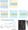
Figure 4. Experimental evidence for the instructive effects of extracellular cues on neuronal polarization in vitro
(A-B) When hippocampal neurons are plated on stripes coated with alternating substrates such as the cell-adhesion molecule NgCAM (blue) and Poly-L-Lysine (PLL) plus laminin (yellow), the first neurite contacting the a new substrate (arrowhead in A and B) is specified to become the axon (red). Note that this axon specification event can occur regardless of the type of substrate interface (see A and B) suggesting that in these conditions, axon specification can occur when unspecified neurites detect a relative change in the substrate composition rather than a specific substrate. Adapted from (Esch et al 1999) (C-E) Hippocampal neurons plated on control stripes coated with PLL (purple) or PLL plus Bovine Serum Albumin (BSA) show no trend for axon specification when neurites encounter a stripe boundary (C), whereas an immature neurite encountering a stripe containing Brain Derived Neurotrophic Factor (BDNF) become an axon (D). This effect is abrogated if neurons express a non-phosphorylatable form of LKB1 (LKB1S431A; E). Adapted from (Shelly et al 2007). (F-G) The slice overlay assay was developed to test if the cortical wall contains extracellular cues that could polarize axon emergence towards the ventricle. In this assay, immature E18 rat cortical neurons are dissociated and fluorescently labeled with DiI before being plated onto isochronic E18 or heterochronic (P3) cortical slices. Only three hours after plating most neurons have a short neurite becoming the axon allowing to test between two hypothesis: in scenario 1, polarized axon emergence is the sole result of intrinsic polarization inherited, for example, from neuroepithelial cell progenitors (see Figures 5 and 6). In this case, the plated neurons should show a randomized direction of axon emergence. According to scenario 2, graded extracellular cues are able to polarize the direction of axon emergence and therefore, neurons plated on the slice should show a directed axon outgrowth towards the ventricle. Polleux et al. (1998) demonstrated that scenario 2 is the most likely one since the overwhelming majority of pyramidal neurons present in the cortical plate show directed axon outgrowth in this assay, only few hours after plating (arrowheads in Fig. 4G, (Polleux et al 1998)).
Figure 5
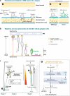
Figure 5. Experimental evidence for instructive effects of extracellular cues on neuronal polarization in vivo
(A) In C. elegans, the immature HSN neurons undergo a series of morphological changes leading to the polarized outgrowth of their axon ventrally at larval stage 4 (L4) (A1). Most noticeably, the polarized lamelipodial outgrowth observed at stage early L2 correlates with a polarized distribution of the attractive Netrin receptor (Unc40) and the intracellular cytoskeletal effector Lamellipodin (MIG-10) (A2) and also requires the presence of Netrin (UNC-6) secreted from the ventral part of the embryo since the Unc6 mutant shows non-directional process outgrowth at stage early L2 accompanied by non-asymmetrical distribution of UNC-40 (A3). Adapted from (Adler et al 2006) (B) Axon polarization during the migration of Xenopus retinal ganglion cells. As described in Figure 1A, retinal ganglion cells inherit their axon-dendrite polarity as their cell body translocate basally to the ganglion cell layer. In these cells, the basal process of the dividing progenitor gives rise to the leading process of the migrating RGC which becomes the axon (red). Using live cell imaging Zolessi et al. demonstrated that the centrosome and the polarity complex protein PAR3 are localized to the apical side of the RGC during translocation. The apical membrane containing atypical Protein Kinase C (aPKC), α-catenin and F-actin is also localized apically in the translocating RGC in the trailing process. Therefore, in RGC, the PAR3/apical polarity complex is localized in the trailing process which becomes the dendritic domain whereas on the basal side, the leading process becomes the axon which grows rapidly along the basal membrane. Adapted from (Zolessi et al 2006). (C) A cellular and molecular model of the function of LKB1 and SAD kinases in the polarization of pyramidal cortical neurons. (C1) Upon asymmetric cell division of radial glial progenitors, early unpolarized post-mitotic neurons show a transient phase of non-directed neurite outgrowth in the subventricular zone before adopting a bipolar morphology in the intermediate zone where they engage radial migration with a leading process directed toward the pial surface and a trailing process directed toward the ventricle. (C2) On the basis of recent reports (Barnes et al 2007, Shelly et al 2007), we propose that in vivo, the trailing process is specified to become the axon in response to putative extracellular cues that preferentially induce phosphorylation of LKB1 on Serine 431. Modified from (Barnes et al 2008).
Figure 6
Figure 6. Relationship between epithelial cell polarity, neuroepithelial progenitor polarity and post-mitotic neuron polarity
(A) Parallel between epithelial cell polarity and neuroepithelial cell polarity. Epithelial cells have two main cell membrane compartments: the apical membrane and the baso-lateral membrane separated by tight junctions (TJ) and adherens junctions (AJ) which act both as cell adhesion sites and strong diffusion barriers preventing direct exchange between the apical and the baso-lateral domains. Neuroepithelial progenitors form a pseudo-epithelium in vivo which is characterized by a basal attachment through their radial process to the pial surface and an apical domain separated by Cadherin-based adherens junctions. (B) Potential model for the molecular control of apical polarity in cortical neuroepithelial progenitors and its potential relationship with post-mitotic neuron polarity. Several protein complexes recently involved in apical polarity of cortical progenitors include (1) the centrosomal proteins Cep120-TACC1-3 and microtubules, (2) Numb-regulated adherens junctions composed of cadherins and catenins, and (3) atypical protein kinase C (aPKC)-cdc42-Par3/6 (see text for details). Question marks point to unresolved issues regarding the functional interactions between specific molecular components of the apical polarity complex. During cell cycle exit, the centrosome might play a role in axon specification (de Anda et al 2005) by leaving an ‘apical trace’ transiently localizing to the base of multipolar neurons which might play a role in specifying the position of the trailing process (future axon) before translocating to the base of the leading process (future apical dendrite) when neurons initiates radial translocation (Tsai et al 2007).

Similar articles

Cited by

References

    1. Adler CE, Fetter RD, Bargmann CI. UNC-6/Netrin induces neuronal asymmetry and defines the site of axon formation. Nat Neurosci. 2006;9:511–8. - PMC - PubMed
    1. Aravamudan B, Broadie K. Synaptic Drosophila UNC-13 is regulated by antagonistic G-protein pathways via a proteasome-dependent degradation mechanism. J Neurobiol. 2003;54:417–38. - PubMed
    1. Arimura N, Kaibuchi K. Neuronal polarity: from extracellular signals to intracellular mechanisms. Nat Rev Neurosci. 2007;8:194–205. - PubMed
    1. Arimura N, Menager C, Fukata Y, Kaibuchi K. Role of CRMP-2 in neuronal polarity. J Neurobiol. 2004;58:34–47. - PubMed
    1. Asada N, Sanada K, Fukada Y. LKB1 regulates neuronal migration and neuronal differentiation in the developing neocortex through centrosomal positioning. J Neurosci. 2007;27:11769–75. - PMC - PubMed

Publication types

Substances

LinkOut - more resources