. 2009 Apr 28:6:44.
doi: 10.1186/1743-422X-6-44.

Vaccinia virus p37 interacts with host proteins associated with LE-derived transport vesicle biogenesis

Affiliations

Vaccinia virus p37 interacts with host proteins associated with LE-derived transport vesicle biogenesis

Yali Chen et al. Virol J. .

Abstract

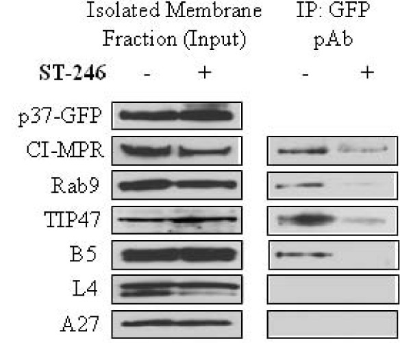
Background: Proteins associated with the late endosome (LE) appear to play a central role in the envelopment of a number of taxonomically diverse viruses. How viral proteins interact with LE-associated proteins to facilitate envelopment is not well understood. LE-derived transport vesicles form through the interaction of Rab9 GTPase with cargo proteins, and TIP47, a Rab9-specific effector protein. Vaccinia virus (VV) induces a wrapping complex derived from intracellular host membranes to envelope intracellular mature virus particles producing egress-competent forms of virus.

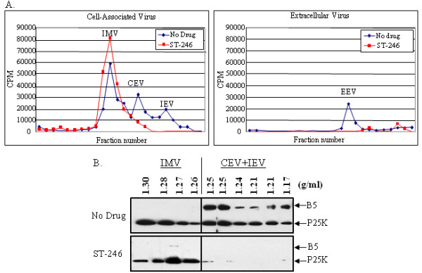
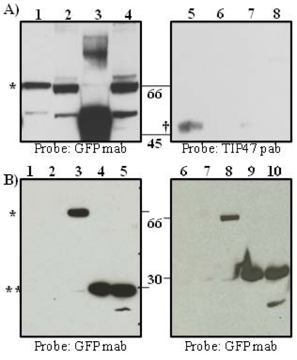
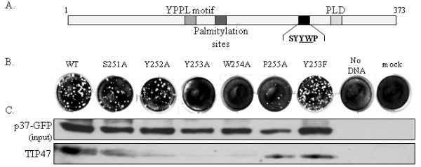
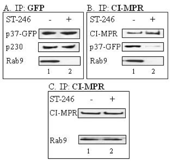
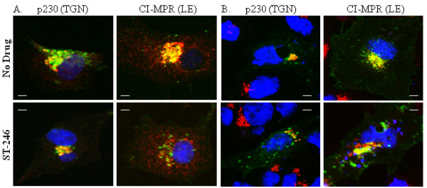
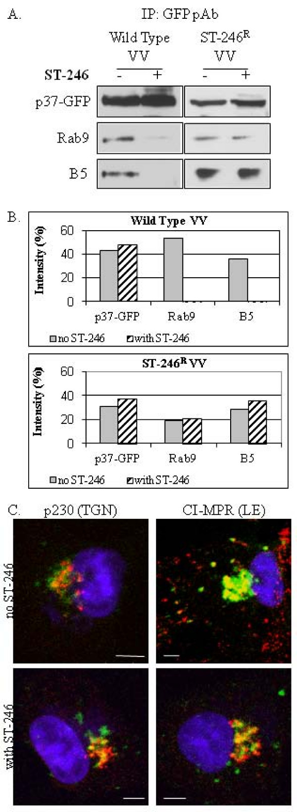
Results: We show that VV p37 protein associates with TIP47-, Rab9-, and CI-MPR-containing membranes. Mutation of a di-aromatic motif in p37 blocks association with TIP47 and inhibits plaque formation. ST-246, a specific inhibitor of p37 function, inhibits these interactions and also blocks wrapped virus particle formation. Vaccinia virus expressing p37 variants with reduced ST-246 susceptibility associates with Rab9 and co-localizes with CI-MPR in the presence and absence of compound.

Conclusion: These results suggest that p37 localizes to the LE and interacts with proteins associated with LE-derived transport vesicle biogenesis to facilitate assembly of extracellular forms of virus.


Figures

Figure 1
Figure 1
Equilibrium centrifugation of radiolabeled virus particles in the presence and absence of ST-246. Intracellular and extracellular virus particles were fractionated by equilibrium centrifugation on preformed cesium chloride gradients. (A) The radiolabeled viral DNA was measured by liquid scintillation counting. (B) The viral proteins in each fraction were detected by immunoblot analysis using antisera against L4 and B5 proteins. IMV, intracellular mature virus; CEV cell-associated enveloped virus; IEV, intracellular enveloped virus.
Figure 2
Figure 2
p37-GFP and TIP47 co-immunoprecipitate in the presence of non-ionic detergent. A. Membrane fractions from BSC-40 cells infected with vvF13LGFP at an MOI of 5 were extracted as described in Materials and Methods. Protein extracts obtained from the high speed pellet were resuspended in a buffer containing a non-ionic detergent (0.5% NP-40, 150 mM NaCl, 20 mM Tris, pH7.4) and immunoprecipitated with antibodies specific for TIP47 or GFP followed by SDS-PAGE analysis and immunoblot. Antibody raised against the trans Golgi marker, TGN46 (Sigma Aldrich) was used as a non-specific control for immunoprecipitation. Lane 1, IP using TIP47 pab; Lane 2, Unbound Fraction; Lane 3, IP using TGN46 pab; Lane 4, Unbound Fraction; Lane 5, IP using GFP pab, Lane 6, Unbound Fraction, Lane 7, IP using TGN46 pab; Lane 8, Unbound Fraction; B. Immunoblot of whole cell extracts and membrane fractions immunoprecipitated with GFP pab from BSC-40 cells mock-infected or infected with vvWR, vvGFP, vvΔF13LGFP, or vvF13LGFP at an MOI of 5 probed with GFP mab. Lane 1 and 6, mock-infected, Lane 2 and 7, vvWR, Lane 3 and 8, vvF13LGFP, Lane 4 and 9, vvΔF13LGFP, Lane 5 and 10, vvGFP. IP, immunoprecipitated; pab, polyclonal antibody; mab, monoclonal antibody; ** pGFP; * p37-GFP fusion protein; † TIP47 protein.
Figure 3
Figure 3
Mutation of the YW motif in F13L blocks association of p37-GFP with TIP47 and prevents complementation of an F13L-deleted virus. (A) Diagram of the VV F13L gene with functional domains indicated. (B) Complementation experiment demonstrating a panel of mutant F13L alleles containing the indicated A or F substitutions. The F13L allelescontained a GFP tag at the C-terminus to facilitate immunoprecipitation of p37. PCR products containing the indicated F13L allele were transfected into BSC-40 cells infected with an F13L-deletion virus. At 3 days post infection/transfection, plaques were visualized by staining with crystal violet, (C) Membrane fractions, prepared as described in Materials and Methods, from infected and transfected cells were immunoprecipitated in hypotonic buffer with anti-GFP antibody and probed with TIP47 antibody (lower panel). The upper panel shows the input controls.
Figure 4
Figure 4
ST-246 affects the co-immunoprecipitation of membrane-associated p37-GFP, Rab9 and CI-MPR but not p230. BSC-40 cells were infected (A and B) with 5 PFU per cell of vvF13LGFP or mock-infected (C) in the absence (column 1) or presence (column 2) of 10 μM ST-246. The membrane fractions were extracted as described in Materials and Methods, resuspended in hypotonic buffer and immunoprecipitated as indicated. Blots were probed with (A) p230 or Rab9 antibody, (B) GFP or Rab9 antibody, and (C) Rab9 antibody. The top row for each are input controls and are probed as indicated.
Figure 5
Figure 5
Co-localization of p37-GFP with p230 and CI-MPR in infected and uninfected cells in the presence and absence of ST-246. Cell monolayers were grown on chamber slides and (A) infected with 8 PFU/cell of vvF13LGFP or (B) transfected with 1 μg of plasmid expressing p37-GFP in the presence or absence of 10 μM ST-246. At 14–16 hpi cells were fixed in 4% paraformaldehyde, permeablized with 0.2% TritonX-100 and stained for 1 hour with anti-p230 antibody or anti-CI-MPR antibody. Proteins were visualized using Alexa 594-conjugated secondary antibody. Samples were mounted in ProLong Gold Antifade Reagent (Invitrogen Molecular Probes) containing DAPI for nuclear staining and analyzed using a Zeiss LSM 510 confocal laser-scanning microscope. Images are representative of 2 separate experiments in which a minimum of 50 infected cells displaying a similar fluorescent phenotype were observed. Images were collected and processed using LSM 510 acquisition and Adobe Photoshop software. Bar, 5 μm.
Figure 6
Figure 6
ST-246 inhibits membrane precursor biogenesis. BSC-40 cells were infected with 5 PFU per cell of vvF13LGFP in the presence or absence of 10 μM ST-246. Membrane extracts were prepared as described in Materials and Methods and analyzed by immunoblot (left panel). The remaining supernatant was then subjected to immunoprecipitation (right panel). Blots were probed with the indicated antibodies. Left panel, input controls; right panel, immunoprecipitated samples.
Figure 7
Figure 7
Co-immunoprecipitation and subcellular localization of Rab9 and B5 protein with p37-GFP expressed from vvF13LGFP or an ST-246 resistant VV virus variant. (A) BSC-40 cells were infected with wild type VV (left) or an ST-246-resistant variant that exhibits reduced susceptibility to ST-246 (right) in the presence or absence of 10 μM compound. The membrane fractions were extracted as described in Materials and Methods, resuspended in hypotonic buffer and immunoprecipitated with anti-GFP polyclonal antibody. Blots were probed with Rab9 antibody or antiserum to B5 as indicated. Top row: input controls; (B) Quantitative comparison of immunoblot intensity in the absence and presence of ST-246; (C) Cell monolayers were grown on chamber slides and infected with 8 PFU/cell of an ST-246-resistant variant in the presence or absence of 10 μM ST-246. At 14–16 hpi cells were fixed in 4% paraformaldehyde, permeablized with 0.2% TritonX-100 and stained for 1 hour with anti-p230 antibody or anti-CI-MPR antibody. Proteins were visualized using Alexa 594-conjugated secondary antibody. Samples were mounted in ProLong Gold Antifade Reagent (Invitrogen Molecular Probes) containing DAPI for nuclear staining and analyzed using a Zeiss LSM 510 confocal laser-scanning microscope. Images are representative of experiments in which a minimum of 50 infected cells displaying a similar fluorescent phenotype were observed. Images were collected and processed using LSM 510 acquisition and Adobe Photoshop software. Bar, 5 μm.

References

    1. Smith GL, Vanderplasschen A, Law M. The formation and function of extracellular enveloped vaccinia virus. J Gen Virol. 2002;83:2915–2931. - PubMed
    1. Moss B. Poxviridae and their replication. In: Knipe B, Howley P, editor. Fields Virology. New York: Raven Press; 2001. pp. 2849–2884.
    1. Schmelz M, Sodeik B, Ericsson M, Wolffe EJ, Shida H, Hiller G, Griffiths G. Assembly of vaccinia virus: the second wrapping cisterna is derived from the trans Golgi network. J Virol. 1994;68:130–147. - PMC - PubMed
    1. Tooze J, Hollinshead M, Reis B, Radsak K, Kern H. Progeny vaccinia and human cytomegalovirus particles utilize early endosomal cisternae for their envelopes. Eur J Cell Biol. 1993;60:163–178. - PubMed
    1. Husain M, Moss B. Similarities in the induction of post-Golgi vesicles by the vaccinia virus F13L protein and phospholipase D. J Virol. 2002;76:7777–7789. doi: 10.1128/JVI.76.15.7777-7789.2002. - DOI - PMC - PubMed

Publication types

MeSH terms

LinkOut - more resources