. 2009 Jul;41(7):1601-12.
doi: 10.1016/j.biocel.2009.02.003. Epub 2009 Feb 20.

A major cathepsin B protease from the liver fluke Fasciola hepatica has atypical active site features and a potential role in the digestive tract of newly excysted juvenile parasites

Affiliations

A major cathepsin B protease from the liver fluke Fasciola hepatica has atypical active site features and a potential role in the digestive tract of newly excysted juvenile parasites

Simone A Beckham et al. Int J Biochem Cell Biol. 2009 Jul.

Abstract

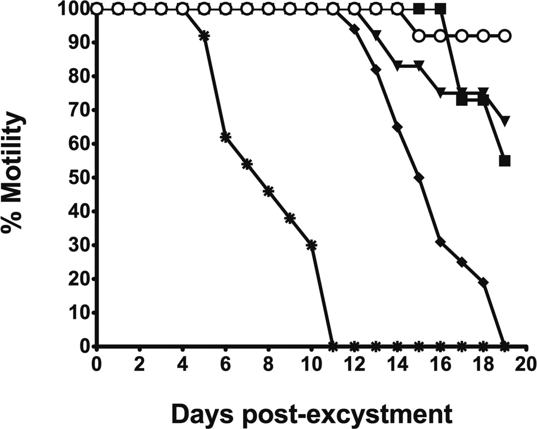
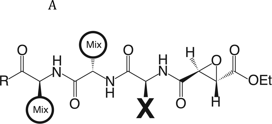
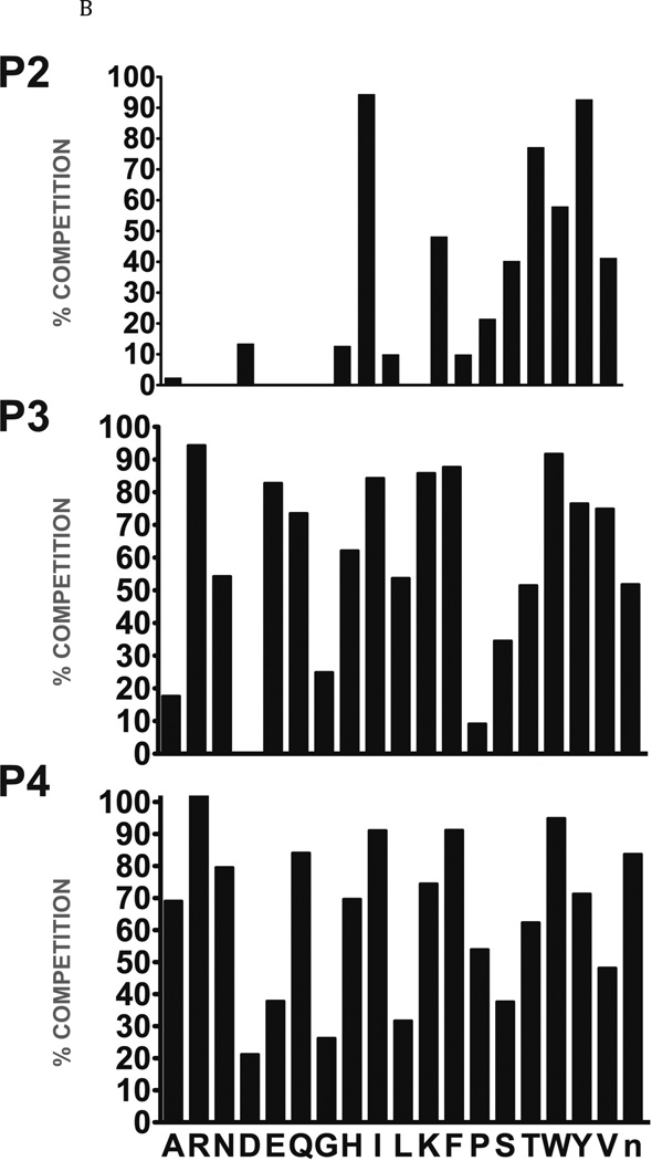
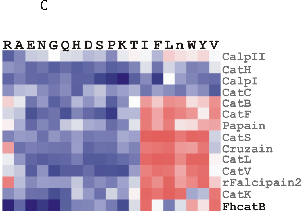
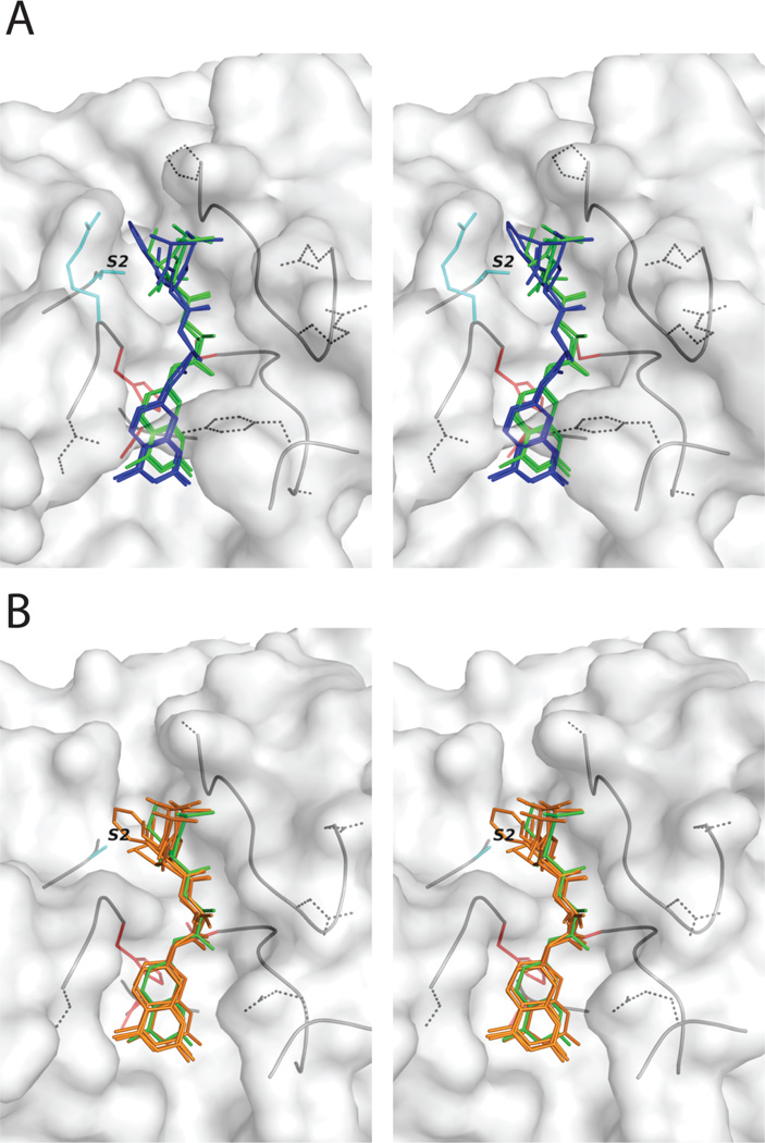
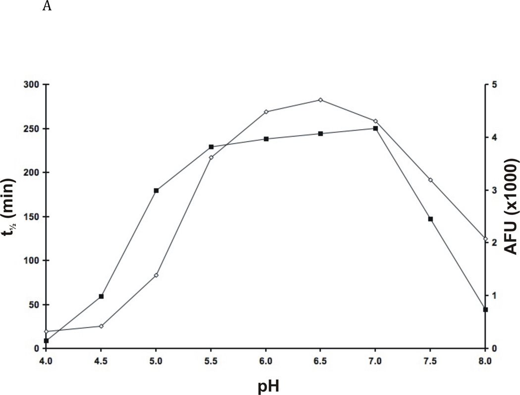
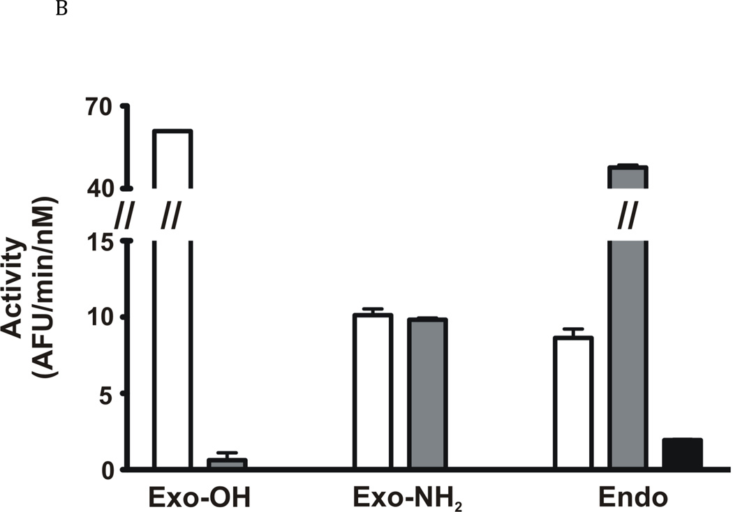
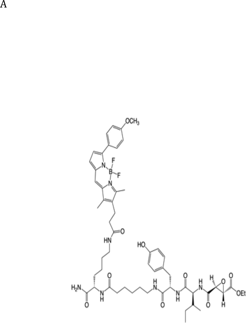
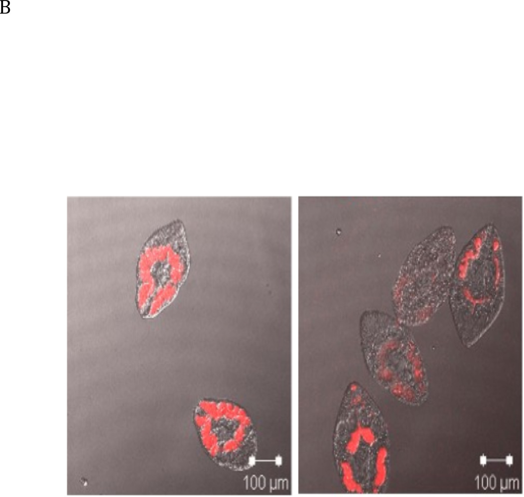
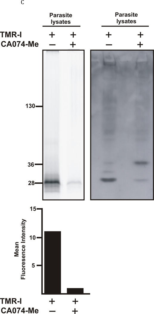
The newly excysted juvenile (NEJ) stage of the Fasciola hepatica lifecycle occurs just prior to invasion into the wall of the gut of the host, rendering it an important target for drug development. The cathepsin B enzymes from NEJ flukes have recently been demonstrated to be crucial to invasion and migration by the parasite. Here we characterize one of the cathepsin B enzymes (recombinant FhcatB1) from NEJ flukes. FhcatB1 has biochemical properties distinct from mammalian cathepsin B enzymes, with an atypical preference for Ile over Leu or Arg residues at the P(2) substrate position and an inability to act as an exopeptidase. FhcatB1 was active across a broad pH range (optimal activity at pH 5.5-7.0) and resistant to inhibition by cystatin family inhibitors from sheep and humans, suggesting that this enzyme would be able to function in extracellular environments in its mammalian hosts. It appears, however, that the FhcatB1 protease functions largely as a digestive enzyme in the gut of the parasite, due to the localization of a specific, fluorescently labeled inhibitor with an Ile at the P(2) position. Molecular modelling and dynamics were used to predict the basis for the unusual substrate specificity: a P(2) Ile residue positions the substrate optimally for interaction with catalytic residues of the enzyme, and the enzyme lacks an occluding loop His residue crucial for exopeptidase activity. The unique features of the enzyme, particularly with regard to its specificity and likely importance to a vital stage of the parasite's life cycle, make it an excellent target for therapeutic inhibitors or vaccination.


Figures

Fig. 1
Fig. 1. Viability of F. hepatica NEJ flukes in the presence of cysteine protease inhibitors
Following excystment, NEJ flukes were maintained in culture in the presence of 50 µM cysteine protease inhibitors. The NEJ flukes were examined and scored daily for loss of motility. ○, E-64; ▼, E-64-c; ◆ E-64-d; ■, CA-074; ✳, CA-074Me. Parasites incubated in DMSO alone displayed no loss of motility (viability) over the assay period.
Fig. 2
Fig. 2. Determination of the specificity of FhcatB1 using a combinatorial peptide inhibitor library
A: Inhibitor scaffold used in libraries utilized to determine the specificity of FhcatB1. The scaffold encompasses a fixed position (indicated by ‘X’) which can be changed to each natural amino acid and norleucine, and two mixed positions (indicated by ‘mix’). B: The substrate preferences of FhcatB1 for the P2, P3 and P4 positions were determined using a combinatorial peptide inhibitor library and are displayed as the percentage of competition for each amino acid substituted at the indicated position. ‘n’ represents norleucine (a substitute for methionine). C: Data from positional scanning of the P2 position of FhcatB1 compared with other papain family proteases. Substrate preferences of the S2 subsite of papain family cysteine proteases were determined using combinatorial libraries. The preferences are presented in red for substrates with greater competition, or blue for those with little or no competition, with 50% competition in white. Amino acids, represented as single letters, are above the chart. The enzymes shown other than FhcatB1 are: CalpII, calpain II purified from porcine kidney (EMD Bioscience); CatH, cathepsin H purified from human liver (EMD Bioscience); CalpI, calpain I purified from porcine kidney (EMD Bioscience); CatC, cathepsin C purified from bovine spleen (Sigma); CatB, cathepsin B purified from bovine spleen (Sigma); CatF, recombinant cathepsin F; Papain, purified from Carica papaya (Sigma); CatS, recombinant human cathepsin S; Cruzain, cruzipain from Trypanosoma cruzi; CatL, recombinant human cathepsin L; rFalcipain 2, recombinant falcipain-2 from Plasmodium falciparum; CatK, recombinant human cathepsin K.
Fig. 3
Fig. 3. Homology modeling of the binding of FhcatB1 showing binding of substrates to the active site in comparison to human cathepsin B
A: A homology model of FhcatB1 showing the predicted positions of tripeptide substrates in the active site. The substrates shown have the general form of Val-P2-Arg-7-amino-4-methylcoumarin, where P2=Val/Ile (green stick) and P2=Leu/Phe (blue stick). The side-chains of the P3 and P1 positions have been omitted for clarity. Residues of the catalytic triad appear in red, the two residues that flank the S2 subsite in FhCatB1 are in cyan, and dotted side-chains indicate substitutions in FhCatB1 with respect to the human enzyme. B: The same procedure was applied, based on the crystal structure of human cathepsin B. All four tripeptide substrates, P2=Ile/Val/Leu/Phe (orange stick), adopt a similar position to P2=Ile in FhCatB1 (shown as green stick for comparison). Stereo figures were prepared using PyMOL (De Lano Scientific).
Fig. 4
Fig. 4. Enzymatic properties of FhcatB1
A: FhcatB1 was incubated in AMT buffers at various pH values, following which the residual activity at pH 4.5 was determined against Z Phe Arg AMC (t½ is represented by the closed squares). The activity of FhcatB1 was measured against FITC-casein at different pH values (activity values represented by open diamonds). B: Activity of human cathepsin B enzymes and FhcatB1 against exopeptidase substrates. The initial velocities for human wild-type (open bars), His110Ala cathepsin B (grey bars) and FhcatB1 (dark bars) are plotted as AFU/min/nM enzyme. To aid visualization, a break has been introduced between 15 and 40 AFU/min/nM enzyme.
Fig. 5
Fig. 5. Incubation of Fasciola hepatica juvenile flukes with a fluorescent active site probe reveals the localisation of active cathepsin B like enzymes
A: Structure of the TMR-I probe used for visualization of the cathepsin B enzymes within the parasites. B: 40 newly excysted juvenile parasites per well were either exposed to the TMR-I (6.25 µM) for 24 hrs (left panel) or exposed to CA074Me (6.25 µM) for 7 days prior to exposure to the TMR-I (6.25 µM) for 24 hrs (right panel). C: Determination of the molecular target of TMR-I and CA074Me. The left hand panel shows a fluorogram of the binding of the TMR-I to proteins in a lysate from F. hepatica NEJ flukes. As indicated, proteins were incubated with the TMR-I in the presence and absence of CA074Me. The intensity of the bands obtained with and without CA074Me is shown below the fluorogram. The right hand panel shows an immunoblot with rabbit anti-FhcatB1 against parasite lysate proteins treated with TMR-I in the presence and absence of CA074Me.

Similar articles

Cited by

References

    1. Abdulla MH, Lim KC, Sajid M, McKerrow JH, Caffrey CR. Schistosomiasis mansoni: novel chemotherapy using a cysteine protease inhibitor. PLoS Med. 2007;4:130–138. - PMC - PubMed
    1. Anon . World Health Organization Technical Series No. 849. Geneva, Switzerland: 1995. Control of Foodborne Trematode Infections. - PubMed
    1. Aronja R, Riancho JA, Aguado JM. Fascioliasis in developed countries: A review of classic and aberrant forms of the disease. Medicine. 1995;74:13–23. - PubMed
    1. Barrett AJ, Kembhavi AA, Brown MA, Kirschke H, Knight CG, Tamai M, et al. L-trans-epoxysuccinyl-leucylamido(4-guanidino)butane (E-64) and its analogues as inhibitors of cysteine proteinases including cathepsins, B. H. and L. Biochem J. 1982;201:189–198. - PMC - PubMed
    1. Beckham SA, Law RHP, Smooker PM, Quinsey NS, Caffrey CR, McKerrow JH, et al. Production and processing of a recombinant Fasciola hepatica cathepsin B-like enzyme (FhcatB1) reveals potential processing mechanisms in the parasite. Biol Chem. 2006;387:1053–1061. - PubMed

Publication types

MeSH terms

LinkOut - more resources