. 2009 Aug 4;162(1):128-47.
doi: 10.1016/j.neuroscience.2009.04.054. Epub 2009 May 3.

Parathyroid hormone 2 receptor and its endogenous ligand tuberoinfundibular peptide of 39 residues are concentrated in endocrine, viscerosensory and auditory brain regions in macaque and human

Affiliations

Parathyroid hormone 2 receptor and its endogenous ligand tuberoinfundibular peptide of 39 residues are concentrated in endocrine, viscerosensory and auditory brain regions in macaque and human

A G Bagó et al. Neuroscience. .

Abstract

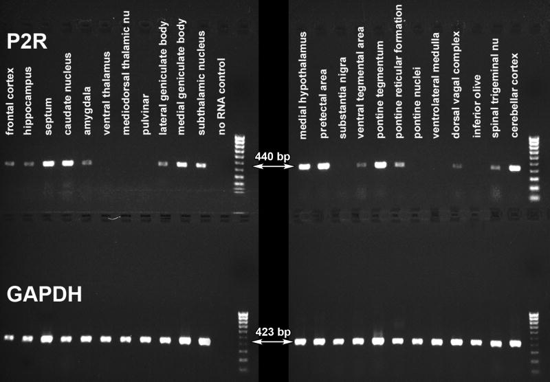
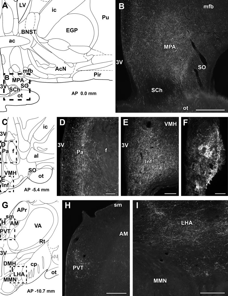
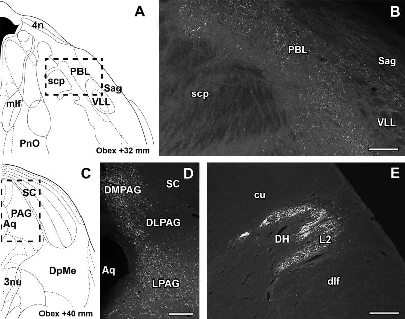
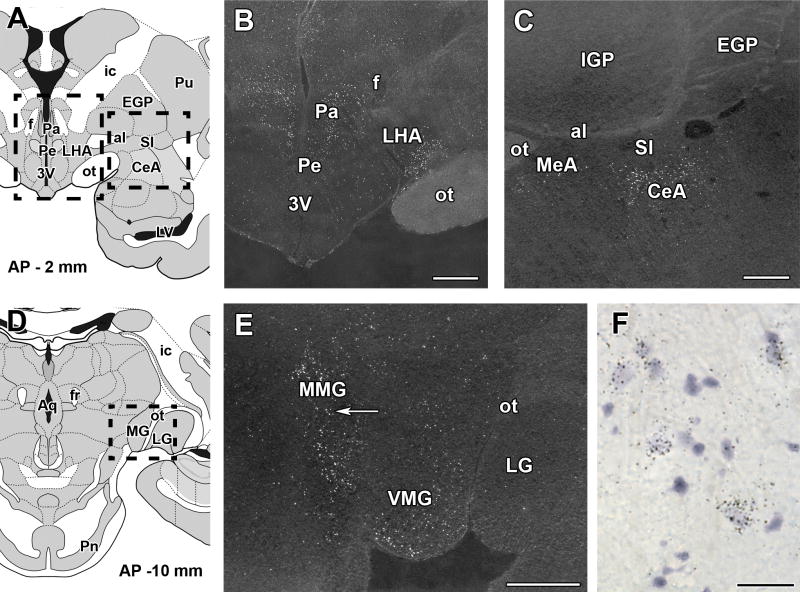
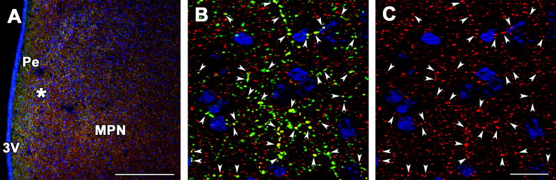
Parathyroid hormone receptor 2 (PTH2R) and its ligand, tuberoinfundibular peptide of 39 residues (TIP39) constitute a neuromodulator system implicated in endocrine and nociceptive regulation. We now describe the presence and distribution of the PTH2R and TIP39 in the brain of primates using a range of tissues and ages from macaque and human brain. In situ hybridization histochemistry of TIP39 mRNA, studied in young macaque brain, due to its possible decline beyond late postnatal ages, was present only in the thalamic subparafascicular area and the pontine medial paralemniscal nucleus. In contrast, in situ hybridization histochemistry in macaque identified high levels of PTH2R expression in the central amygdaloid nucleus, medial preoptic area, hypothalamic paraventricular and periventricular nuclei, medial geniculate, and the pontine tegmentum. PTH2R mRNA was also detected in several human brain areas by RT-PCR. The distribution of PTH2R-immunoreactive fibers in human, determined by immunocytochemistry, was similar to that in rodents, including dense fiber networks in the medial preoptic area, hypothalamic paraventricular, periventricular and infundibular (arcuate) nuclei, lateral hypothalamic area, median eminence, thalamic paraventricular nucleus, periaqueductal gray, lateral parabrachial nucleus, nucleus of the solitary tract, sensory trigeminal nuclei, medullary dorsal reticular nucleus, and dorsal horn of the spinal cord. Co-localization suggested that PTH2R fibers are glutamatergic, and that TIP39 may directly influence hypophysiotropic somatostatin containing and indirectly influence corticotropin releasing-hormone containing neurons. The results demonstrate that TIP39 and the PTH2R are expressed in the brain of primates in locations that suggest involvement in regulation of fear, anxiety, reproductive behaviors, release of pituitary hormones, and nociception.


Figures

Fig. 1
Fig. 1
Expression of the PTH2R in the human brain detected by RT-PCR. Products of PCR reactions performed using cDNA templates from different human brain regions were run on gels. In the upper line, PTH2R-specific 440 bp PCR products are visible. The positive bands in several brain areas suggest a widespread expression of the PTH2R. In the lower line, bands of 423 bp from control PCR reactions performed using primers specific for the housekeeping gene GAPDH are present.
Fig. 2
Fig. 2
Immunolabeling of the PTH2R in the human diencephalon. Photomicrographs demonstrate PTH2R immunoreactivity visualized by FITC-tyramide fluorescent amplification immunocytochemistry. The presence of PTH2R-ir fibers is demonstrated in coronal sections of the human diencephalon. A - A schematic drawing shows a part of the human brain at the level of the preoptic area in the coronal plane. The framed area indicates the field in B. B - A dense network of PTH2R-ir fibers is present in the medial preoptic area. In contrast, the density of PTH2R-ir fibers is very low in the supraoptic nucleus, and PTH2R-ir fibers are absent in the suprachiasmatic nucleus. C - A schematic drawing shows a part of the human brain at the level of the paraventricular nucleus in the coronal plane. The framed areas correspond to fields in D, and E, respectively. D - A high density of PTH2R-ir fibers is present in the hypothalamic paraventricular nucleus. E - The infundibular nucleus contains a high density of PTH2R-ir fibers. F - A very high level of PTH2R immunoreactivity is present in fiber terminals of the median eminence. G - A schematic drawing shows a part of the human brain at the level of the mamillary body in the coronal plane. H - PTH2R-ir fibers are abundant in the paraventricular thalamic nucleus but not in the adjacent anteromedial thalamic nucleus. I - The density of PTH2R-ir fibers is high in the lateral hypothalamic area. In contrast, PTH2R-ir fibers are absent from the medial mamillary nucleus. Schematic drawings (A, C, and G) are modifications from an atlas of the human brain (Mai et al., 1997). In agreement with the scaling of this atlas, antero-posterior coordinates (AP) are indicated from the level of the anterior commissure set as zero. Scale bars: 1 mm for B, H, and I, and 500 μm for D, E, and F.
Fig. 3
Fig. 3
Immunolabeling of the PTH2R in the human brainstem and spinal cord. Photomicrographs demonstrate PTH2R immunoreactivity visualized by FITC-tyramide fluorescent amplification immunocytochemistry in coronal sections. A - A schematic drawing shows a section of the human pons in coronal plane. The framed area indicates the field in B. B - A high density of PTH2R-ir fibers is present in the lateral parabrachial nucleus whereas the adjacent sagulum nucleus and the ventral nucleus of the lateral lemnsicus do not contain PTH2R-ir fibers. C - A schematic drawing shows a mesencephalic section of the human brain in coronal plane. The framed area indicates the field in D. D - A dense network of PTH2R-ir fibers is present in the periaqueductal gray. A particularly high density of PTH2R-ir fibers is present in the dorsomedial and lateral subdivisions of the periaqueductal gray. E - A particularly dense network of PTH2R-ir fibers is present in lamina 2 of the dorsal horn as demonstrated at cervical segment 2. In contrast, the cuneate and dorsolateral fasciculi, and the deeper laminae of the dorsal horn do not contain more than few, sparse PTH2R-ir fibers. Schematic drawings (A and C) are modifications from an atlas of the human brainstem (Paxinos and Huang, 1995). Antero-posterior coordinates are indicated in agreement with the scaling of this atlas, with the level of the obex set as zero. Scale bars: 500 μm for B, D, and E.
Fig. 4
Fig. 4
PTH2R-immunoreactive fibers in relation to somatostatin and CRH immunoreactivities in the human hypothalamic periventricular and paraventricular nuclei and in the median eminence. Photomicrographs demonstrate PTH2R-ir fibers in coronal sections labeled for somatostatin (A–C) and CRH (D–E). A - Somatostatin-ir cell bodies and fibers (red), PTH2R-ir fibers (green) indicated by black arrowheads, as well as double-labeled fibers (yellow) indicated by white arrowheads are present in the hypothalamic periventricular nucleus. The co-localization of somatostatin and the PTH2R suggests that a portion of somatostatin neurons contains PTH2R. B - The framed area in A is shown in a high magnification confocal image. C - Fibers containing only somatostatin immunoreactivity (red) as well as fibers, in which somatostatin and the PTH2R co-localize (yellow), are demonstrated in the median eminence in a high magnification confocal image. White arrowheads indicate some of the double-labeled fiber terminals. D - CRH-ir cell bodies and fibers are located in the hypothalamic paraventricular nucleus. The framed area indicates the field in E. E - CRH-ir cell bodies and fibers (red) and PTH2R-ir fibers and fiber terminals (green) are shown in a high magnification confocal image. The lack of double-labeled yellow structures suggest no co-localization between CRH and the PTH2R. In turn, PTH2R-ir fiber terminals seem to closely appose CRH-ir cell bodies as indicated by white arrows. Scale bars: 200 μm for A, 50 μm for B, and E, 30 μm for C, and 1 mm for D.
Fig. 5
Fig. 5
Expression of the PTH2R in diencephalic and amygdaloid regions of the macaque. The presence of mRNA encoding the PTH2R is shown by in situ hybridization histochemistry in coronal sections of the macaque brain. Positive autoradiography signals appear as white dots demonstrating the presence of TIP39 mRNA in the dark-field photomicrographs of emulsion dipped material. A - A schematic drawing shows a coronal section of the macaque brain through the hypothalamus and the amygdala. The framed areas indicate the fields in B and C. B - The dark-field photomicrograph shows a high density of PTH2R-expressing neurons in the hypothalamic paraventricular nucleus and in the ventral part of the lateral hypothalamic area. C - The dark-field photomicrograph shows a very high density of PTH2R-expressing neurons in the central amygdaloid nucleus and a lower density of PTH2R-expressing neurons in the medial amygdaloid nucleus. D - A schematic drawing shows a coronal section of the macaque brain at the level of the medial and lateral geniculate bodies. The framed area corresponds to the field in E. E - The dark-field photomicrograph shows a very high density of PTH2R-expressing neurons in the medial nucleus of the medial geniculate body while the PTH2R-expressing neurons are also abundant in the ventral nucleus of the medial geniculate body. In contrast, only few scattered PTH2R expressing cells are present in the lateral geniculate body. The arrow points to the field in F. F - A high magnification bright-field photomicrograph demonstrates the accumulation of individual autoradiography grains above cell bodies containing PTH2R mRNA in the medial geniculate body. Schematic drawings in all macaque brain figures are modifications from an atlas of the macaque brain (Martin and Bowden, 1996), and antero-posterior coordinates (AP) are indicated in agreement with the scaling of this atlas, with the level of the anterior commissure set as zero. Scale bars: 1 mm for B, C, and E, and 30 μm for F.
Fig. 6
Fig. 6
Expression of the PTH2R in the macaque midbrain. The presence of PTH2R encoding mRNA is exhibited by in situ hybridization histochemistry in coronal sections of the macaque midbrain. Positive autoradiography signals appear as white dots to demonstrate the presence of PTH2R mRNA in the dark-field photomicrographs. A - A schematic drawing shows a coronal section of the macaque midbrain. The framed areas indicate the fields in the dark-field photomicrographs in B-E. B - The dark-field photomicrograph shows that PTH2R-expressing neurons are abundant in the superior colliculus. The density of labeled neurons is especially high in the superficial layers. C -The dark-field photomicrograph shows PTH2R-expressing neurons in the periaqueductal gray. The density of labeled neurons is especially high in the ventrolateral subdivision. D - PTH2R-expressing neurons are abundant in the dorsal raphe nucleus. E - A high density of PTH2R-expressing neurons is present in the parabigeminal nucleus lateral to the lateral lemniscus. The framed area corresponds to the field in F. F - A high magnification bright-field photomicrograph demonstrates the accumulation of individual autoradiography grains above cell bodies containing PTH2R mRNA in the parabigeminal nucleus. Scale bars: 1 mm for B, and C, 500 μm for D, and E, and 100 μm for F.
Fig. 7
Fig. 7
PTH2R-immunoreactive fibers, in relation to vesicular glutamate transporter 2 (VGLUT2)-immunoreactivity in a coronal section of the macaque medial preoptic area. A – A photomicrograph demonstrates that PTH2R-ir (green) fibers as well as VGLUT2 -ir (red) fibers and fiber terminals are present in the hypothalamic periventricular nucleus, the medial preoptic nucleus, and the adjacent preoptic regions. The nuclei of cell bodies are labeled blue with DAPI counterstaining. Star indicates the position of the field in B and C. B - The co-localization of PTH2R- and VGLUT2 -immunoreactivities (yellow) in the high magnification confocal image demonstrates that the majority of PTH2R-ir fiber terminals also co-localize VGluT2. Several of these double-labeled terminals are indicated by white arrowheads. In contrast, there are numerous VGluT2-containing fiber terminals that do not contain PTH2R as indicated by their red color. C - The same field as in B is shown without the green channel to demonstrate the presence of VGLUT2 immunoreactivity (red) in fiber terminals co-localizing PTH2R and VGLUT2 as indicated by white arrowheads. Scale bars: 300 μm for A, and 30 μm for B, and C.
Fig. 8
Fig. 8
Expression of TIP39 in the macaque brain. The presence of mRNA encoding TIP39 is exhibited by in situ hybridization histochemistry in coronal sections of the macaque brain. Positive autoradiography signals appear as white dots to demonstrate the presence of TIP39 mRNA in the dark-field photomicrographs. A - A schematic drawing shows a coronal section of the macaque brain at the diencephalon-mesencephalon junction. The framed area indicates the field in B. B - The dark-field photomicrograph shows TIP39-expressing neurons located between the 3rd ventricle and the white matter of the fasciculus retroflexus in the subparafascicular area, as well as in the parvicellular subparafascicular nucleus lateral to the fasciculus retroflexus. C - A schematic drawing shows a coronal section of the macaque brain at the level of the ponto-mesencephalic junction. The framed area corresponds to the field in D. D - The dark-field photomicrograph shows TIP39-expressing neurons located in the medial paralemniscal nucleus immediately medial to the ventral nucleus of the lateral lemniscus. The framed area corresponds to the field in E. E - A high magnification bright-field photomicrograph demonstrates the accumulation of individual autoradiography grains above cell bodies containing TIP39 mRNA. Scale bars: 1 mm for B, and D, and 200 μm for E.

Similar articles

Cited by

References

    1. Bago AG, Palkovits M, Usdin TB, Seress L, Dobolyi A. Evidence for the expression of parathyroid hormone 2 receptor in the human brainstem. Ideggyogy Sz. 2008;61:123–126. - PMC - PubMed
    1. Behar V, Pines M, Nakamoto C, Greenberg Z, Bisello A, Stueckle SM, Bessalle R, Usdin TB, Chorev M, Rosenblatt M, Suva LJ. The human PTH2 receptor: binding and signal transduction properties of the stably expressed recombinant receptor. Endocrinology. 1996;137:2748–2757. - PubMed
    1. Bluet-Pajot MT, Epelbaum J, Gourdji D, Hammond C, Kordon C. Hypothalamic and hypophyseal regulation of growth hormone secretion. Cellular and molecular neurobiology. 1998;18:101–123. - PMC - PubMed
    1. Brenner D, Bago AG, Gallatz K, Palkovits M, Usdin TB, Dobolyi A. Tuberoinfundibular peptide of 39 residues in the embryonic and early postnatal rat brain. J Chem Neuroanat. 2008;36:59–68. - PMC - PubMed
    1. Cole RL, Sawchenko PE. Neurotransmitter regulation of cellular activation and neuropeptide gene expression in the paraventricular nucleus of the hypothalamus. J Neurosci. 2002;22:959–969. - PMC - PubMed

Publication types

Substances