. 2009 Jul;58(7):1659-67.
doi: 10.2337/db08-1008. Epub 2009 Apr 28.

The transforming growth factor-beta pathway is a common target of drugs that prevent experimental diabetic retinopathy

Affiliations

The transforming growth factor-beta pathway is a common target of drugs that prevent experimental diabetic retinopathy

Chiara Gerhardinger et al. Diabetes. 2009 Jul.

Abstract

Objective: Prevention of diabetic retinopathy would benefit from availability of drugs that preempt the effects of hyperglycemia on retinal vessels. We aimed to identify candidate drug targets by investigating the molecular effects of drugs that prevent retinal capillary demise in the diabetic rat.

Research design and methods: We examined the gene expression profile of retinal vessels isolated from rats with 6 months of streptozotocin-induced diabetes and compared it with that of control rats. We then tested whether the aldose reductase inhibitor sorbinil and aspirin, which have different mechanisms of action, prevented common molecular abnormalities induced by diabetes. The Affymetrix GeneChip Rat Genome 230 2.0 array was complemented by real-time RT-PCR, immunoblotting, and immunohistochemistry.

Results: The retinal vessels of diabetic rats showed differential expression of 20 genes of the transforming growth factor (TGF)-beta pathway, in addition to genes involved in oxidative stress, inflammation, vascular remodeling, and apoptosis. The complete loop of TGF-beta signaling, including Smad2 phosphorylation, was enhanced in the retinal vessels, but not in the neural retina. Sorbinil normalized the expression of 71% of the genes related to oxidative stress and 62% of those related to inflammation. Aspirin had minimal or no effect on these two categories. The two drugs were instead concordant in reducing the upregulation of genes of the TGF-beta pathway (55% for sorbinil and 40% for aspirin) and apoptosis (74 and 42%, respectively).

Conclusions: Oxidative and inflammatory stress is the distinct signature that the polyol pathway leaves on retinal vessels. TGF-beta and apoptosis are, however, the ultimate targets to prevent the capillary demise in diabetic retinopathy.


Figures

FIG. 1.
FIG. 1.
Diabetes alters the expression of multiple genes of the TGF-β pathway (left side of the panel) and BMPs pathway (right side) in rat retinal vessels. Upregulated genes are shown in red, downregulated in blue. The expected effect of overexpression of genes on Smad signaling is shown by arrowed lines (stimulation) or blunt lines (inhibition). The expected effect of downregulation of Hoxc8 is release of inhibition, and thus activation of signaling (dotted blunt line). BMPR1a, BMP receptor 1a; HSP47, heat shock protein 47; IFNγR2, γ-interferon receptor 2; Mxi1, MAX interactor 1; Soat1, sterol O-acyltransferase 1; TIMP1, tissue inhibitor of metalloproteinase 1; Ubc9, ubiquitin conjugating enzyme 9.
FIG. 2.
FIG. 2.
Upregulation of the TGF-β1 signaling loop in retinal vessels of diabetic rats. Total RNA and protein lysates were prepared from retinal vessels isolated by hypotonic lysis from the retina of diabetic (D) and age-matched control (C) rats. A: TGF-β1 mRNA levels assayed by quantitative real-time PCR. Relative expression of TGF-β1 mRNA was calculated by the comparative CT method using β-actin as endogenous control. Values are the means ± SD of n = 6 rats per group. *P = 0.04. B: Representative Western blot of TGFβR1 (ALK-5) and bar plot of the quantitative analysis. Blots were probed first for TGFβR1 followed by Ras GTPase activating protein (RasGAP) as control for loading. Values are the means ± SD of n = 3–5 rats per group. *P < 0.04. C: Representative Western blots of phosphorylated Smad2 (P-Smad2) and total Smad2 (Smad2) and bar plot of the P-Smad2–to–Smad2 ratio. The P-Smad2–to–Smad2 ratio was examined in the retinal vessels of rats with 6 months (left panel) and 3 months of diabetes (right panel). The 58–60-kDa bands detected in both retinal microvessels and the positive control (+; TGF-β–treated HepG2 cells) corresponds to Smad2; the 50-kDa band present in the positive control only corresponds to Smad3. The bar plot presents the pooled results obtained in rats with 6 and 3 months of diabetes. Values are the means ± SD of n = 5–9 rats per group. *P = 0.05.
FIG. 3.
FIG. 3.
TGF-β1 signaling is not increased in the neural retina of diabetic rats. Total RNA and protein lysates were prepared from the whole retina of diabetic (D) and age-matched control (C) rats. A: TGF-β1 mRNA levels assayed by quantitative real-time PCR. Values are the means ± SD of n = 4–5 rats per group. B: Representative Western blot of TGFβR1 (ALK-5) and bar plot of the quantitative analysis. Blots were probed first for TGFβR1 followed by β-actin as control for loading. Values are the means ± SD of five rats per group. C: Representative Western blots of phosphorylated Smad2 (P-Smad2) and total Smad2 (Smad2) and bar plot of the P-Smad2–to–Smad2 ratio. Values are the means ± SD of 8–14 rats per group.
FIG. 4.
FIG. 4.
Effects of sorbinil and aspirin treatments on the gene expression changes induced by diabetes in rat retinal vessels. Each functional category includes all genes pertinent to that category, as indicated in Table 2. Data were analyzed by Fisher's exact test. □, the number of genes differentially expressed in the retinal vessels of diabetic rats (untreated or treated) compared with control rats; ■ the number of genes whose expression is normalized by the drug (i.e., the expression in the retinal vessels of treated diabetic rats was not different from that in control rats). D, diabetic rats; D+Sor, diabetic rats treated with sorbinil; D+Asa, diabetic rats treated with aspirin; ns, nonsignificant.
FIG. 5.
FIG. 5.
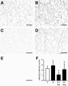
Increased TGFβRI immunoreactivity in retinal vessels of diabetic rats is prevented by sorbinil and aspirin treatments. TGFβRI was detected by immunohistochemistry in retinal trypsin digests from diabetic rats (8 months of diabetes duration), diabetic rats treated with sorbinil or aspirin, and age-matched control rats. The effect of diabetes and of the two drugs on TGFβRI immunostaining was quantitated by four masked observers. AC: Representative photographs of midretina fields showing TGFβRI immunostaining of retinal capillaries. A: Control, score 2.0 ± 0.7 (mean ± SD of scores by the different masked observers). B: Diabetes, score 3.8 ± 0.04. C: Diabetes treated with sorbinil, score 1 ± 0.0. D: Diabetes treated with aspirin, score 2.3 ± 0.7. E: Negative control. Scale bar = 100 microns. F: Bar plot of the quantitative analysis of staining intensity. Values are the means ± SD of the final scores computed for each individual rat. C, control rats, n = 11; D, diabetic rats, n = 10; D+Sor, diabetic rats treated with sorbinil, n = 6; D+Asa, diabetic rats treated with aspirin, n = 6. *P = 0.05 vs. control rats; †P < 0.02 versus control rats, diabetic rats, and diabetic rats treated with aspirin, ‡P < 0.04 versus diabetic rats.

References

    1. Davis M: Diabetic retinopathy: a clinical overview. Diabetes Care 1992; 15: 1844– 1874 - PubMed
    1. Roy S, Maiello M, Lorenzi M: Increased expression of basement membrane collagen in human diabetic retinopathy. J Clin Invest 1994; 93: 438– 442 - PMC - PubMed
    1. Mizutani M, Kern TS, Lorenzi M: Accelerated death of retinal microvascular cells in human and experimental diabetic retinopathy. J Clin Invest 1996; 97: 2883– 2890 - PMC - PubMed
    1. The Writing Team for the Diabetes Control and Complications Trial/Epidemiology of Diabetes Interventions and Complications Research Group: Effect of intensive therapy on the microvascular complications of type 1 diabetes mellitus. JAMA 2002; 287: 2563– 2569 - PMC - PubMed
    1. Lorenzi M: Mechanisms and strategies for prevention in diabetic retinopathy. Curr Diab Rep 2006; 6: 102– 107 - PubMed

Publication types

MeSH terms