

Pig embryonic pancreatic tissue as a source for transplantation in diabetes: transient treatment with anti-LFA1, anti-CD48, and FTY720 enables long-term graft maintenance in mice with only mild ongoing immunosuppression
- PMID: 19401429
- PMCID: PMC2699862
- DOI: 10.2337/db09-0112
Pig embryonic pancreatic tissue as a source for transplantation in diabetes: transient treatment with anti-LFA1, anti-CD48, and FTY720 enables long-term graft maintenance in mice with only mild ongoing immunosuppression
Abstract
Objective: Defining an optimal costimulatory blockade-based immune suppression protocol enabling engraftment and functional development of E42 pig embryonic pancreatic tissue in mice.
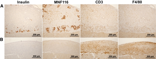
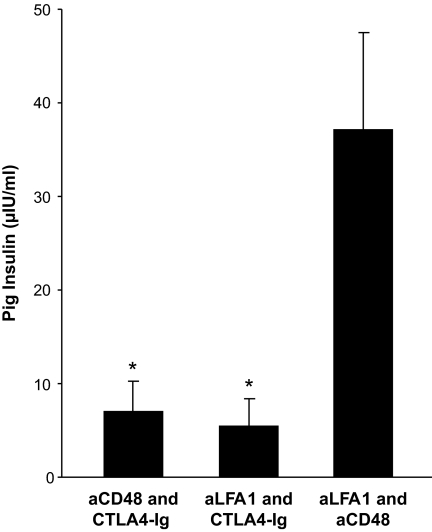
Research design and methods: Considering that anti-CD40L was found to be thrombotic in humans, we sought to test alternative costimulatory blockade agents already in clinical use, including CTLA4-Ig, anti-LFA1, and anti-CD48. These agents were tested in conjunction with T-cell debulking by anti-CD4 and anti-CD8 antibodies or with conventional immunosuppressive drugs. Engraftment and functional development of E42 pig pancreatic tissue was monitored by immunohistology and by measuring pig insulin blood levels.
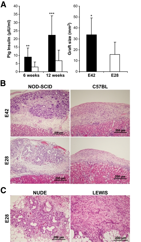
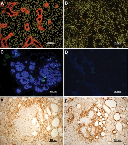
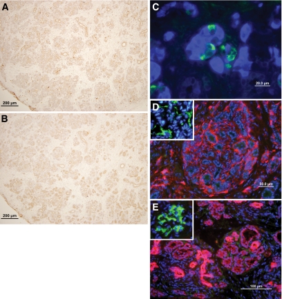
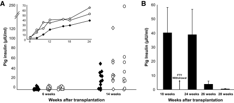
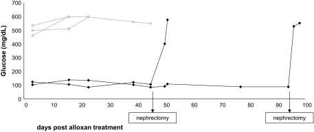
Results: Fetal pig pancreatic tissue harvested at E42, or even as early as at E28, was fiercely rejected in C57BL/6 mice and in Lewis rats. A novel immune suppression comprising anti-LFA1, anti-CD48, and FTY720 afforded optimal growth and functional development. Cessation of treatment with anti-LFA1 and anti-CD48 at 3 months posttransplant did not lead to graft rejection, and graft maintenance could be achieved for >8 months with twice-weekly low-dose FTY720 treatment. These grafts exhibited normal morphology and were functional, as revealed by the high pig insulin blood levels in the transplanted mice and by the ability of the recipients to resist alloxan induced diabetes.
Conclusions: This novel protocol, comprising agents that simulate those approved for clinical use, offer an attractive approach for embryonic xenogeneic transplantation. Further studies in nonhuman primates are warranted.
Figures
Similar articles
-
Embryonic pig pancreatic tissue transplantation for the treatment of diabetes.PLoS Med. 2006 Jul;3(7):e215. doi: 10.1371/journal.pmed.0030215. PLoS Med. 2006. PMID: 16768546 Free PMC article.
-
Embryonic pig pancreatic tissue for the treatment of diabetes: potential role of immune suppression with "off-the-shelf" third-party regulatory T cells.Transplantation. 2011 Feb 27;91(4):398-405. doi: 10.1097/TP.0b013e318204be15. Transplantation. 2011. PMID: 21192322
-
Immunosuppression with FTY720 and cyclosporine A inhibits rejection of adult porcine islet xenografts in rats.Transplantation. 2003 Apr 27;75(8):1409-14. doi: 10.1097/01.TP.0000061770.39569.7F. Transplantation. 2003. PMID: 12717240
-
Current perspectives on FTY720.Expert Opin Investig Drugs. 2007 Apr;16(4):505-18. doi: 10.1517/13543784.16.4.505. Expert Opin Investig Drugs. 2007. PMID: 17371198 Review.
-
FTY720: A new kid on the block for transplant immunosuppression.Expert Opin Biol Ther. 2003 Jul;3(4):665-81. doi: 10.1517/14712598.3.4.665. Expert Opin Biol Ther. 2003. PMID: 12831371 Review.
Cited by
-
Pig-islet xenotransplantation: recent progress and current perspectives.Front Surg. 2014 Mar 24;1:7. doi: 10.3389/fsurg.2014.00007. eCollection 2014. Front Surg. 2014. PMID: 25593932 Free PMC article. Review.
-
Development of a novel xenotransplantation strategy for treatment of diabetes mellitus in rat hosts and translation to non-human primates.Organogenesis. 2012 Apr-Jun;8(2):41-8. doi: 10.4161/org.20930. Epub 2012 Apr 1. Organogenesis. 2012. PMID: 22699748 Free PMC article. Review.
-
Engraftment of cells from porcine islets of Langerhans and normalization of glucose tolerance following transplantation of pig pancreatic primordia in nonimmune-suppressed diabetic rats.Am J Pathol. 2010 Aug;177(2):854-64. doi: 10.2353/ajpath.2010.091193. Epub 2010 Jun 25. Am J Pathol. 2010. PMID: 20581052 Free PMC article.
-
Engraftment of cells from porcine islets of Langerhans following transplantation of pig pancreatic primordia in non-immunosuppressed diabetic rhesus macaques.Organogenesis. 2011 Jul-Sep;7(3):154-62. doi: 10.4161/org.7.3.16522. Epub 2011 Jul 1. Organogenesis. 2011. PMID: 21654197 Free PMC article.
-
Feasibility of large experimental animal models in testing novel therapeutic strategies for diabetes.World J Diabetes. 2021 Apr 15;12(4):306-330. doi: 10.4239/wjd.v12.i4.306. World J Diabetes. 2021. PMID: 33889282 Free PMC article. Review.
References
-
- Dekel B, Burakova T, Arditti FD, Reich-Zeliger S, Milstein O, Aviel-Ronen S, Rechavi G, Friedman N, Kaminski N, Passwell JH, Reisner Y: Human and porcine early kidney precursors as a new source for transplantation. Nat Med 2003; 9: 53– 60 - PubMed
-
- Dekel B, Burakova T, Marcus H, Shezen E, Polack S, Canaan A, Passwell J, Reisner Y: Engraftment of human kidney tissue in rat radiation chimera: I. A new model of human kidney allograft rejection. Transplantation 1997; 64: 1541– 1550 - PubMed
-
- Statter MB, Foglia RP, Parks DE, Donahoe PK: Fetal and postnatal testis shows immunoprivilege as donor tissue. J Urol 1988; 139: 204– 210 - PubMed
-
- Metzger R, Parasta A, Joppich I, Till H: Organ-specific maturation of the major histocompatibility antigens in rats. Pediatr Surg Int 2002; 18: 640– 647 - PubMed
Publication types
MeSH terms
Substances
Grants and funding
LinkOut - more resources
Full Text Sources
Other Literature Sources
Research Materials
Miscellaneous
