

Majority of children with type 1 diabetes produce and deposit anti-tissue transglutaminase antibodies in the small intestine
- PMID: 19401430
- PMCID: PMC2699874
- DOI: 10.2337/db08-0962
Majority of children with type 1 diabetes produce and deposit anti-tissue transglutaminase antibodies in the small intestine
Abstract
Objective: Anti-tissue transglutaminase (TG2) antibodies are the serological marker of celiac disease. Given the close association between celiac disease and type 1 diabetes, we investigated the production and deposition of anti-TG2 antibodies in the jejunal mucosa of type 1 diabetic children.
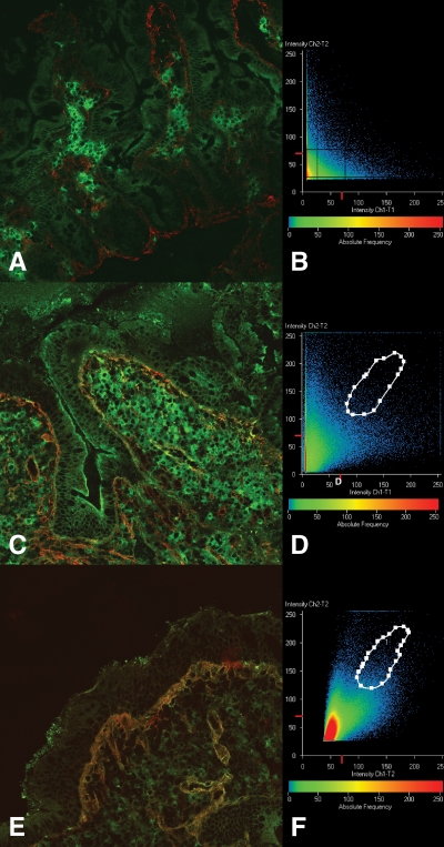
Research design and methods: Intestinal biopsies were performed in 33 type 1 diabetic patients with a normal mucosal architecture: 14 had high levels (potential celiac disease patients) and 19 had normal levels of serum anti-TG2 antibodies. All biopsy specimens were investigated for intestinal deposits of IgA anti-TG2 antibodies by double immunofluorescence. In addition, an antibody analysis using the phage display technique was performed on the intestinal biopsy specimens from seven type 1 diabetic patients, of whom four had elevated and three had normal levels of serum anti-TG2 antibodies.
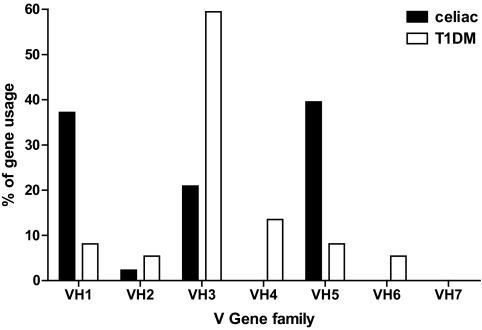
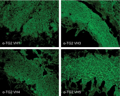
Results: Immunofluorescence studies showed that 11 of 14 type 1 diabetic children with elevated levels and 11 of 19 with normal serum levels of anti-TG2 antibodies presented with mucosal deposits of such autoantibodies. The phage display analysis technique confirmed the intestinal production of the anti-TG2 antibodies; however, whereas the serum-positive type 1 diabetic patients showed a preferential use of the VH5 antibody gene family, in the serum-negative patients the anti-TG2 antibodies belonged to the VH1 and VH3 families, with a preferential use of the latter.
Conclusions: Our findings demonstrate that there is intestinal production and deposition of anti-TG2 antibodies in the jejunal mucosa of the majority of type 1 diabetic patients. However, only those with elevated serum levels of anti-TG2 antibodies showed the VH usage that is typical of the anti-TG2 antibodies that are produced in patients with celiac disease.
Figures
References
-
- Mallone R, van Ender P: T cells in the pathogenesis of type 1 diabetes. Curr Diab Rep 2008; 8: 101– 106 - PubMed
-
- Westerholm-Ormio M, Vaarala O, Pihkala P, Ilonen J, Savilahti E: Immunologic activity in the small intestinal mucosa of pediatric patients with type 1 diabetes. Diabetes 2003; 52: 2287– 2295 - PubMed
-
- Carratù R, Secondulfo M, De Magistris L, Iafusco D, Urio A, Cardone MG, Pontoni G, Cartenì M, Prisco F: Altered intestinal permeability to mannitol in diabetes mellitus type 1. J Pediatr Gastroenterol Nutr 1999; 28: 264– 269 - PubMed
-
- Auricchio R, Paparo F, Maglio M, Franzese A, Lombardi F, Valerio G, Nardone G, Percopo S, Greco L, Troncone R: In vitro-deranged intestinal immune response to gliadin in type 1 diabetes. Diabetes 2004; 53: 1680– 1683 - PubMed
Publication types
MeSH terms
Substances
LinkOut - more resources
Full Text Sources
Medical
Miscellaneous
