. 2009 May 6;101(9):663-77.
doi: 10.1093/jnci/djp063. Epub 2009 Apr 28.

Clusterin, a haploinsufficient tumor suppressor gene in neuroblastomas

Affiliations

Clusterin, a haploinsufficient tumor suppressor gene in neuroblastomas

Olesya Chayka et al. J Natl Cancer Inst. .

Abstract

Background: Clusterin expression in various types of human cancers may be higher or lower than in normal tissue, and clusterin may promote or inhibit apoptosis, cell motility, and inflammation. We investigated the role of clusterin in tumor development in mouse models of neuroblastoma.

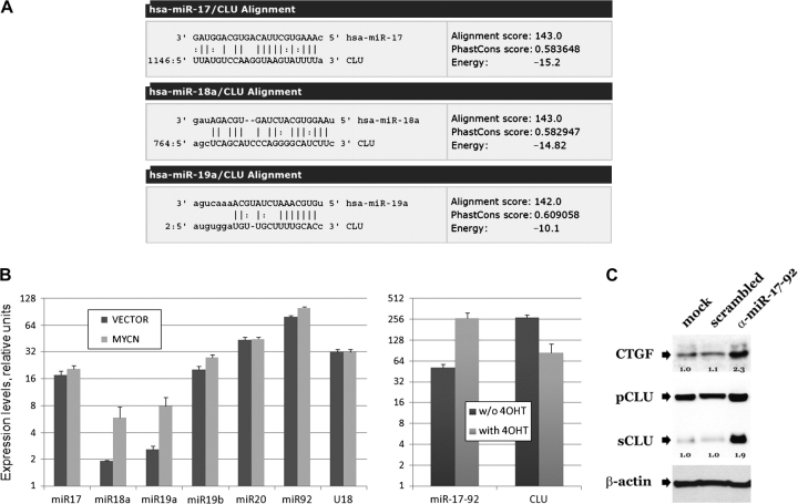
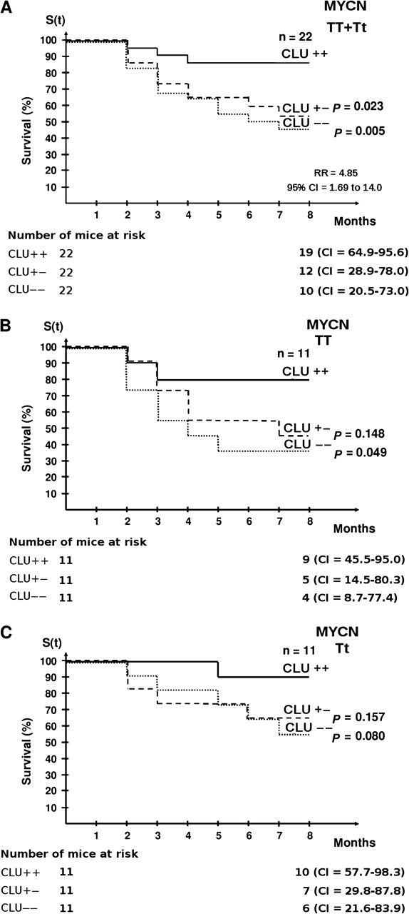
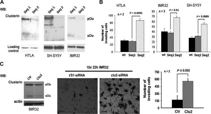
Methods: We assessed expression of microRNAs in the miR-17-92 cluster by real-time reverse transcription-polymerase chain reaction in MYCN-transfected SH-SY5Y and SH-EP cells and inhibited expression by transfection with microRNA antisense oligonucleotides. Tumor development was studied in mice (n = 66) that were heterozygous or homozygous for the MYCN transgene and/or for the clusterin gene; these mice were from a cross between MYCN-transgenic mice, which develop neuroblastoma, and clusterin-knockout mice. Tumor growth and metastasis were studied in immunodeficient mice that were injected with human neuroblastoma cells that had enhanced (by clusterin transfection, four mice per group) or reduced (by clusterin short hairpin RNA [shRNA] transfection, eight mice per group) clusterin expression. All statistical tests were two-sided.

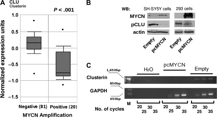
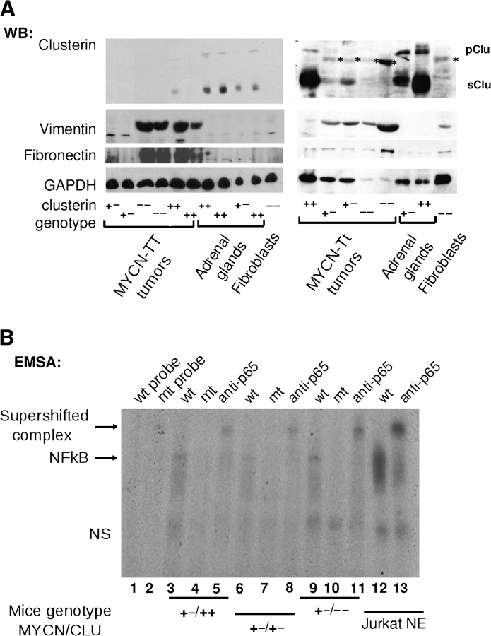
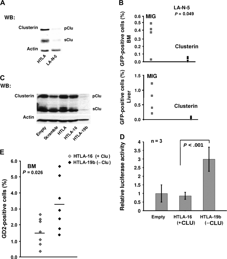
Results: Clusterin expression increased when expression of MYCN-induced miR-17-92 microRNA cluster in SH-SY5Y neuroblastoma cells was inhibited by transfection with antisense oligonucleotides compared with scrambled oligonucleotides. Statistically significantly more neuroblastoma-bearing MYCN-transgenic mice were found in groups with zero or one clusterin allele than in those with two clusterin alleles (eg, 12 tumor-bearing mice in the zero-allele group vs three in the two-allele group, n = 22 mice per group; relative risk for neuroblastoma development = 4.85, 95% confidence interval [CI] = 1.69 to 14.00; P = .005). Five weeks after injection, fewer clusterin-overexpressing LA-N-5 human neuroblastoma cells than control cells were found in mouse liver or bone marrow, but statistically significantly more clusterin shRNA-transfected HTLA230 cells (3.27%, with decreased clusterin expression) than control-transfected cells (1.53%) were found in the bone marrow (difference = 1.74%, 95% CI = 0.24% to 3.24%, P = .026).

Conclusions: We report, to our knowledge, the first genetic evidence that clusterin is a tumor and metastasis suppressor gene.


Figures

Figure 1
Figure 1
Expression of clusterin and MYCN in neuroblastoma. A) Box plot of the expression of clusterin mRNA in primary human neuroblastomas with MYCN amplification (Positive) or without MYCN amplification (Negative). The figure was generated with tools found in the Oncomine Web site (www.oncomine.org), where the information regarding the neuroblastoma samples can also be found. Statistical significance was assessed by Student t test. In the Oncomine Web site, all mRNA datasets are normalized by being log2-transformed, with the median set to 0 and SD set to 1. All statistical tests were two-sided. B) WB analysis. Whole-cell lysates were prepared from SH-SY5Y and HEK-293 cells that were stably (SH-SY5Y cells) or transiently (HEK-293 cells) transfected with an MYCN (pcMYCN) or control (Empty) vector and subjected to western blot analysis with antibodies specific for MYCN, the precursor form of clusterin (pCLU), or actin (as a loading control). C) Semiquantitative reverse transcription–PCR. Clusterin mRNA expression was assessed in SH-SY5Y cells stably transfected with MYCN (pcMYCN) or with control (Empty) vector. H2O indicates PCR amplification without reverse transcription (negative control). Samples were analyzed after completion of the indicated number of PCR cycles. Amplification of glyceraldehyde-3-phosphate dehydrogenase served as a loading control. M indicates molecular weight marker. PCR = polymerase chain reaction; WB = Western blot.
Figure 2
Figure 2
MicroRNAs of the 17-92 cluster and clusterin in neuroblastoma cells that overexpress MYCN. A) miRanda algorithm–predicted alignment of miR-17a, -18a, and -19a RNAs with clusterin (CLU) mRNA. The closely related miR-19b aligns with the same sequence as miR-19a. Alignment scores reflect sequence complementarity by using a position-weighted local alignment algorithm. Phastcons scores are a measure of evolutionary conservation of sequence blocks across multiple vertebrate species. Energy is an estimate of the free energy of formation of the microRNA–mRNA duplex. Perfect base pairing and G:U wobble base pairing are indicated by the vertical bars and colons, respectively. The numbers 1146, 764, and 2 are nucleotide positions in clusterin mRNA. B) Real-time polymerase chain reaction analysis. Left) Mature miR-17-92 cluster members in SH-SY5Y cells transfected with MYCN or control (vector) plasmids. The small nucleolar RNA U18 was used as control for RNA quality and/or input. Values on the y-axis equal 2(30Ct), where Ct is the threshold cycle (ie, the cycle at which fluorescence rises statistically significantly above the baseline). The ABI sequence detection system software was set to automatically generate baseline and threshold values. Error bars = 95% confidence intervals. Right) miR-17-92 and clusterin transcripts in SH-EP cells expressing the MYCN–estrogen receptor fusion protein before and 48 hours after treatment with 4-hydroxytamoxifen (4OHT). A 24-hour treatment yielded essentially identical results. Expression levels were adjusted for β-actin. C) Western blot analysis. Blots were probed with antibodies against precursor (pCLU) or secreted (sCLU) clusterin, connective tissue growth factor (CTGF), and β-actin in MYCN-expressing SH-SY5Y cells transfected with anti-microRNA 17-92 (α-miR-17-92) or scrambled 2′-O-methyl oligoribonucleotides. Numbers refer to expression levels relative to those observed in mock-transfected cells. For clusterin, values represent the sum of the precursor and secreted forms under each condition. This assay was repeated twice with similar results.
Figure 3
Figure 3
Tumor-free survival in MYCN-transgenic mice with different dosages of clusterin alleles. Kaplan–Meier survival curves of mice with different MYCN-transgene dosages (homozygous = TT or heterozygous = Tt) and clusterin genotypes (homozygously deleted = − −; heterozygously deleted = + −; wild type = + +) are shown. A) Cumulative tumor-free survival of the entire population of homozygous (MYCN TT) plus heterozygous MYCN-transgenic mice (MYCN Tt) by clusterin expression. There were 22 mice in each MYCN-transgenic group. B) Tumor-free survival of homozygous MYCN-transgenic mice (MYCN TT) by clusterin expression. There were 11 mice in each MYCN-transgenic group. C) Tumor-free survival of hemizygous MYCN-transgenic mice (MYCN Tt) by clusterin expression. There were 11 mice in each MYCN-transgenic group. The log-rank test was used to assess whether the difference in cumulative tumor-free survival between groups was statistically significant (RR for neuroblastoma development in clusterin +/+ vs clusterin −/− for all MYCN-transgenic mice = 4.85, 95% CI = 1.69 to 14.00). The number of mice at risk at 0 and 8 months and the 95% CI for the survival at 8 months are also shown. All statistical tests were two-sided. CI = confidence interval; RR = relative risk.
Figure 4
Figure 4
MYCN overexpression, clusterin expression, NF-κB activation, and the epithelial-to-mesenchymal transition. A) Western blot (WB) analysis of lysates of samples obtained from neuroblastoma tumors, normal adrenal glands, or embryonic fibroblasts from mice homozygous (MYCN TT) or hemizygous (MYCN Tt) for the MYCN transgene with wild-type or heterozygously or homozygously deleted clusterin alleles (indicated by + +, + −, or − −, respectively). Blots were incubated with antibodies against clusterin, vimentin, fibronectin, or glyceraldehyde-3-phosphate dehydrogenase (as a loading control), as indicated. Nonspecific bands in some of the lanes are indicated by an asterisk. B) EMSA of tumor protein lysates obtained from MYCN-transgenic mice with various MYCN and clusterin genotypes. Tumor extracts were incubated with a 32P-labeled oligonucleotide probe containing a wild-type (wt; lanes 3, 6, and 9) or mutated (mt; lanes 4, 7, and 10) NF-κB consensus motif. Lanes 1 and 2 contain free wt and mt probes, respectively. Supershifted complexes of the oligonucleotide and p65-NF-κB were identified by use of anti-p65-NF-κB antibody (lanes 5, 8, 11, and 13). Nuclear extract of Jurkat cells that had been stimulated with 12-O-tetradecanoylphorbol-13-acetate (Jurkat NE) was used as a positive control for NF-κB activation. The mouse genotypes for MYCN and clusterin are indicated at the bottom. pClu = precursor clusterin; sClu = secreted clusterin; NS = nonspecific DNA–protein complex; NF-κB = nuclear factor κB; EMSA = electrophoretic mobility shift assay.
Figure 5
Figure 5
Immunohistochemical staining for clusterin expression in sections from neuroblastoma tumors. Representative histological sections stained with hematoxylin and anti-clusterin (CLU) antibodies (brown color). A) Extracellular or cytoplasmic staining for clusterin. Clusterin staining in the neuropil = arrows. Original magnification was ×200. Scale bar = 0.05 mm. B) Necrotic tissue. Thin arrow = clusterin staining in necrotic tissue; thick arrow = nuclear clusterin staining. Original magnification was ×100. Scale bar = 0.1 mm. C) Ganglion cells. Thick arrow = differentiated ganglion cells; thin arrow = immature neuroblasts. Original magnification was ×200. Scale bar = 0.05 mm. D) Apoptotic cells. Thick arrow = normal nucleus; thin arrow = pyknotic nucleus. Note that only cells with fragmented or pyknotic, but not normal, nuclei are stained by the antibody against clusterin. Original magnification was ×400. Scale bar = 0.001 mm.
Figure 6
Figure 6
Clusterin and in vitro invasion of neuroblastoma. A) WB analysis of clusterin expression in neuroblastoma cells after transfection with siRNA against clusterin. Cell lysates were collected 24 hours after transfection of control sequence 1 (Seq 1) or clusterin sequence 2 (Seq 2) and then subjected to WB analysis with antibodies against clusterin to detect precursor (pClu) and secreted (sClu) clusterin. Expression of housekeeping genes, as a loading control, was monitored by WB with actin (HTLA cells) or keratin antibodies (IMR32 and SH-SY5Y cells). B) In vitro invasion assay with cells transfected with siRNAs against clusterin. The numbers of IMR32, HTLA230, and SH-SY5Y cells that migrated to the bottom chamber after mock transfection (wt), transfection of control sequence 1 (Seq1), or clusterin sequence 2 (Seq2) siRNAs are indicated. C) In vitro invasion assay with cells transfected with other siRNAs against clusterin. Left) WB analysis with a clusterin antibody of IMR-32 cells transfected with clusterin (Clu2) or control (Ctr) siRNAs. Middle) Crystal violet staining of invading IMR32 cells. Scale bars = 0.05 mm. Right) In vitro invasion assay results with cells transfected with the control (Ctr) or clusterin (Clu2) siRNAs. Statistical significance was calculated with a Student t test (n = number of independent assays). Error bars = 95% confidence intervals. All statistical tests were two-sided. siRNA = small interfering RNA; WB = Western blot.
Figure 7
Figure 7
Clusterin and an experimental model of metastasis in mice bearing xenotransplanted human neuroblastoma. A) Western blot analysis of clusterin expression in human neuroblastoma cell lines LA-N-5 and HTLA230. Cells were lysed and subjected to Western blot analysis with a clusterin antibody. Positions of the human 60-kDa clusterin precursor (pClu) and the mature 36-kDa secreted (sClu) clusterin are shown. Actin was used as the loading control. B) Quantification of neuroblastoma cells recovered from BM or liver of mice after 5 weeks that had been injected with LA-N-5 cells transduced with empty or clusterin MIGR1 (Clusterin) vectors (n = 4 mice per group). Tumor cells which were positive for green fluorescent protein expression from the MIGR1 vector were counted by flow cytometry. The statistical significance between the clusterin-transduced and untransduced groups was assessed with Student t test. C) Western blot analysis of clusterin expression in HTLA clones. HTLA cells were stably transfected with control vector (Empty), a plasmid containing short hairpin RNA control sequence 1 (termed HTLA-16 cells), or a plasmid containing clusterin short hairpin RNA sequence 2 (termed HTLA-19b cells), and then subjected to Western blot analysis for clusterin with a clusterin antibody. Actin was used as the loading control. D) Luciferase assays and the activity of NF-κB in HTLA cells with (HTLA-16 cells) or without (HTLA-19b cells) clusterin expression. A NF-κB luciferase reporter construct was transiently transfected into HTLA-16 and -19b cells. Firefly luciferase activity was measured and normalized to that of Renilla luciferase. Statistical significance was assessed with the two-sample Welch t test (n = 3 independent experiments). Error bars = 95% confidence intervals. E) Quantification of neuroblastoma cells recovered from BM of mice injected with HTLA clones HTLA-16 or -19b (n = 8 mice per group). Grey diamonds = HTLA-16 cells; black diamonds = HTLA-19b cells. Statistical significance was assessed with Student t test. All statistical tests were two-sided. NF-κB = nuclear factor κB; BM = bone marrow.

Similar articles

Cited by

References

    1. Brodeur GM. Neuroblastoma: biological insights into a clinical enigma. Nat Rev Cancer. 2003;3(3):203–216. - PubMed
    1. Lonergan GJ, Schwab CM, Suarez ES, Carlson CL. Neuroblastoma, ganglioneuroblastoma, and ganglioneuroma: radiologic-pathologic correlation. Radiographics. 2002;22(4):911–934. - PubMed
    1. Shannan B, Seifert M, Leskov K, et al. Challenge and promise: roles for clusterin in pathogenesis, progression and therapy of cancer. Cell Death Differ. 2006;13(1):12–19. - PubMed
    1. Trougakos IP, Gonos ES. Clusterin/apolipoprotein J in human aging and cancer. Int J Biochem Cell Biol. 2002;34(11):1430–1448. - PubMed
    1. Nizard P, Tetley S, Le Drean Y, et al. Stress-induced retrotranslocation of clusterin/ApoJ into the cytosol. Traffic. 2007;8(5):554–565. - PubMed

Publication types

MeSH terms