. 2009 Jul 2;114(1):187-94.
doi: 10.1182/blood-2008-07-170712. Epub 2009 Apr 28.

SCF induces gamma-globin gene expression by regulating downstream transcription factor COUP-TFII

Affiliations

SCF induces gamma-globin gene expression by regulating downstream transcription factor COUP-TFII

Wulin Aerbajinai et al. Blood. .

Abstract

Increased fetal hemoglobin expression in adulthood is associated with acute stress erythropoiesis. However, the mechanisms underlying gamma-globin induction during the rapid expansion of adult erythroid progenitor cells have not been fully elucidated. Here, we examined COUP-TFII as a potential repressor of gamma-globin gene after stem cell factor (SCF) stimulation in cultured human adult erythroid progenitor cells. We found that COUP-TFII expression is suppressed by SCF through phosphorylation of serine/threonine phosphatase (PP2A) and correlated well with fetal hemoglobin induction. Furthermore, down-regulation of COUP-TFII expression with small interfering RNA (siRNA) significantly increases the gamma-globin expression during the erythroid maturation. Moreover, SCF-increased expression of NF-YA associated with redox regulator Ref-1 and cellular reducing condition enhances the effect of SCF on gamma-globin expression. Activation of Erk1/2 plays a critical role in SCF modulation of downstream transcriptional factor COUP-TFII, which is involved in the regulation of gamma-globin gene induction. Our data show that SCF stimulates Erk1/2 MAPK signaling pathway, which regulates the downstream repressor COUP-TFII by inhibiting serine/threonine phosphatase 2A activity, and that decreased COUP-TFII expression resulted in gamma-globin reactivation in adult erythropoiesis. These observations provide insight into the molecular pathways that regulate gamma-globin augmentation during stress erythropoiesis.


Figures

Figure 1
Figure 1
SCF-suppressed COUP-TFII expression inversely correlates with increase in γ-globin during erythroid differentiation. CD34+ cells cultured in EPO-containing medium for 6 days were stimulated with SCF (50 ng/mL) from day 6 to day 14, and RNA extracted from cells harvested on days 6, 8, 10, 12, and 14 was analyzed by quantitative RT-PCR. (A) Expression levels of COUP-TFII mRNA in EPO versus EPO plus SCF. (B) Expression levels of γ-globin mRNA in EPO versus EPO plus SCF. (C) Expression levels of β-globin mRNA in EPO versus EPO plus SCF. (D) The ratio of γ/(γ + β) globin percentages are shown in EPO versus EPO plus SCF. Results are shown as mean ± SD from 3 independent donors that were analyzed in separate experiments. *P < .05 versus SCF-stimulated cells. **P < .01 versus SCF-stimulated cells.
Figure 2
Figure 2
SCF-induced phosphorylation of PP2A correlates with increased fetal hemoglobin during erythroid differentiation. CD34+ cells were cultured in EPO-containing medium for 6 days before the addition of SCF. (A) Cells were incubated with SCF (50 ng/mL) for the indicated times, and whole cell lysates (30 μg) were analyzed by immunoblot for phosphorylation of PP2A. (B) Cells were incubated with SCF (50 ng/mL) from day 6 to day 14, and total cell lysates (30 μg) harvested on the indicated culture days were analyzed by immunoblot for phosphorylation of PP2A, COUP-TFII, or fetal hemoglobin expression. The lowest panel shows the same blot stripped and reprobed with total β-actin antibody to confirm that similar amounts of protein extracts were analyzed in each lane. Representative immunoblots from 3 independent experiments are shown.
Figure 3
Figure 3
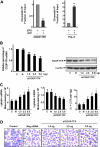
SCF suppresses COUP-TFII binding to the γ-globin promoter and knockdown of endogenous COUP-TFII induces γ-globin expression in erythroblasts. (A) Antibodies against COUP-TFII and RNA polymerase II (PoL II) were used to immunoprecipitate chromatin. Precipitated DNA was amplified and quantitated by real-time PCR with primers flanking the γ-globin gene promoter. Results are expressed as relative proportions of immunoprecipitated DNA (ratios of immunoprecipitated versus input DNA) normalized to the ratio obtained for the γ-globin promoter in unstimulated cells (arbitrarily set at 1). (B) Effect of different doses of silencing COUP-TFII mRNA or protein levels was evaluated by quantitative RT-PCR or immunoblot analysis on day 12 cultured cells after transiently transfected with either COUP-TFII or control non–targeting siRNA by Amaxa electroporation. (C) Quantitative RT-PCR analysis of γ-, β-globin mRNA levels, and the ratio of γ/(γ + β) globin percentages in negative control or knockdown COUP-TFII (different doses) cells. (D) Representative fields of Giemsa-stained erythroid cell at day 12 after knockdown of COUP-TFII at different doses. Results are shown as mean ± SD from 3 independent donors that were analyzed in separate experiments. *P < .05 versus negative control siRNA.
Figure 4
Figure 4
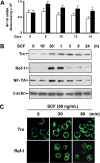
Expression of NF-YA in response to SCF during erythroid differentiation. (A) Quantitative real-time PCR was performed to determine NF-YA expression levels for the indicated days after CD34+ cells were added with SCF (50 ng/mL) from day 6 to day 14. Results are shown as mean ± SD from 3 different donors. *P < .05 versus unstimulated cells. (B) CD34+ cells grown in EPO-containing medium for 6 days were incubated with SCF (50 ng/mL) for the indicated times, and whole cell lysates (30 μg) were analyzed by immunoblot with thioredoxin (Trx), Ref-1, or NF-YA antibodies. The lowest panel shows the same blot stripped and reprobed with anti–β-actin antibody to confirm that similar amounts of protein extracts were analyzed in each lane. (C) Immunofluorescence analysis of Trx and Ref-1 localization after cells grown in EPO-containing medium for 6 days were stimulated with SCF (50 ng/mL) for the indicated times. Representative immunoblots from 3 independent experiments are shown.
Figure 5
Figure 5
Cellular reducing condition enhances the effect of SCF. CD34+ cells cultured with or without SCF (50 ng/mL) in the presence or absence of the indicated concentration of NAC or 2-ME from day 6 to day 14. Total RNA was isolated, and quantitative real-time PCR was performed to analyze the expression of γ-globin mRNA under the reducing conditions (A), the expression of β-globin mRNA under the reducing conditions (B), the ratios of γ/(γ + β) globin percentages under the reducing conditions (C). Results are shown as mean ± SD from 3 independent donors that were analyzed in separate experiments. *P < .05 versus unstimulated cells.
Figure 6
Figure 6
SCF-induced activation of Erk1/2 and p38 MAPK. CD34+ cells were cultured in EPO-containing medium for 6 days before the addition of SCF. (A) Cells were incubated with SCF (50 ng/mL) for the indicated times, and whole cell lysates (30 μg) were analyzed by immunoblot for phosphorylation of Erk1/2 and p38. The lowest panel shows the same blot stripped and reprobed with total p38 antibody to confirm that similar amounts of protein extracts were analyzed in each lane. (B) Cells were stimulated with SCF (50 ng/mL) for 10 minutes with and without preincubation with PD98059 (20 μM), SB202109 (5 μM), 2-ME (500 μM), or NAC (100 μM) for 30 minutes, and whole cell lysates were analyzed by immunoblot for phosphorylation of Erk1/2 and p38. The lowest panel shows the same blot stripped and reprobed with total Erk2 antibody to confirm that similar amounts of protein extracts were analyzed in each lane. Representative immunoblots from 3 independent experiments are shown.
Figure 7
Figure 7
SCF-induced γ-globin reactivation is mediated by activation of Erk1/2 through down-regulation of COUP-TFII. CD34+ cells were cultured with or without SCF (50 ng/mL) in the presence or absence of the indicated inhibitors from day 6 to day 14. On day 14, whole cell lysates (30 μg) were analyzed by immunoblot for phosphorylation of PP2A, COUP-TFII, γ-hemoglobin, and β-actin (A), total RNA was analyzed by quantitative real-time PCR to determine the expression of γ-globin mRNA level (B), the expression of β-globin mRNA level (C), the ratios of γ/(γ + β) globin percentages (D). Results are shown as mean ± SD from 3 independent donors that were analyzed in separate experiments. *P < .05 versus SCF-stimulated cells.

References

    1. Bank A. Regulation of human fetal hemoglobin: new players, new complexities. Blood. 2006;107:435–443. - PMC - PubMed
    1. Patrinos GP, de Krom M, de Boer E, et al. Multiple interactions between regulatory regions are required to stabilize an active chromatin hub. Genes Dev. 2004;18:1495–1509. - PMC - PubMed
    1. Vakoc CR, Letting DL, Gheldof N, et al. Proximity among distant regulatory elements at the beta-globin locus requires GATA-1 and FOG-1. Mol Cell. 2005;17:453–462. - PubMed
    1. Boyer SH, Belding TK, Margolet L, Noyes AN. Fetal hemoglobin restriction to a few erythrocytes (F cells) in normal human adults. Science. 1975;188:361–363. - PubMed
    1. Alter BP. Fetal erythropoiesis in stress hematopoiesis. Exp Hematol. 1979;7:200–209. - PubMed

MeSH terms