. 2009 Apr 28;2(68):ra18.
doi: 10.1126/scisignal.2000188.

Ligand binding to LRP1 transactivates Trk receptors by a Src family kinase-dependent pathway

Affiliations

Ligand binding to LRP1 transactivates Trk receptors by a Src family kinase-dependent pathway

Yang Shi et al. Sci Signal. .

Abstract

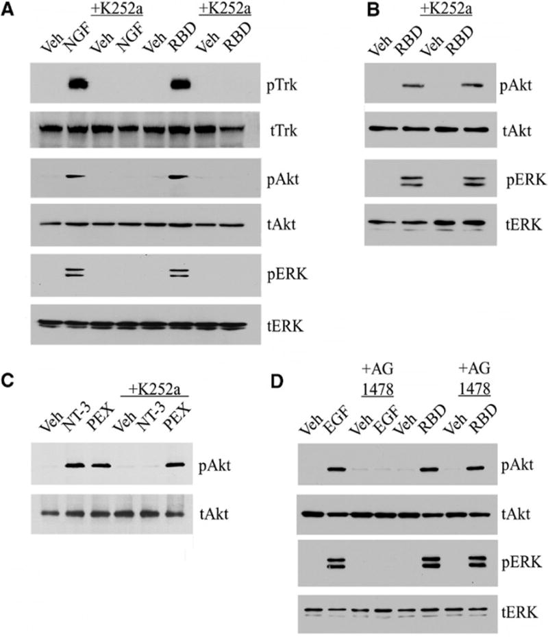
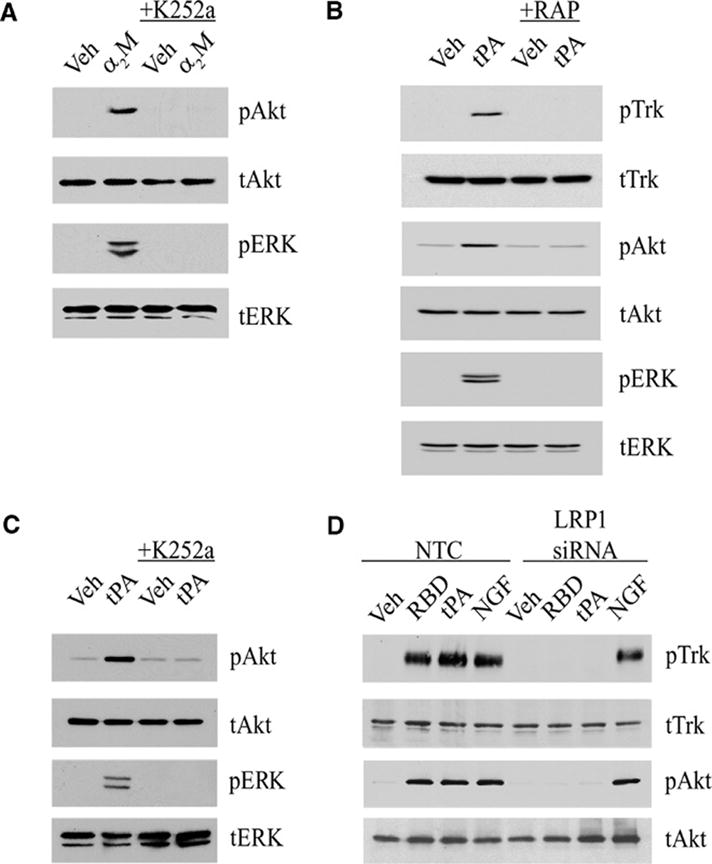
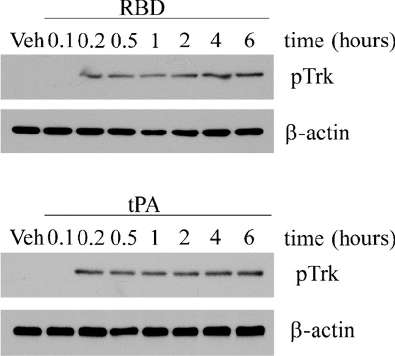
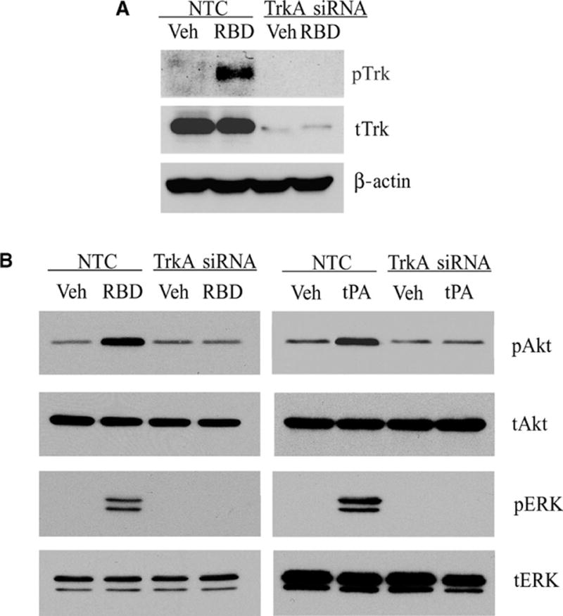
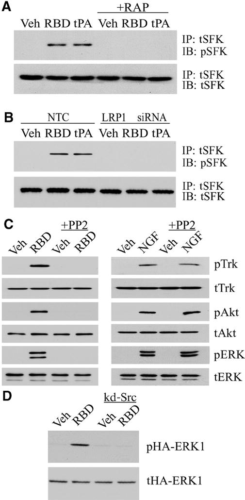
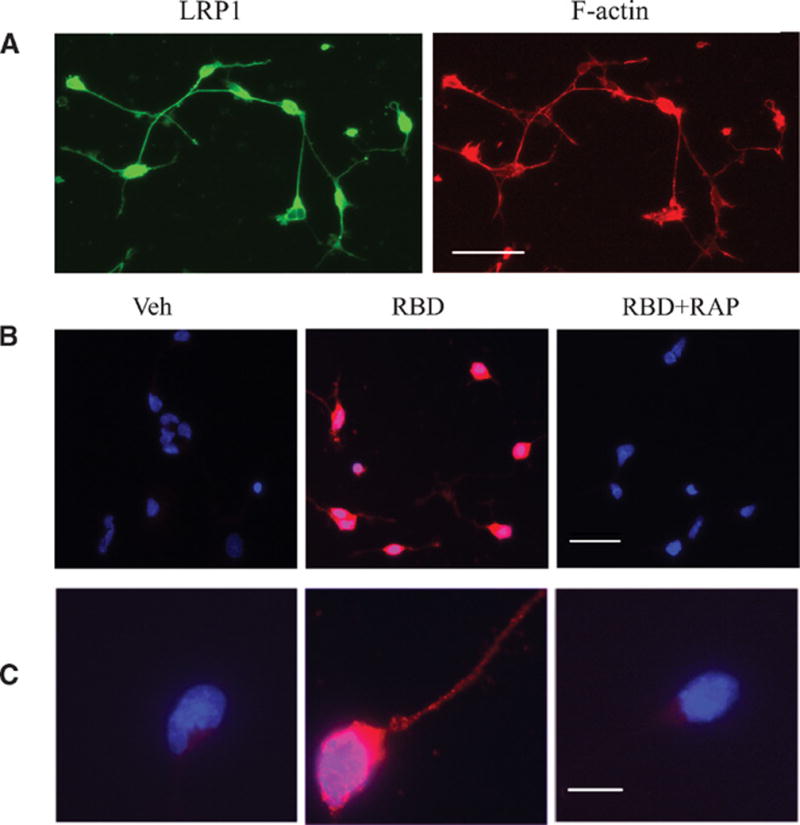
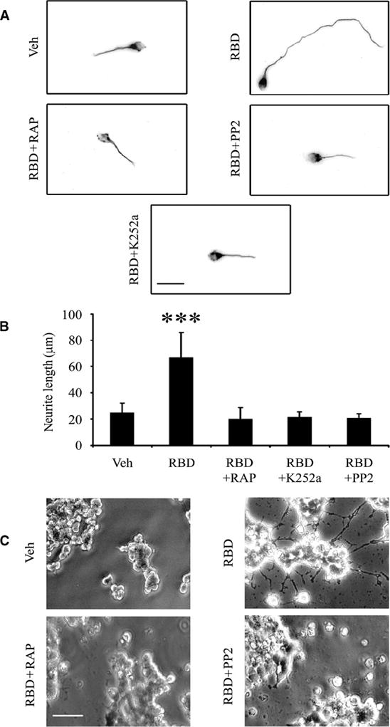
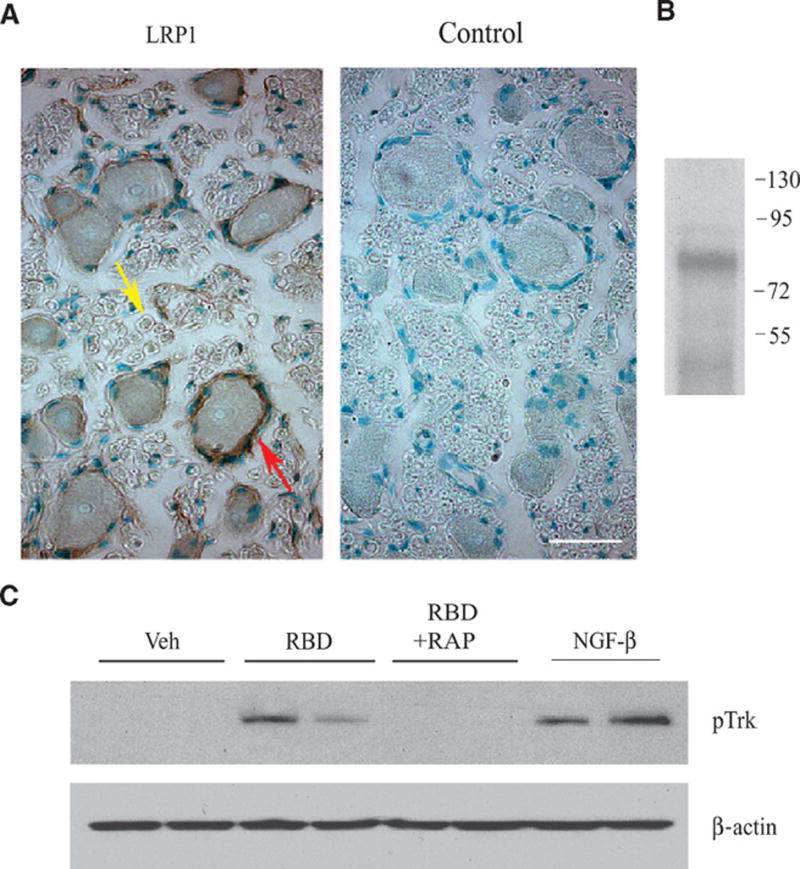
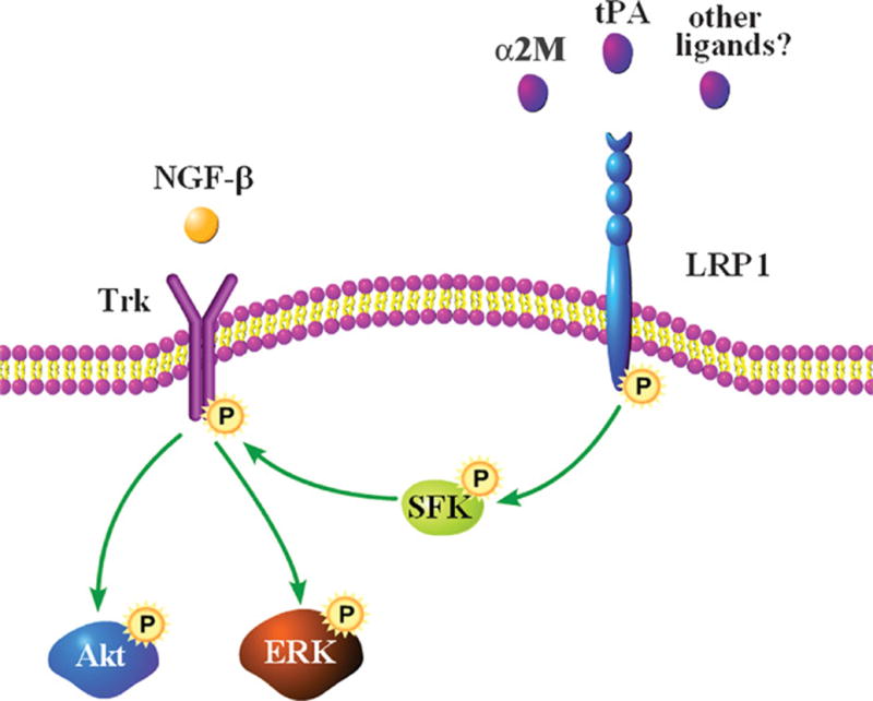
Low-density lipoprotein receptor-related protein 1 (LRP1) functions in endocytosis and intracellular signaling for a variety of structurally diverse ligands. Although LRP1 has been implicated in several aspects of neuronal function, molecular mechanisms underlying the activity of neuronal LRP1 remain unclear. Here, we describe a signaling pathway whereby LRP1 transactivates Trk receptors. Binding of tissue-type plasminogen activator or alpha(2)-macroglobulin (alpha(2)M) to LRP1 resulted in Src family kinase (SFK) activation and SFK-dependent Trk receptor transactivation in PC12 cells and neurons. Trk receptor transactivation was necessary for activation of Akt and extracellular signal-regulated kinase and for neurite outgrowth downstream of LRP1. Injection of the LRP1-binding domain of alpha(2)M into rat dorsal root ganglia induced Trk receptor phosphorylation, which was blocked by receptor-associated protein, an antagonist of ligand binding to LRP1. Trk receptor transactivation provides a mechanism by which diverse LRP1 ligands may show neurotrophic activity.


Figures

Fig. 1
Fig. 1
The α2M RBD induces Trk receptor phosphorylation. (A) PC12 cells were treated with serum-free medium (veh), NGF-β (100 ng/ml), or the α2M RBD (100 nM) for 10 min. Cultures were pre-treated with K252a (10 nM) for 2 hours as indicated. (B) Schwann cells were treated with the α2M RBD or vehicle after pre-treatment with K252a as indicated. (C) Schwann cells were treated with vehicle, NT-3 (10 nM), or the hemopexin domain of MMP-9 (PEX, 10 nM) for 10 min, with or without pretreatment of K252a (10 nM). (D) PC12 cells were pre-treated with AG1478 (50 nM) for 2 hours as indicated and then with EGF (10 ng/ml) or the α2M RBD (100 nM). For each experiment, cell extracts were immunoblotted for indicated proteins with antibodies specific for phosphorylated Trk (pTrk), phosphorylated Akt (pAkt), phosphorylated ERK1/2 (pERK), total Trk (tTrk), total Akt (tAkt), or total ERK1/2 (tERK).
Fig. 2
Fig. 2
Trk receptors, Akt, and ERK1/2 are activated by two different LRP1 ligands. (A) PC12 cells were treated with 50 nM activated full-length α2M (α2M) or with vehicle (veh) for 10 min, with or without K252a pre-treatment. (B) PC12 cells were treated for 10 min with non-enzymatic tPA (10 nM) in the presence of GST-RAP (100 nM) or GST (100 nM) as a control. (C) PC12 cells were treated with non-enzymatic tPA (10 nM) after pretreatment with K252a as indicated. (D) PC12 cells transfected with LRP1-specific or NTC siRNA were treated for 10 min with vehicle, α2M RBD (100 nM), non-enzymatic tPA (10 nM) or NGF-β (100 ng/ml). For each experiment, cell extracts were immunoblotted with antibodies specific for pTrk, tTrk, pAkt, tAkt, pERK, and tERK.
Fig. 3
Fig. 3
Trk receptor phosphorylation is sustained in PC12 cells treated with LRP1 ligands. Cells were treated for the indicated times with the α2M RBD (100 nM) or with tPA (10 nM). Cell extracts prepared at different time points were immunoblotted for pTrk. Blots were reprobed for β-actin as a loading control.
Fig. 4
Fig. 4
TrkA gene silencing blocks LRP1-initiated signaling. (A) PC12 cells transfected with TrkA or NTC siRNAs were stimulated with the α2M RBD or treated with vehicle. (B) PC12 cells transfected with TrkA or NTC siRNAs were treated with the α2M RBD, non-enzymatic tPA, or vehicle. For each experiment, cell extracts were immunoblotted with antibodies specific for pTrk, tTrk, pERK, tERK, pAkt, tAkt, and β-actin.
Fig. 5
Fig. 5
SFKs mediate Trk receptor transactivation by LRP1. (A) PC12 cells were treated with vehicle, the α2M RBD (100 nM), or non-enzymatic tPA (10 nM), with or without RAP pre-treatment. SFKs were immunoprecipitated (IP) from cell extracts, and precipitates were immunoblotted (IB) for phosphorylated SFK (pSFK) and total SFK (tSFK). (B) PC12 cells transfected with LRP1 or NTC siRNAs were treated with the α2M RBD, tPA, or vehicle. pSFK and tSFK were determined as described in A. (C) PC12 cells were pre-treated for 2 hours with PP2 (1 μM) in the indicated lanes. The cells were then stimulated with the α2M RBD (100 nM) or NGF-β (100 ng/ml). Cell extracts were immunoblotted for pTrk, tTrk, pAkt, tAkt, pERK, and tERK. (D) PC12 cells that were transfected with hemagglutinin (HA)-tagged ERK1 alone or with kinase defective (kd)-Src were stimulated with the α2M RBD or treated with vehicle. Cell extracts were immunoblotted for phosphorylated HA-ERK1 (pHA-ERK1) and total HA-ERK1 (tHA-ERK1).
Fig. 6
Fig. 6
(A) LRP1 is present in cultured cerebellar neurons. Cerebellar neurons were labeled with an LRP1 antibody (green) and with phalloidin to detect actin (red). Scale bar: 40 μm. (B) Phosphorylation of Trk in α2M RBD-treated CGNs. Cultured cerebellar neurons were treated with the α2M RBD (100 nM) or with vehicle for 10 min. In the panels labeled RBD+RAP, the cells were pre-treated with 100 nM RAP for 1 h. Fluorescence microscopy was performed to detect phosphorylated Trk (pTrk; red). Nuclei were stained with DAPI (blue). Merged images are presented. Scale bar: 40 μm. (C) Merged images of a single representative neuron are shown. Scale bar: 10 μm.
Fig. 7
Fig. 7
The α2M RBD promotes neurite outgrowth in CGNs and PC12 cells. (A) Representative CGNs are shown after culturing for 48 hours in the presence of the α2M RBD (100 nM) alone or with RAP, K252a, or PP2 as indicated. The neurons were stained with anti-tubulin antibody and imaged by fluorescence microscopy. Scale bar, 20 μm. (B) Neurite length for 50 CGNs in each condition was measured (***, p<0.001). (C) PC12 cells were treated with the α2M RBD (100 nM) for 48 hours, in the presence of K252a or PP2 as indicated. Phase contrast images are shown. Scale bar, 40 μm.
Fig. 8
Fig. 8
Trk receptors are phosphorylated by the α2M RBD in vivo. (A) Immunohistochemistry for LRP1 in rat DRGs detected immunopositive neuronal cell bodies, satellite cells (red arrow) surrounding neuronal cell bodies, and axons (yellow arrow). The panel labeled “control” was not stained with primary antibody. Scale bar: 10 μm. (B) DRG extracts were immunoblotted for LRP1 β-chain. Numbers to the right of the immunoblot indicate molecular mass markers in kilodaltons. (C) Vehicle, the α2M RBD, the α2M RBD+RAP, or NGF-β were injected directly into DRGs, which were collected 10 min later. Extracts were immunoblotted for pTrk, and then probed for β-actin as a loading control. Each lane shows extracts from a different animal.
Fig. 9
Fig. 9
Proposed model for Trk receptor transactivation by LRP1. Although we show Trk receptor in the plasma membrane, transactivated Trk may be localized to different subcellular compartments, such as the Golgi. Future work will be needed to determine which LRP1 ligands transactivate Trk receptors.

References

    1. Strickland DK, Gonias SL, Argraves WS. Diverse roles for the LDL receptor family. Trends Endocrinol Metab. 2002;13:66–74. - PubMed
    1. Bacskai BJ, Xia MQ, Strickland DK, Rebeck GW, Hyman BT. The endocytic receptor protein LRP also mediates neuronal calcium signaling via N-methyl-D-aspartate receptors. Proc Natl Acad Sci U S A. 2000;97:11551–11556. - PMC - PubMed
    1. Hu K, Yang J, Tanaka S, Gonias SL, Mars WM, Liu Y. Tissue-type plasminogen activator acts as a cytokine that triggers intracellular signal transduction and induces matrix metalloproteinase-9 gene expression. J Biol Chem. 2006;281:2120–2127. - PubMed
    1. Hayashi H, Campenot RB, Vance DE, Vance JE. Apolipoprotein E-containing lipoproteins protect neurons from apoptosis via a signaling pathway involving low-density lipoprotein receptor-related protein-1. J Neurosci. 2007;27:1933–1941. - PMC - PubMed
    1. Mantuano E, Inoue G, Li X, Takahashi K, Gaultier A, Gonias SL, Campana WM. The hemopexin domain of matrix metalloproteinase-9 activates cell signaling and promotes migration of schwann cells by binding to low-density lipoprotein receptor-related protein. J Neurosci. 2008;28:11571–11582. - PMC - PubMed

MeSH terms