. 2009 May 19;100(10):1638-46.
doi: 10.1038/sj.bjc.6605055. Epub 2009 Apr 28.

CXCR1 and CXCR2 enhances human melanoma tumourigenesis, growth and invasion

Affiliations

CXCR1 and CXCR2 enhances human melanoma tumourigenesis, growth and invasion

S Singh et al. Br J Cancer. .

Abstract

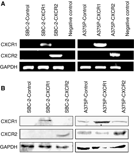
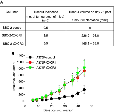
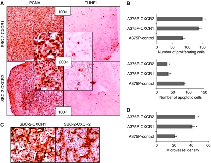
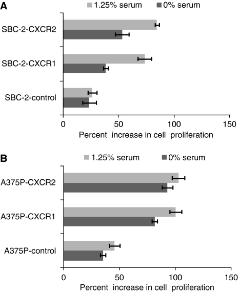
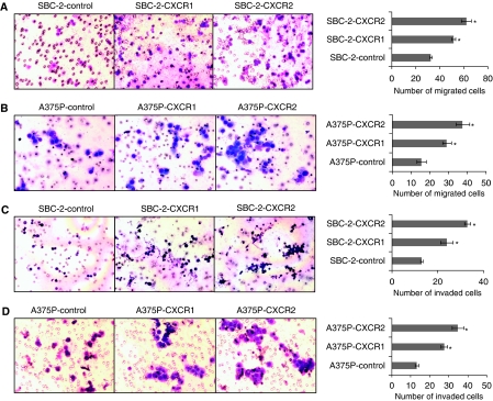
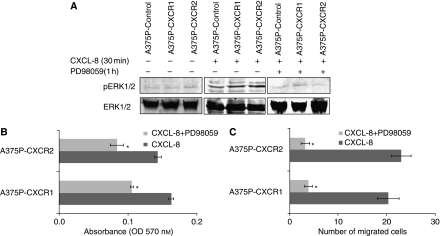
The aggressiveness of malignant melanoma is associated with differential expression of CXCL-8 and its receptors, CXCR1 and CXCR2. However, the precise functional role of these receptors in melanoma progression remains unclear. In this study, we investigate the precise functional role of CXCR1 and CXCR2 in melanoma progression. CXCR1 or CXCR2 were stably overexpressed in human melanoma cell lines, SBC-2 (non-tumourigenic) and A375P (low-tumourigenic) exhibiting low endogenous expression of receptors. Functional assays were performed to study the resulting changes in cell proliferation, motility and invasion, and in vivo tumour growth using a mouse xenograft model. Our data demonstrated that CXCR1- or CXCR2-overexpressing SBC-2 and A375P melanoma cells had enhanced proliferation, chemotaxis and invasiveness in vitro. Interestingly, CXCR1 or CXCR2 overexpression in SBC-2 cells induced tumourigenicity, and A375P cells significantly enhanced tumour growth as examined in vivo. Immunohistochemical analyses showed significantly increased tumour cell proliferation and microvessel density and reduced apoptosis in tumours generated from CXCR1- or CXCR2-overexpressing melanoma cells. CXCR1- or CXCR2-induced modulation of melanoma cell proliferation and migration was observed to be mediated through the activation of ERK1/2 phosphorylation. Together, these studies demonstrate that CXCR1 and CXCR2 play essential role in growth, survival, motility and invasion of human melanoma.


Figures

Figure 1
Figure 1
CXCR1 and CXCR2 expression in SBC-2 and A375P melanoma cells. Cells were stably transfected with vector control, or with vector containing CXCR1 or CXCR2 cDNA insert. (A) RT–PCR analysis shows increased expression of CXCR1 or CXCR2 mRNA in selected pooled clones. GAPDH was used as a control. (B) Western blotting showing increased expression of CXCR1 and CXCR2 in overexpressing melanoma cells as compared with control cells. GAPDH was used as a loading control. This is a representative gel picture of at least three experiments with similar results.
Figure 2
Figure 2
CXCR1 and CXCR2 expression enhances melanoma tumour growth. Stably transfected SBC-2 (3 × 10⁶ cells in 0.1 ml of HBSS) or A375P (1 × 10⁶ cells in 0.1 ml of HBSS) cells were subcutaneously (s.c.) injected into the flank of nude mice. In SBC-2-group, tumour incidence represents the number of tumours formed per number of injected mice and tumour volume on day 75 after tumour injection (A). For A375P-group tumour volume from day 0 to day 45 (n=5) (B). Growth of A375P-CXCR1 and A375P-CXCR2 increased significantly compared with A375P-control. Results are shown as mean±s.e.m. *Significantly different from controls (P<0.05).
Figure 3
Figure 3
CXCR1 and CXCR2 expression enhances in vivo melanoma cell proliferation, survival and tumour neovascularisation. Immunohistochemical staining for PCNA and TUNEL were analysed based on DAB staining as described in Materials and Methods. In SBC-2-group, tumours showed an imbalance of PCNA to TUNEL-positive cells (A). For A375P-group PCNA (B, upper panel) and TUNEL (B, lower panel) positive cells were counted in 10 arbitrarily selected fields at 200 × magnification in a double-blinded manner and expressed as average number of cells per field view±s.e.m. Immunohistochemical staining for microvessel using anti-GS-IB4 was analysed as described in Materials and Methods. Tumours from SBC-2 group were highly vascular (C). For A375P tumours, quantification of microvessel density at 200 × magnification in 10 random fields was examined. The values are average number of immunostained positive cells±s.e.m. (D). *Significantly different from control (P<0.05).
Figure 4
Figure 4
Overexpression of CXCR1 and CXCR2 affect in vitro melanoma cell growth. SBC-2 (5000 cells per well) (A) and A375P cells (1000 cells per well) (B) in a 96-well plate were cultured in media with (1.25%) or without serum. Cellular growth was determined at 72 h by MTT assay. Growth increase was calculated as percent (%)=[{(A/B)–1} × 100], where A and B are the absorbance of treated (CXCL-8 stimulated) and untreated cells (media alone), respectively. The values are mean percent inhibition of growth±s.e.m.
Figure 5
Figure 5
CXCR1 and CXCR2 overexpression enhances cell motility and invasion. SBC-2 and A375P cells expressing CXCR1 or CXCR2 were seeded on non-coated or Matrigel-coated membranes for motility (A and B) and invasion (C and D) assays, respectively, and incubated for 24 h. Serum-free medium containing 10 ng ml−1 CXCL-8 was added to the lower chamber. The cells that did not migrate through the Matrigel and/or pores in the membrane were removed, and cells on the other side of the membrane were stained and photographed at 200 × magnification. Cells were counted in 10 random fields (200 × ) and expressed as the average number of cells per field of view. The values are number of migrated cells±s.e.m. This is a representative of three experiments done in triplicate. *Significantly different from control CXCL-8-treated cells (P<0.05).
Figure 6
Figure 6
CXCR1 and CXCR2 activate ERK1/2 phosphorylation and mediated melanoma cell growth and motility. (A) For ERK phosphorylation, A375P cells overexpressing CXCR1 or CXCR2 were stimulated with CXCL-8 (10 ng ml−1) for 0 or 30 min (left and middle panels). Effect of ERK1/2 inhibitor was examined by treating the cells for 1 h and stimulated with CXCL-8 for 30 min (right panel). Cells were lysed, and equal amounts of protein were analysed by western blot analysis using antibodies against pERK1/2 and ERK1/2 antibody. Bound immunocomplexes were detected using ECL Plus chemiluminescence detection reagent. (B) For Growth, A375P-transfected cells were seeded (1000 cells per well) in 96-well plates and incubated with serum-free medium containing CXCL-8 (10 ng ml−1) with or without PD98059 (20 μM). Cellular growth was determined at 72 h by MTT assay. The values are average absorbance±s.e.m. (C) A375P-transfected cells treated for 1 h with 20 μM PD98059 were used for migration assay as described in Materials and Methods. Migrated cells were counted in 10 random fields (200 × ). The values are number of migrated cells±s.e.m. This is a representative of three experiments done in triplicate. *Significantly different from control cells (P<0.05).

References

    1. Addison CL, Daniel TO, Burdick MD, Liu H, Ehlert JE, Xue YY, Buechi L, Walz A, Richmond A, Strieter RM (2000) The CXC chemokine receptor 2, CXCR2, is the putative receptor for ELR+ CXC chemokine-induced angiogenic activity. J Immunol 165(9): 5269–5277 - PubMed
    1. Bar-Eli M (1999) Role of interleukin-8 in tumor growth and metastasis of human melanoma. Pathobiology 67(1): 12–18 - PubMed
    1. Brat DJ, Bellail AC, Van Meir EG (2005) The role of interleukin-8 and its receptors in gliomagenesis and tumoral angiogenesis. Neuro Oncol 7(2): 122–133 - PMC - PubMed
    1. Clark-Lewis I, Dewald B, Geiser T, Moser B, Baggiolini M (1993) Platelet factor 4 binds to interleukin 8 receptors and activates neutrophils when its N terminus is modified with Glu-Leu-Arg. Proc Natl Acad Sci USA 90(8): 3574–3577 - PMC - PubMed
    1. Dunn KL, Espino PS, Drobic B, He S, Davie JR (2005) The Ras-MAPK signal transduction pathway, cancer and chromatin remodeling. Biochem Cell Biol 83(1): 1–14 - PubMed

Publication types

MeSH terms

Substances