. 2009 Jul;29(7):1251-61.
doi: 10.1038/jcbfm.2009.46. Epub 2009 Apr 29.

Therapeutic neutralization of the NLRP1 inflammasome reduces the innate immune response and improves histopathology after traumatic brain injury

Affiliations

Therapeutic neutralization of the NLRP1 inflammasome reduces the innate immune response and improves histopathology after traumatic brain injury

Juan Pablo de Rivero Vaccari et al. J Cereb Blood Flow Metab. 2009 Jul.

Abstract

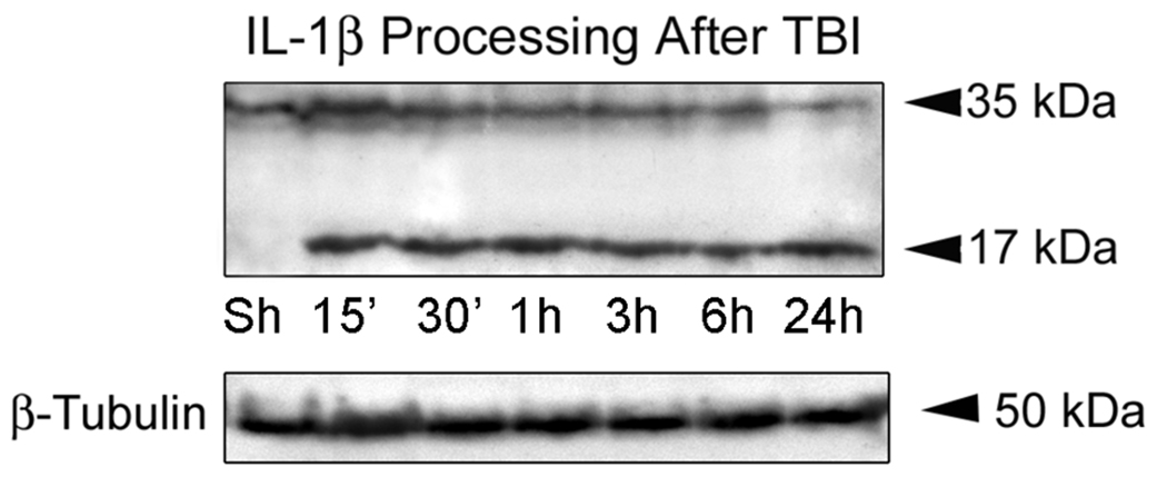
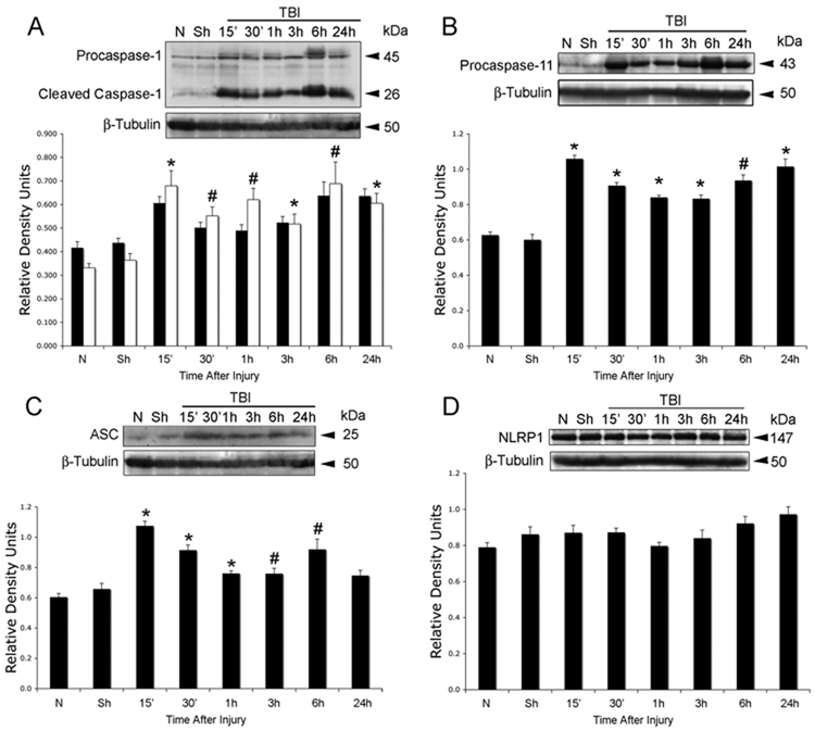
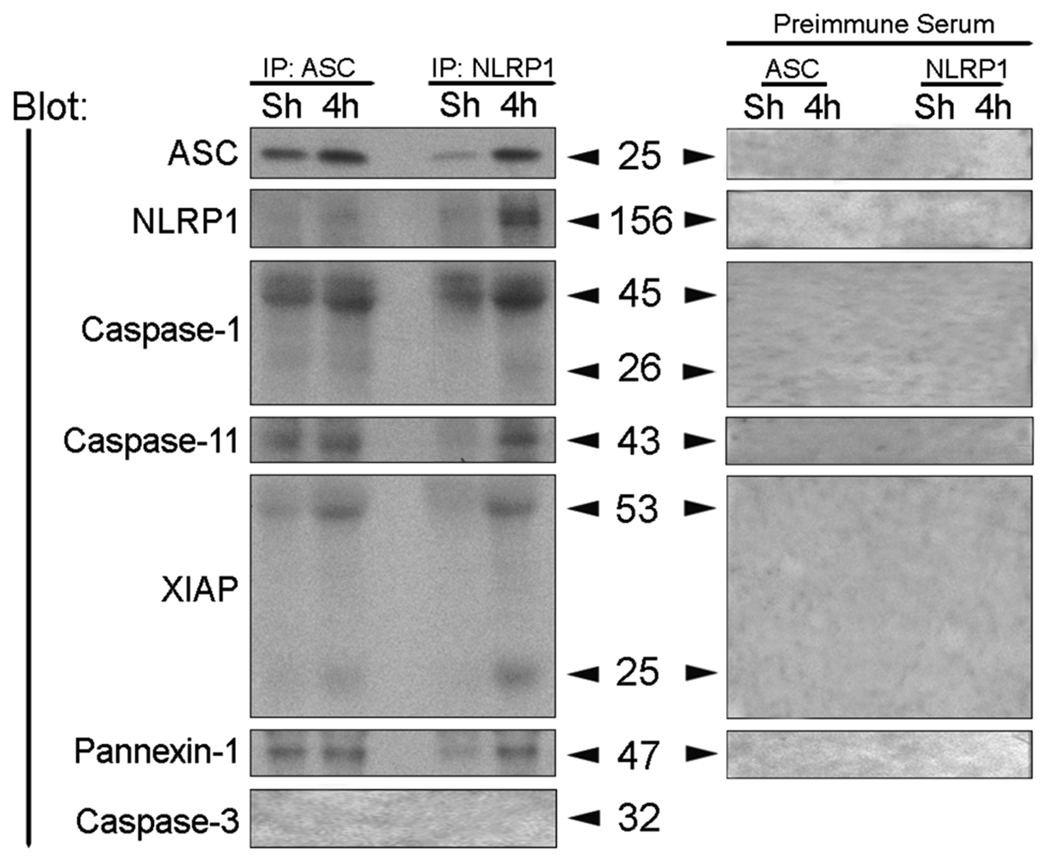
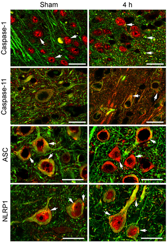
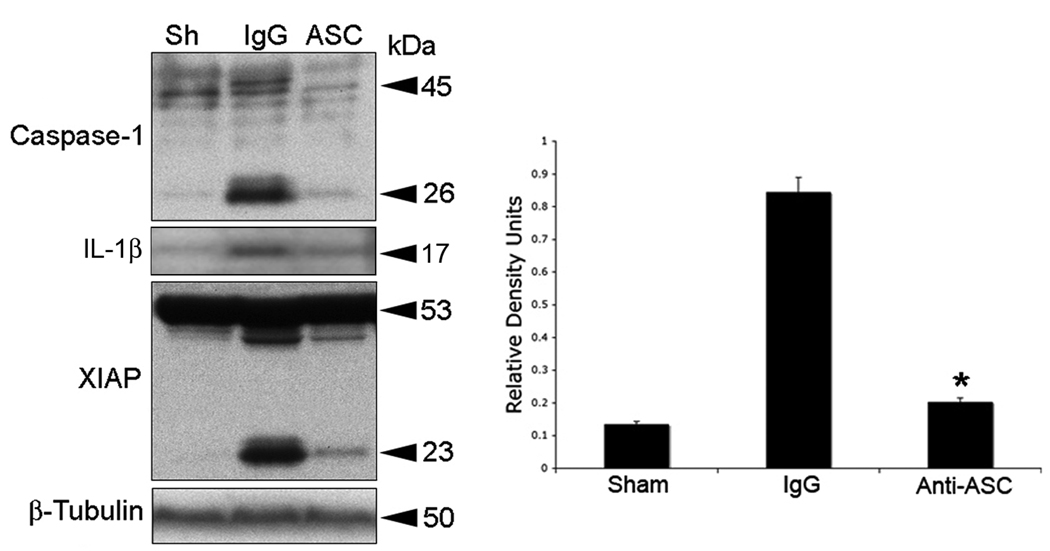
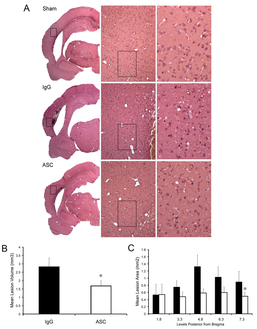
Traumatic brain injury elicits acute inflammation that in turn exacerbates primary brain damage. A crucial part of innate immunity in the immune privileged central nervous system involves production of proinflammatory cytokines mediated by inflammasome signaling. Here, we show that the nucleotide-binding, leucine-rich repeat pyrin domain containing protein 1 (NLRP1) inflammasome consisting of NLRP1, caspase-1, caspase-11, apoptosis-associated speck-like protein containing a caspase recruitment domain (ASC), the X-linked inhibitor of apoptosis protein, and pannexin 1 is expressed in neurons of the cerebral cortex. Moderate parasagittal fluid-percussion injury (FPI) induced processing of interleukin-1beta, activation of caspase-1, cleavage of X-linked inhibitor of apoptosis protein, and promoted assembly of the NLRP1 inflammasome complex. Anti-ASC neutralizing antibodies administered immediately after fluid-percussion injury to injured rats reduced caspase-1 activation, X-linked inhibitor of apoptosis protein cleavage, and processing of interleukin-1beta, resulting in a significant decrease in contusion volume. These studies show that the NLRP1 inflammasome constitutes an important component of the innate central nervous system inflammatory response after traumatic brain injury and may be a novel therapeutic target for reducing the damaging effects of posttraumatic brain inflammation.


Figures

Figure 1
Figure 1. TBI induces IL-1β processing in the injured cortex
Immunoblot analysis of IL-1β in cortical lysates of sham-operated animals (Sh) and traumatized rat cortices at 15, 30 min, 1, 3, 6 and 24 hr after injury. β-tubulin was used as internal standard and control for protein loading. N = 5 per group.
Figure 2
Figure 2. TBI induces activation and processing of caspase-1 and increases levels of ASC and caspase-11, but not NLRP1
Representative immunoblot analysis of caspase-1 (A), caspase-11 (B), ASC (C) and NLRP1 (D) in cortical lysates of sham (Sh), naïve (N) and traumatized cortices at indicated times after injury. β-tubulin was used as internal standard and control for protein loading. (A) caspase-1 proform (solid bars), caspase-1 cleaved form (open bars). Data are presented as mean ± s.e.m. *p < 0.05; #p < 0.10; compared to sham. N = 5 per group.
Figure 3
Figure 3. TBI induces association of NLRP1 inflammasome proteins, processing of caspase-1, and cleavage of XIAP
Coimmunoprecipitation with anti-ASC, anti-NLRP1 and preimmune serum of cortical lysates obtained from sham animals (Sh) and traumatized animals at 4 h after TBI. Immunoprecipitates were blotted for ASC, NLRP1, caspase-1, caspase-11, XIAP, pannexin 1 and caspase-3 (control). Anti-NLRP1 and anti-ASC immunoprecipitated NLRP1, processed caspase-1, caspase-11, pannexin 1, and the pro- and cleaved forms of XIAP, thus indicating association of these proteins in a multiprotein complex. Preimmune serum did not immunoprecipitate inflammasome proteins and was used as control.
Figure 4
Figure 4. NLRP1 inflammasome proteins are present in cortical neurons and TBI induces alterations in protein expression patterns
Confocal images of cortical neurons in sham and injured brains at 4 h post-trauma. Sections were stained for caspase-1, caspase-11, ASC and NLRP1 (red) and the neuronal marker MAP2 (Fisher et al.). In sham animals, caspase-1 immunoreactivity was seen in the nucleus (arrow). By 4 h after injury, increased caspase-1 staining was present in neuronal nuclei and patchy staining was present in the cell cytoplasm and processes near the plasma membrane. Caspase-11 immunoreactivity showed diffuse punctate staining confined to the neuronal soma and processes (arrow). Increased caspase-11 staining was present by 4 h post-trauma in the neuronal soma in a patchy distribution (arrow). Intense ASC and NLRP1 staining was detected in the soma of cortical neurons and exhibited a patchy distribution pattern in the cytoplasm (arrow). Both inflammasome proteins showed increased expression as evidenced by intense patchy staining located near or associated with the plasma membrane (arrows) by 4 h post-trauma. Bar = 20 µm.
Figure 5
Figure 5. ASC neutralization decreases TBI-induced activation and processing of caspase-1, IL-1β, and XIAP cleavage
Representative immunoblots of injured cortices from animals subjected to TBI and treated intracerebroventricularly with antibodies to ASC (ASC), IgG controls (IgG) or left untreated (Sh) after injury. Animals were sacrificed 24 h after treatment. Treatment resulted in inhibition of inflammasome activation as detected by a decrease in the processing of procaspase-1, IL-1β, and cleavage of XIAP. Anti-ASC treatment also resulted in a significant reduction in caspase-1 activity (right panel). *p < 0.05 vs sham, **p < 0.05 vs IgG.
Figure 6
Figure 6. ASC neutralization decreases brain contusion volume
(A) Representative coronal sections of brains of antibody-treated (anti-ASC), IgG treated and sham animals at 3 d after TBI. Hematoxylin & eosin stained sections at bregma level 4.8 represent the injury epicenter. (B) Administration of anti-ASC significantly reduced the lesion volume at 3 d after injury. Significance was determined by comparing average contusion volume of antibody-treated animals to control groups using Student’s t-test. (C) Effect of anti-ASC on lesion size. Solid bars represent mean lesion area of rats that received IgG, whereas white bars represents areas of rats that received anti-ASC. Significance was determined by two-way paired comparisons ANOVA followed by Tukey’s multiple comparison tests (N=5 per group). Data are presented as mean ± s.e.m. *p<0.05 compared to nontreated animals. Bar = 50 µm.

References

    1. Abulafia DP, de Rivero Vaccari JP, Lozano JD, Lotocki G, Keane RW, Dietrich WD. Inhibition of the inflammasome complex reduces the inflammatory response after thromboembolic stroke in mice. J Cereb Blood Flow Metab. 2009;29:534–544. - PubMed
    1. Allan SM, Tyrrell PJ, Rothwell NJ. Interleukin-1 and neuronal injury. Nat Rev Immunol. 2005;5:629–640. - PubMed
    1. Arend WP, Palmer G, Gabay C. IL-1, IL-18, and IL-33 families of cytokines. Immunol Rev. 2008;223:20–38. - PubMed
    1. Bhat RV, DiRocco R, Marcy VR, Flood DG, Zhu Y, Dobrzanski P, Siman R, Scott R, Contreras PC, Miller M. Increased expression of IL-1beta converting enzyme in hippocampus after ischemia: selective localization in microglia. J Neurosci. 1996;16:4146–4154. - PMC - PubMed
    1. Braddock M, Quinn A. Targeting IL-1 in inflammatory disease: new opportunities for therapeutic intervention. Nat Rev Drug Discov. 2004;3:330–339. - PubMed

Publication types

LinkOut - more resources