. 2009;4(4):e5391.
doi: 10.1371/journal.pone.0005391. Epub 2009 Apr 29.

Peptide Bbeta(15-42) preserves endothelial barrier function in shock

Affiliations

Peptide Bbeta(15-42) preserves endothelial barrier function in shock

Marion Gröger et al. PLoS One. 2009.

Erratum in

  • PLoS One. 2009;4(6) doi: 10.1371/annotation/9ae032a2-c48d-46d9-9f8f-d3f401714e42

Abstract

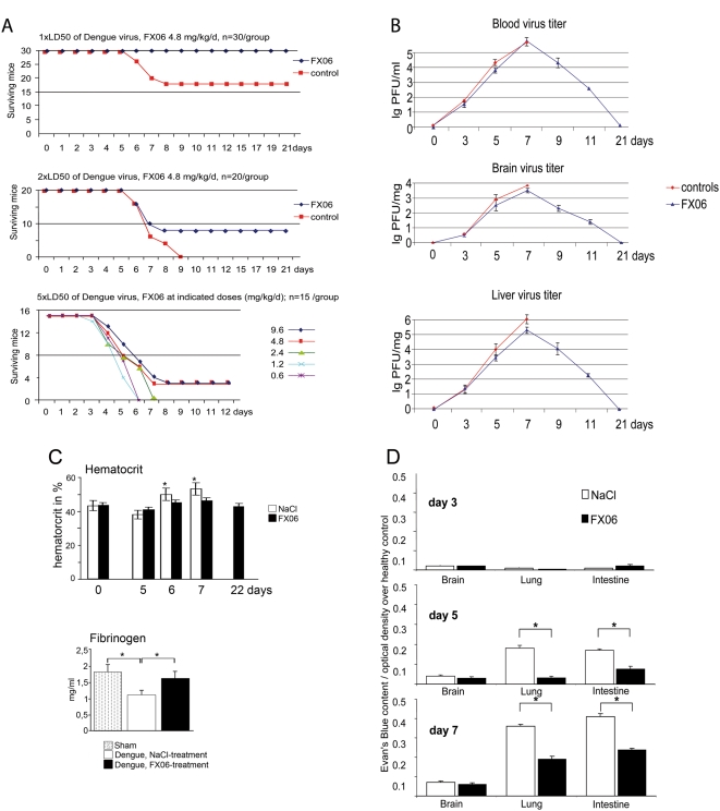
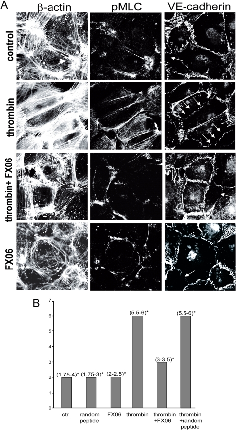
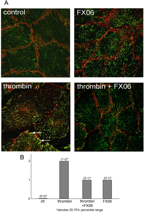
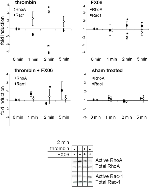
Loss of vascular barrier function causes leak of fluid and proteins into tissues, extensive leak leads to shock and death. Barriers are largely formed by endothelial cell-cell contacts built up by VE-cadherin and are under the control of RhoGTPases. Here we show that a natural plasmin digest product of fibrin, peptide Bbeta15-42 (also called FX06), significantly reduces vascular leak and mortality in animal models for Dengue shock syndrome. The ability of Bbeta15-42 to preserve endothelial barriers is confirmed in rats i.v.-injected with LPS. In endothelial cells, Bbeta15-42 prevents thrombin-induced stress fiber formation, myosin light chain phosphorylation and RhoA activation. The molecular key for the protective effect of Bbeta15-42 is the src kinase Fyn, which associates with VE-cadherin-containing junctions. Following exposure to Bbeta15-42 Fyn dissociates from VE-cadherin and associates with p190RhoGAP, a known antagonists of RhoA activation. The role of Fyn in transducing effects of Bbeta15-42 is confirmed in Fyn(-/-) mice, where the peptide is unable to reduce LPS-induced lung edema, whereas in wild type littermates the peptide significantly reduces leak. Our results demonstrate a novel function for Bbeta15-42. Formerly mainly considered as a degradation product occurring after fibrin inactivation, it has now to be considered as a signaling molecule. It stabilizes endothelial barriers and thus could be an attractive adjuvant in the treatment of shock.


Conflict of interest statement

Competing Interests: PP, SR are founders of Fibrex Medical Inc. www.fibrexmedical.com and own shares, KZ owns stock options of Fibrex, SR, PF and WP are employed by Fibrex.

Figures

Figure 1
Figure 1. FX06 improved survival following dengue-infection.
A. Dengue virus was inoculated i.p. at day 0. Starting with day 3, animals were treated with NaCl (100 µl) or indicated doses of FX06 (diluted in 100 µl NaCl). In 1×LD50 and 2×LD50 groups, differences in survival rates between the NaCl and the FX06 groups were significant (p<0.05). B. Virus titers following Dengue infection. Mice were infected with 2×LD50 of Dengue virus i.p and treated with NaCl (controls) or FX06 (2.4 mg/kg bodyweight twice daily). The virus load was quantified by titration of serum or brain lysates onto Vero E6 cell cultures as described Diamond and plaque forming units (PFU) per ml serum or per mg organ were determined. n = 3/data point. All control treated animals were dead at day 9. C. Hematocrit and fibrinogen following Dengue infection. Mice were infected with 2×LD50 of Dengue virus i.p and treated with NaCl (controls) or FX06 (2.4 mg/kg bodyweight twice daily). n = 3/data point, all control mice were dead at day 9, * denotes p<0.05 as compared with controls. D. Organs of 15 animals per time point and group infected with 2×LD50 were analyzed. Treatment started at day 3, mice received NaCl or FX06 (daily dose was 4.8 mg/kg bodyweight in 100 µl NaCl i.p.). Samples at day 3 were taken before treatment was initiated. Evan's blue extravasation was measured as described in Methods and data are presented as optical density above that of healthy controls. At days 5 and 7, in lungs and the intestine, the difference in Evan's blue extravasation between the NaCl and the FX06 group was significant (*p<0.05, mean+/−SD).
Figure 2
Figure 2. FX06 decreased capillary leak following LPS injection in rats.
One hour after LPS injection (i.v., 12 mg/kg), FX06 (2.4 mg/kg) or an equal volume of NaCl (100 µl) were injected i.v.; 350 min after LPS injection, FluoSpheres® were injected i.v.; 360 min. after LPS injection, rats were sacrificed. The right lung was used for histology and photographed on a laser scan microscope. Examples of images from LPS (A) or LPS+FX06-treated animals (B). From the entire left lung, FluoSpheres® were recovered and fluorescence measured by ELISA as described in Methods (C). The difference between LPS and LPS plus FX06 was significant (n = 11 per group; * denotes p<0.05, mean+/−SD).
Figure 3
Figure 3. FX06 prevented thrombin-induced actin-bundling and myosin light chain phosphorylation (pMLC).
A. Examples of laser scan images of an immunofluorescence triple staining: actin (left) pMLC (middle), and VE-cadherin (right column). Endothelial cells were exposed to thrombin (1 U/ml), FX06 (50 µg/ml) or thrombin+FX06 for 5 min. Following thrombin, actin formed parallel bundles, pMLC co-localized with parallel actin bundles and VE-cadherin formed a discontinuous band. Arrows denote discontinuous VE-cadherin at the cell margins. FX06 (50 µg/ml) prevented these thrombin-induced changes; staining patterns were comparable to those seen in control cells or FX06-treated cells. B. The amount and localization of pMLC, the amount of actin bundling and the continuity of VE-cadherin as exemplified in (A) were quantified in 15 randomly photographed LSM images derived from 3 independent experiments by 2 observers blinded to the condition as described in Methods. Thrombin induced a significant raise in scores as compared to controls (p<0.05) and this was significantly reduced by FX06 (p<0.05, median and interquartile ranges are shown (* denotes the 25% and 75% ranges).
Figure 4
Figure 4. FX06 reduced thrombin-induced FAK activation.
A. Immunofluorescence images (anti-ß-catenin Ab in red, anti-phospho-FAK (p397FAK) Ab in green). Endothelial cells were treated as indicated for 5 min. Thrombin induced a robust increase in p397FAK and relocation to cell margins producing a pattern of parallel stripes (arrows) which was not seen in thrombin plus FX06-treated cells. B. The amount and localization of pFAK as exemplified in (A) was scored in 3 independent experiments by 2 observers blinded to the condition as described in Methods. Thrombin induced a significant raise in scores as compared to random peptide-treated cells (*p<0.05) and this was significantly reduced by FX06 (p<0.05), median and interquartile ranges are shown (* denotes the 25% and 75% ranges).
Figure 5
Figure 5. FX06 prevents thrombin-induced RhoA activation.
Endothelial cells were exposed to thrombin (1 U/ml), FX06 (50 µg/ml) or thrombin plus FX06 for indicated times. Pull down experiments were performed with GST-coupled Rhotekin (for activated RhoA) or with GST-coupled Pak-1 (for activated Rac1). Graphs depict the mean+/−SD of 4 independent experiments. Thrombin induced significant elevations of RhoA and reductions of Rac1 activity as indicated by asterisks (p<0.05 compared to sham). FX06 alone had the reverse effect (asterisks denote p<0.05 compared to sham). In thrombin plus FX06-treated cells, RhoA activation was completely inhibited (asterisks denote p<0.05 compared to cells treated with thrombin alone). The Western blot is an example of a pull down experiment 2 minutes after stimulation.
Figure 6
Figure 6. FX06 dissociates Fyn from VE-cadherin and associates Fyn to FAK and p190RhoGAP.
Endothelial cells were treated with thrombin, FX06 or thrombin+FX06 for 1 min. Following lysis, proteins were immunoprecipitated (IP) and co-precipitated proteins detected by western blots (WB) as indicated in (A–D). Mean+/−SD of three independent experiments; *p<0.05 compared to medium control, **p<0.05 compared to thrombin; representative Western blots are shown at the right side.
Figure 7
Figure 7. FX06 reduces leak in wild-type but not in Fyn−/− mice.
LPS (10 µg) or solvent were instilled intranasally (i.n.). FX06 (2×2.4 µg/kg), random peptide or NaCl were injected i.p., the first dose immediately after LPS challenge, the second dose 60 min later. 330 min after LPS challenge, Evan's Blue was injected i.v. 30 min later mice were sacrificed and lungs processed as described in the Methods and analyzed for Evan's blue content; mean+/−SD, n = 8 per group. The difference between LPS and LPS+FX06 in wild-type mice was significant, *p<0.05.

Similar articles

Cited by

References

    1. van Hinsbergh V, van Nieuw Amerongen GP. Intracellular signalling involved in modulating human endothelial barrier function. J Anat. 2002;200:549–560. - PMC - PubMed
    1. Faust SN, Levin M, Harrison OB, Goldin RD, Lockhart MS, et al. Dysfunction of endothelial protein C activation in severe meningococcal sepsis. N Engl J Med. 2001;345:408–416. - PubMed
    1. Matsuda N, Hattori Y. Vascular biology in sepsis: pathophysiological and therapeutic significance of vascular dysfunction. J Smooth Muscle Res. 2007;43:117–137. - PubMed
    1. Petzelbauer P, Zacharowski PA, Miyazaki Y, Friedl P, Wickenhauser G, et al. The fibrin-derived peptide Bbeta15-42 protects the myocardium against ischemia-reperfusion injury. Nat Med. 2005;11:298–304. - PubMed
    1. Sohn RH, Deming CB, Johns DC, Champion HC, Bian C, et al. Regulation of endothelial thrombomodulin expression by inflammatory cytokines is mediated by activation of nuclear factor-kappa B. Blood. 2005;105:3910–3917. - PubMed

Publication types

MeSH terms