. 2009 Apr 28;7(4):e92.
doi: 10.1371/journal.pbio.1000092.

Structural studies of the giant mimivirus

Affiliations

Structural studies of the giant mimivirus

Chuan Xiao et al. PLoS Biol. .

Abstract

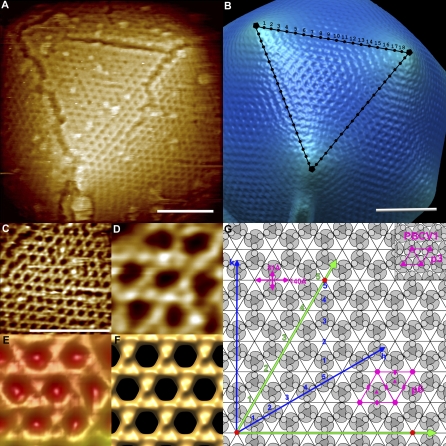
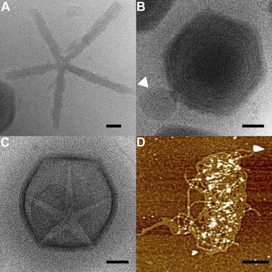
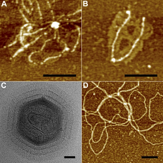
Mimivirus is the largest known virus whose genome and physical size are comparable to some small bacteria, blurring the boundary between a virus and a cell. Structural studies of Mimivirus have been difficult because of its size and long surface fibers. Here we report the use of enzymatic digestions to remove the surface fibers of Mimivirus in order to expose the surface of the viral capsid. Cryo-electron microscopy (cryoEM) and atomic force microscopy were able to show that the 20 icosahedral faces of Mimivirus capsids have hexagonal arrays of depressions. Each depression is surrounded by six trimeric capsomers that are similar in structure to those in many other large, icosahedral double-stranded DNA viruses. Whereas in most viruses these capsomers are hexagonally close-packed with the same orientation in each face, in Mimivirus there are vacancies at the systematic depressions with neighboring capsomers differing in orientation by 60 degrees . The previously observed starfish-shaped feature is well-resolved and found to be on each virus particle and is associated with a special pentameric vertex. The arms of the starfish fit into the gaps between the five faces surrounding the unique vertex, acting as a seal. Furthermore, the enveloped nucleocapsid is accurately positioned and oriented within the capsid with a concave surface facing the unique vertex. Thus, the starfish-shaped feature and the organization of the nucleocapsid might regulate the delivery of the genome to the host. The structure of Mimivirus, as well as the various fiber components observed in the virus, suggests that the Mimivirus genome includes genes derived from both eukaryotic and prokaryotic organisms. The three-dimensional cryoEM reconstruction reported here is of a virus with a volume that is one order of magnitude larger than any previously reported molecular assembly studied at a resolution of equal to or better than 65 Angstroms.


Conflict of interest statement

Competing interests. The authors have declared that no competing interests exist.

Figures

Figure 1
Figure 1. Comparison of Virus MCPs
The N-terminal and C-terminal jelly-roll domains are colored green and red, respectively. Top left is a ribbon diagram of the adenovirus capsid protein. Diagrammatic representation of the arrangement of the β strands (arrows) within each jelly-roll are given for adenovirus, PBCV1, and Mimivirus at the top right, bottom left, and bottom right, respectively. The β strands within each domain are labelled A to H. This gives rise to the two opposing BIDG and CHEF β sheets in each jelly-roll as indicated in the ribbon diagram. Occasional α helices are represented as bars.
Figure 2
Figure 2. Sequence Alignments of Virus MCPs
Sequence alignments of Mimivirus (gene product L425 [5]), PBCV1, PRD1, and adenovirus MCPs based on structural superpositions in all cases except Mimivirus. The β strands in the first and second jelly-roll motifs are labelled in green and red, respectively, with the corresponding sequences highlighted in yellow.
Figure 3
Figure 3. Capsomer Arrangement of Mimivirus
(A) AFM image of defibered virus showing one face of the virion with irregular cracks along the edges of the face. (B) Shaded surface rendering of the cryoEM reconstruction computed from untreated Mimivirus, orientated similarly as in (A). The surface fibers are not visible because of their disorder. Depressions at the edges of a face are marked with dots. The dots along one edge are labelled from 1 to 18. Three 5-fold vertices are labelled with black pentagons. (C) An AFM image of defibered particle at high magnification shows a honeycomb array of the depressions. (D) Enlargement of a portion of (C). (E) CryoEM reconstruction of fibered particles on the same scale as (D). (F) Mimivirus surface simulated using the homologous PBCV1 capsomer structure and the observed spatial arrangement of the capsomers in (D) and (E). (G) Diagram showing the p6 plane group lattice of capsomers as found in (D) and (E). Each monomer of the MCP consists of two consecutive jelly-rolls indicated by a kidney-shaped outline with a circle at one end representing the inserted “tower” loop. Three monomers constitute one capsomer. The depressions along one edge are shown in green. Three 5-fold vertices at the corners of a face are shown with red pentagons. As an example the 5-fold vertices are placed 5 depressions apart. This would correspond to h = 5 and k = 5 in terms of the Caspar-Klug p3 plane group hexagonal array of capsomers (see blue axes). For Mimivirus, there are 19 depressions along an edge. One unit cell of the p6 (Mimivirus) and p3 (PBCV1) are outlined in red. The distance between depressions (140 Å) and between capsomers (81 Å) is shown in red. The scale bars in (A), (B), and (C) represent 1,000 Å.
Figure 4
Figure 4. AFM Images of Starfish-shaped Features on Defibered Mimiviruses
(A) Defibered Mimivirus at low magnification showing that the majority of the particles have starfish-shaped features. Presumably, the few particles that do not show the starfish-shaped feature are orientated with their special vertex towards the substrate. The fibers on a particle in the middle of the micrograph were not fully digested, as is evident by its larger diameter. (B) Partially digested Mimivirus at median magnification. One particle on the upper-left corner is fully digested and shows a starfish-shaped feature. The other three particles have retained their fibers and have a larger diameter with star-shaped crevices between the fibers, which demonstrates that the starfish-shaped feature has no attached fibers. (C) A high-magnification image of a starfish-shaped feature on a defibered virus. (D) A defibered particle treated with proteinase K showing the arm of a starfish-shaped feature wedged into the 2-fold edge creating a 500 Å separation between arrays of capsomers. The scale bars in (A) and (B) represent 1 μm, and those in (C) and (D) are 1,000 Å.
Figure 5
Figure 5. CryoEM Reconstruction of Mimivirus Applying Only 5-fold Symmetry Averaging
(A–C) Surface-shaded rendering of cryoEM reconstruction of untreated Mimivirus. (A) Looking down the starfish-shaped feature associated vertex, (B) looking from one side, and (C) looking from the opposite side of the “starfish”-associated vertex. (D) The “starfish”-associated vertex was removed to show the internal nucleocapsid with its concave surface facing the special vertex. (E) Central slice of the reconstruction looking from the side of the particle showing the concave face of the nucleocapsid and the low density space beneath the “starfish”-associated vertex. A perfectly icosahedral particle is outlined in gray to show the extension of the unique vertex. (F) Central slice of the reconstruction looking along the 5-fold axis from the starfish-shaped feature showing the enveloped nucleocapsid surrounded by a lower density space. The coloring is based on radial distance from the center of the virus. Gray is from 0 to 1,800 Å, red from 1,800 to 2,100 Å, and rainbow coloring from red to blue between 2,100 and 2,500 Å. The scale bars in all panels represent 1,000 Å.
Figure 6
Figure 6. Function of the Starfish-Shaped Feature
The features shown here have been observed reproducibly and frequently in numerous images. (A) CryoEM image of a starfish-shaped feature that has become detached from the virus. (B) CryoEM image of a defibered Mimivirus that has lost its starfish-shaped feature. A “puff” (white arrow), surrounded by a membrane-like envelope [17,25] is observed at the special vertex. (C) CryoEM image of a Mimivirus showing a gap (less density represented by lighter gray shading) between five fibered faces surrounding the open unique vertex. The actual starfish-shaped feature and probably the viral nucleocapsid are missing. (D) AFM image of DNA ejected from Mimivirus. No other molecules appear to be protecting the ejected DNA (see Materials and Methods). Scale bar represents 1,000 Å in all panels.
Figure 7
Figure 7. Mimivirus Fibers
(A) AFM image of several surface fibers attached to a common central feature. Each of the fibers has a globular density at its free end. (B) AFM image of two detached surface fibers of Mimivirus. Murky material surrounding the fibers might be peptidoglycans [5] consistent with the uptake of Gram stain [1,4]. (C) CryoEM image of a Mimivirus that has been digested with lysozyme and then partially digested with bromelain. There are two successive rings of densities on the fibers. (D) AFM image of internal fibers of Mimivirus. The repeat distance between subunit is ∼70 Å, different to the repeating unit of DNA. Furthermore, the organization of the fibers is different to that of viral DNA as seen in Figure 6D. Scale bar represents 1,000 Å in all panels.

References

    1. La Scola B, Audic S, Robert C, Jungang L, de Lamballerie X, et al. A giant virus in amoebae. Science. 2003;299:2033. - PubMed
    1. La Scola B, Desnues C, Pagnier I, Robert C, Barrassi L, et al. The virophage, a unique parasite of the giant Mimivirus . Nature. 2008;455:100–104. - PubMed
    1. Xiao C, Chipman PR, Battisti AJ, Bowman VD, Renesto P, et al. Cryo-electron microscopy of the giant Mimivirus. J Mol Biol. 2005;353:493–496. - PubMed
    1. Raoult D, La Scola B, Birtles R. The discovery and characterization of Mimivirus, the largest known virus and putative pneumonia agent. Clin Infect Dis. 2007;45:95–102. - PubMed
    1. Raoult D, Audic S, Robert C, Abergel C, Renesto P, et al. The 1.2-megabase genome sequence of Mimivirus. Science. 2004;306:1344–1350. - PubMed

Publication types

Substances