Comparative Study
. 2009 Jun;136(7):2325-33.
doi: 10.1053/j.gastro.2009.02.051. Epub 2009 Feb 21.

Intrahepatic bile ducts develop according to a new mode of tubulogenesis regulated by the transcription factor SOX9

Affiliations
Comparative Study

Intrahepatic bile ducts develop according to a new mode of tubulogenesis regulated by the transcription factor SOX9

Aline Antoniou et al. Gastroenterology. 2009 Jun.

Abstract

Background & aims: A number of diseases are characterized by defective formation of the intrahepatic bile ducts. In the embryo, hepatoblasts differentiate to cholangiocytes, which give rise to the bile ducts. Here, we investigated duct development in mouse liver and characterized the role of the SRY-related HMG box transcription factor 9 (SOX9).

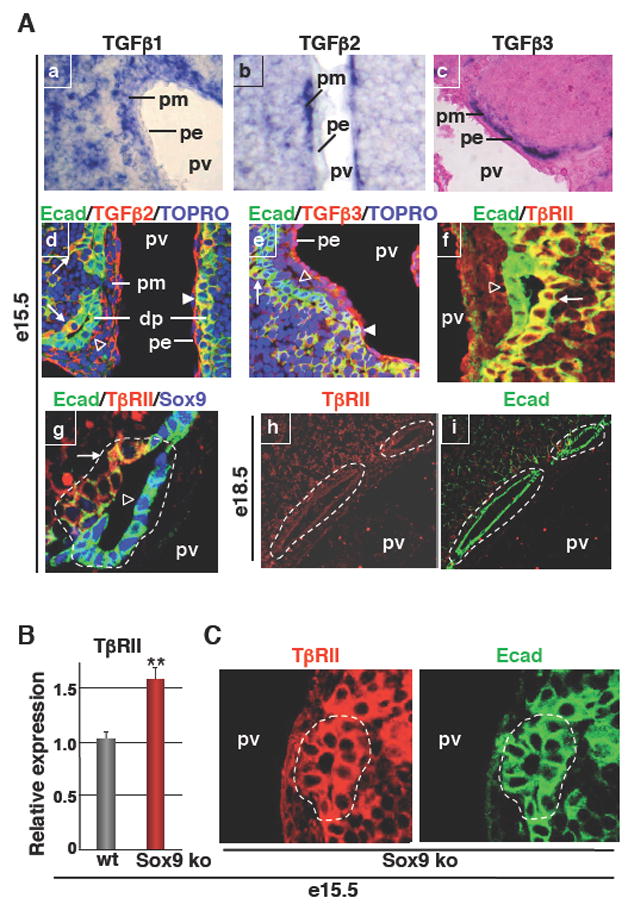
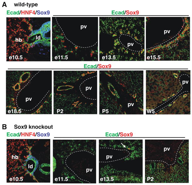
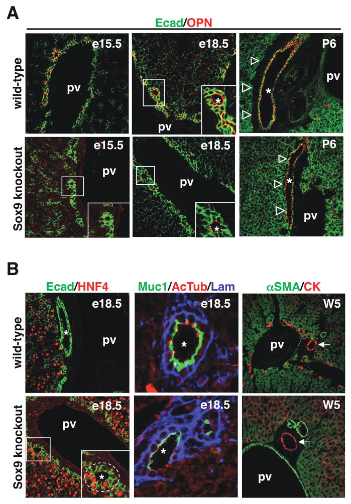
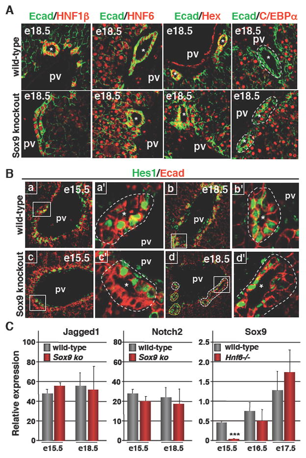
Methods: We identified SOX9 as a new biliary marker and used it in immunostaining experiments to characterize bile duct morphogenesis. The expression of growth factors was determined by in situ hybridization and immunostaining, and their role was studied on cultured hepatoblasts. SOX9 function was investigated by phenotyping mice with a liver-specific inactivation of Sox9.

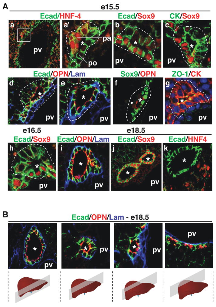
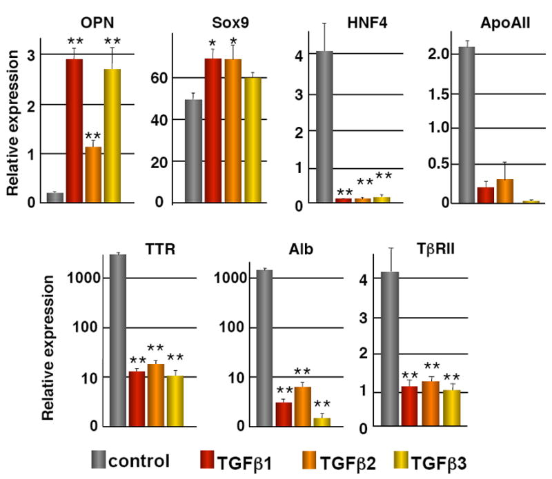
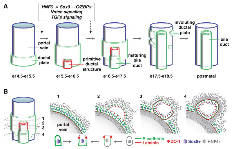
Results: Biliary tubulogenesis started with formation of asymmetrical ductal structures, lined on the portal side by cholangiocytes and on the parenchymal side by hepatoblasts. When the ducts grew from the hilum to the periphery, the hepatoblasts lining the asymmetrical structures differentiated to cholangiocytes, thereby allowing formation of symmetrical ducts lined only by cholangiocytes. We also provide evidence that transforming growth factor-beta promotes differentiation of the hepatoblasts lining the asymmetrical structures. In the absence of SOX9, the maturation of asymmetrical structures into symmetrical ducts was delayed. This was associated with abnormal expression of CCAAT/Enhancer Binding Protein alpha and Homolog of Hairy/Enhancer of Split-1, as well as of the transforming growth factor-beta receptor type II, which are regulators of biliary development.

Conclusions: Our results suggest that biliary development proceeds according to a new mode of tubulogenesis characterized by transient asymmetry and whose timing is controlled by SOX9.


Figures

Figure 1
Figure 1
Development of bile ducts. (A) Immunostaining analysis showed that bile duct development is initiated by the formation of asymmetrical primitive ductal structures (PDS). a′ is a magnified view of the region delineated in a. By the end of gestation, bile ducts had become radially symmetrical (ik). (B) Serial sections shows the hilum-to-periphery development of bile ducts, with symmetrical ducts near the hilum and asymmetrical PDS towards the periphery. The position of the portal vein relative to the bile ducts is indicated (pv). pa, parenchymal side of PDS; po, portal side; *, lumens of developing bile ducts.
Figure 2
Figure 2
TGFβ signaling and development of bile ducts. Hepatoblasts (BMEL cells) were cultured in the presence or absence of TGFβ1, TGFβ2, or TGFβ3 (200 pg/ml). The expression (measured by Q-PCR) of the hepatocyte markers HNF4, Albumin (Alb), Transthyretin (TTR), Apolipoprotein AII (ApoAII) and TβRII was downregulated, and that of the biliary markers Osteopontin (OPN) and Sox9 was upregulated. Cells were treated for 24h (HNF4, ApoAII, Alb, TTR, TβRII, OPN) or for 6h (Sox9) with the TGFβ ligands. Data are means ± SEM; n ≥ 3; *, P < 0.05; **, P < 0.01. Quantification was performed as described in the Materials and Methods section.
Figure 3
Figure 3
TGFβ signaling and development of bile ducts in wild-type and SOX9-deficient livers. (A) In situ hybridization showing wide expression of TGFβ1 in the liver, while TGFβ2 and TGFβ3 mRNAs were predominantly found in the portal mesenchyme (ac). Immunostaining showed binding of TGFβ2 and TGFβ3 to the parenchymal side of PDS (d, e). Immunostaining showing expression of TβRII on the parenchymal side of PDS and lack of expression in mature ducts (fi). Arrows: TGFβ2, TGFβ3 or TβRII staining on parenchymal side of PDS; Arrowheads: TGFβ2 or TGFβ3 staining on single-layered ductal plate; open arrowheads: lack of TGFβ2, TGFβ3 or TβrII staining on portal side of PDS. (B) Q-PCR showing overexpression of TβRII in e15.5 Alfp-Cre-Sox9loxP/loxP (Sox9 ko) livers. (C) Immunostainings showed that primitive ductal structures detected by E-cadherin (E-cad) labeling expressed TβRII on both the portal side and parenchymal side in Alfp-Cre-Sox9loxP/loxP livers, in contrast to wild-type primitive ductal structures which express TβRII only on the parenchymal side (see panels Af, g). dp, single-layered ductal plate; pe, portal endothelium; pm, portal mesenchyme; pv, portal vein; *, lumens of developing bile ducts.
Figure 4
Figure 4
Expression of SOX9 in wild-type livers and in livers with conditional inactivation of Sox9 alleles. (A) Expression profiling by immunostaining showed that SOX9 is an early and specific marker of the developing bile ducts. (B) In Alfp-Cre-Sox9loxP/loxP (Sox9 knockout) embryos, the livers were depleted of SOX9 starting at e11.5, except for one individual showing residual SOX9+ cells at e13.5 (arrow). hb, hepatoblasts; ld, liver diverticulum; pv, portal vein.
Figure 5
Figure 5
SOX9 controls the timing of bile duct development. (A) SOX9-deficient livers showed delayed onset of OPN expression, and (B) persistence of asymmetrical PDS until e18.5 (Ecad/HNF4 and Muc1/AcTub/Lam stainings). (A) After birth, the SOX9-deficient ducts bile ducts were adjacent to the parenchyme, while wild-type embryos were surrounded by periportal mesenchyme. (B) At five weeks of age SOX9-deficient and wild-type mice had normal bile ducts, surrounded by periportal mesenchyme (αSMA/CK stainings). pv, portal vein; *, lumens of developing bile ducts.
Figure 6
Figure 6
SOX9 and the transcriptional cascade regulating bile duct development. (A) Expression of HNF1β, HNF6 and Hex was normal in SOX9-deficient livers. In contrast to wild-type livers, C/EBPα expression was not repressed in the biliary cells of SOX9-deficient livers. (B) HES1 was expressed in the ductal plate and on the portal side of wild-type PDS, and was found at later stages in cells lining the parenchymal and portal sides of symmetrical ducts. In SOX9-deficient livers, expression of HES1 at e15.5 was normal, and by the end of gestation its expression pattern reflected the delayed bile duct morphogenesis. In panel Bd, PDS showing asymmetrical expression of HES1 were delineated by white dotted lines, while ducts which had reached symmetry and which expressed HES1 symmetrically were delineated by a yellow dotted line. a′, b′, c′ and d′ are magnified views of a, b, c and d. (C) The expression of Jagged1 and Notch2 was not significantly different between wild-type and SOX9-deficient livers. Expression of SOX9, measured by Q-PCR, was transiently downregulated in HNF6 knockout livers. Data are means ± SEM; n ≥ 3; *** P < 0.001. pv, portal vein; *, lumens of developing bile ducts.
Figure 7
Figure 7
(A) Model for the formation of bile ducts in mouse liver. The transition from single layered ductal plate to formation and maturation of primitive ductal structure is controlled by a HNF6-SOX9-C/EBPα cascade and by Notch and TGFβ signaling. (B) Schematic representation of the transient asymmetry in developing bile ducts.

Similar articles

Cited by

References

    1. Kamath BM, Piccoli DA. Heritable disorders of the bile ducts. Gastroenterol Clin North Am. 2003;32:857–875. - PubMed
    1. Lemaigre F, Zaret KS. Liver development update: new embryo models, cell lineage control, and morphogenesis. Curr Opin Genet Dev. 2004;14:582–590. - PubMed
    1. Zhao R, Duncan SA. Embryonic development of the liver. Hepatology. 2005;41:956–967. - PubMed
    1. Shiojiri N. The origin of intrahepatic bile duct cells in the mouse. J Embryol Exp Morphol. 1984;79:25–39. - PubMed
    1. Van Eyken P, Sciot R, Callea F, et al. The development of the intrahepatic bile ducts in man: A keratin-immunohistochemical study. Hepatology. 1988;8:1586–1595. - PubMed

Publication types

MeSH terms

Substances