. 2009 Aug 28;284(35):23682-96.
doi: 10.1074/jbc.M109.011700. Epub 2009 Apr 29.

Dynamically driven ligand selectivity in cyclic nucleotide binding domains

Affiliations

Dynamically driven ligand selectivity in cyclic nucleotide binding domains

Rahul Das et al. J Biol Chem. .

Abstract

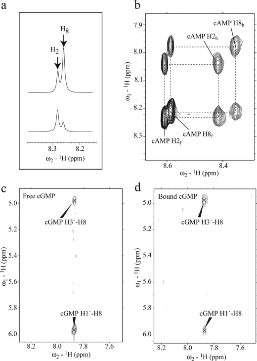
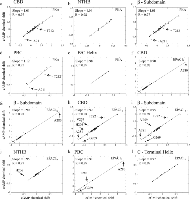
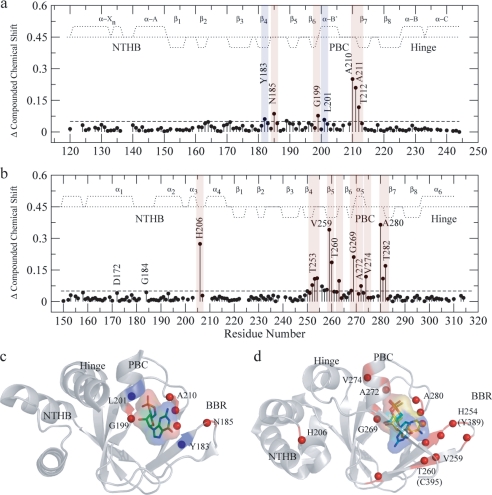
One of the mechanisms that minimize the aberrant cross-talk between cAMP- and cGMP-dependent signaling pathways relies on the selectivity of cAMP binding domains (CBDs). For instance, the CBDs of two critical eukaryotic cAMP receptors, i.e. protein kinase A (PKA) and the exchange protein activated by cAMP (EPAC), are both selectively activated by cAMP. However, the mechanisms underlying their cAMP versus cGMP selectivity are quite distinct. In PKA this selectivity is controlled mainly at the level of ligand affinity, whereas in EPAC it is mostly determined at the level of allostery. Currently, the molecular basis for these different selectivity mechanisms is not fully understood. We have therefore comparatively analyzed by NMR the cGMP-bound states of the essential CBDs of PKA and EPAC, revealing key differences between them. Specifically, cGMP binds PKA preserving the same syn base orientation as cAMP at the price of local steric clashes, which lead to a reduced affinity for cGMP. Unlike PKA, cGMP is recognized by EPAC in an anti conformation and generates several short and long range perturbations. Although these effects do not alter significantly the structure of the EPAC CBD investigated, remarkable differences in dynamics between the cAMP- and cGMP-bound states are detected for the ionic latch region. These observations suggest that one of the determinants of cGMP antagonism in EPAC is the modulation of the entropic control of inhibitory interactions and illustrate the pivotal role of allostery in determining signaling selectivity as a function of dynamic changes, even in the absence of significant affinity variations.


Figures

FIGURE 1.
FIGURE 1.
Schematic representation of the domain organization in the regulatory subunit I-α of PKA (a) and in EPAC (b).The black circles indicate cAMP. a, D/D is the dimerization docking domain; the inhibitory site is shown in orange, and the two tandem cAMP binding domains, CBD-A and CBD-B, are highlighted in different shades of green. b, DEP, disheveled-egl-10-pleckstrin domain; REM, Ras exchange motif; RA, Ras-associated module, and the CDC25HD catalytic domain are represented in gray, green, orange, blue, and yellow, respectively. The black dashed line and the empty cAMP circle in EPAC2 indicate that this domain is not strictly necessary for the cAMP-dependent GEF activity. The module with question mark in EPAC1 denotes an unknown function for this domain. a and b, the CBD in light green shown below the full-length protein represents the construct used for the NMR studies. c, sequence alignment of the CBDs of bovine RIα- domain A and human EPAC1. Fully conserved residues are highlighted in green; cyan denotes conservation for the functional group only, and yellow indicates the residues present only in one of the CBDs. The secondary structure of apo-EPAC2m (PDB ID 1O7F) is shown in red.
FIGURE 2.
FIGURE 2.
a, expansions from the one-dimensional spectra of cAMP (top) and of d8-cAMP (bottom). Replacement of the proton at the 8th position of the adenine base (supplemental Fig. S1) by deuterium significantly reduces the H8 signal in d8-cAMP. b, expansion from the ROESY spectrum of EPAC1h-(149–318)-bound cAMP. Black dashed lines connect the cross-peaks between free cAMP (suffixed with f) and EPAC-bound cAMP (suffixed with b). c, expansion of the off-resonance ROESY spectrum of free cGMP. d, same expansion as in c but for the transfer 13C15N double-filtered nuclear Overhauser effect spectroscopy spectrum of cGMP bound to EPAC1h-(149–318). The cross-peaks are labeled with the assigned proton pairs.
FIGURE 3.
FIGURE 3.
Correlations between cAMP- and cGMP-induced chemical shift changes in RIα–A-(119–244) (a–e) and EPAC1h-(149–318) (f–l). The horizontal axes report the differences in chemical shifts between the cGMP-bound and the apo states calculated as δω = 0.2Δδ15N + Δδ1H (28). The vertical axes report the differences in chemical shifts between the cAMP-bound and the apo states calculated using the same equation (28). a and f, all the assigned CBD residues were used in the linear correlations, whereas in the other panels the correlations were confined to subsets of residues to better identify possible outliers. Specifically, b and j are for the N-terminal helical bundle; c, g, and i are for the β-subdomain (including the PBC); d and k are for the PBC; and e and l are for the B/C helix. Because of the dominant chemical shift changes observed for Ala-280 of EPAC1h-(149–318), the correlations of f and g were recalculated in h and i, respectively, without Ala-280. Similarly, the correlation for the N-terminal helical-bundle region in j was calculated excluding His-206.
FIGURE 4.
FIGURE 4.
Compounded chemical shift changes between the cAMP- and cGMP-bound states of RIα-A-(119–244) (a) and of EPAC1h-(149–318) (b). The chemical shifts observed for Ala-210 in a and for A280 in b are downscaled by a factor of 2 to obtain a better visual dynamic range. The average chemical shift is indicated by the dashed horizontal lines. The secondary structure for RIα-A (according to PDB 1RGS) and EPAC1h (according to PDB 1O7F) is indicated by dotted lines. The positive and negative values indicate α-helical and β-sheet probabilities, respectively. The residues with compounded chemical shift changes above the average chemical shift are highlighted in red, and the residues with compounded chemical shift changes comparable with the average are highlighted in blue. The compounded chemical shifts between the cAMP- and cGMP-bound states are mapped into the ribbon diagrams of RIα-A (c) (PDB 1RL3) and EPAC (PDB- 3CF6) (d). d, cAMP is shown in syn and cGMP is shown in anti conformations (as discussed in the text) along with their van der Waals surfaces. The anti conformation of cGMP was created based on the structure of anti-cAMP in cAMP-bound HCN (PDB code 1VP6), and the cAMP syn conformation was obtained from (Sp)-cAMPS-bound EPAC2m (PDB code 3CF6). The backbone NHs showing chemical shift changes > average are indicated by red spheres. The amino acid residues are labeled according to the EPAC1h sequence, and the nonconserved EPAC2m residues are shown within parentheses.
FIGURE 5.
FIGURE 5.
PFs of RIα-A-(119–244) (a) and EPAC1h-(149–318) (b). a, PFs measured with 10-fold excess cAMP (black), cGMP (green) and without 10-fold excess cAMP (orange) (15) are plotted against the residue number. b, EPAC1h-(149–318) PFs are measured with cAMP (black), cGMP (green) and apo states (orange) are plotted against the residue number. The filled circles at the base line refer to the fast exchanging residues within the dead time (20 min) of the experiment or by the first or second HSQCs. The residues affected by overlap are denoted as squares, and the residues for which no data are reported are either ambiguous or proline. The backbone amides that exchange in the cGMP-bound state faster than the cAMP-bound state are highlighted with a pink background, and the residues exchanging more slowly relative to the cAMP-bound state are highlighted in blue. The secondary structures are denoted by the dotted line as explained in Fig. 4.
FIGURE 6.
FIGURE 6.
Changes in the PFs between the cAMP- and cGMP-bound states mapped into the ribbon diagrams of RIα-A (PDB 1RL3) (a) and EPAC (PDB- 3CF6) (b). a, cGMP is shown as stick bonds with its van der Waals surface. The backbone ribbon is highlighted according to the color coding of Fig. 5. Residues exchanging fast (within the dead time of the H/D experiment or by the second HSQC) are shown as spheres and the dotted line represents hydrogen bonds between the ligand and the protein backbone. b, cyclic nucleotide conformations are as described in Fig. 4, and the backbone ribbon is highlighted according to the color coding of Fig. 5. The amino acid residues are labeled according to the EPAC1h sequence, and the nonconserved EPAC2m residues are shown within parentheses. Selected hydrogen bond networks destabilized by the replacement of cAMP with cGMP are shown in c and d.
FIGURE 7.
FIGURE 7.
Correlation plot of the normalized HSQC intensities for the cAMP-bound and cGMP-bound states of RIα-A-(119–244) (a) and EPAC1h-(149–318) (b). a, in RIα-A-(119–244) the HSQC intensities were normalized against residue Tyr-244. The high correlation (R = 0.94) between the cAMP- and cGMP-bound states indicates that no significant change in dynamics occurs between the two states. b, intensities were normalized against that of the Leu-159 peak in both cAMP- and cGMP-bound states. The linear fit between the cAMP and cGMP intensities is indicated by a solid straight line. The amino acid residues showing significant changes in intensity between the cAMP-bound and cGMP-bound states are labeled. The low correlation (R = 0.65) observed for EPAC1h-(149–318) points to significant changes in dynamics upon substitution of cAMP with cGMP.
FIGURE 8.
FIGURE 8.
Backbone 15N relaxation rates for EPAC1h-(149–318) in its apo (yellow) (14), cAMP-bound (black) (14), and cGMP-bound (green) states are plotted against residue numbers. The experimental relaxation rates were measured at a field of 700 MHz. a, spin-spin relaxation rate R2; b, spin-lattice relaxation rate R1; c, product of the R1 and R2 relaxation rates; d, 15N{1H} NOE calculated as Isat/Inonsat. c and d, residues experiencing quenching or enhancement of dynamics upon cGMP binding relative to the cAMP-bound state of EPAC1h-(149–318) are highlighted with blue and red, respectively. In all panels, the rates calculated based on hydrodynamic bead models of EPAC1h-(149–309), -(161–309), and -(173–309), respectively, are represented by pink horizontal lines, as described previously (14). These values provide an assessment of the contribution to each rate from overall tumbling. The secondary structure is also shown in c similarly to Fig. 4. The residues for which an apparent quenching in millisecond to microsecond relaxation dynamics may also be due to concurrently enhanced picosecond to nanosecond relaxation rates are highlighted in blue dashed lines. Residues for which no relaxation data are available are prolines or are overlapped and/or broadened beyond detection.
FIGURE 9.
FIGURE 9.
Reduced spectral densities for EPAC1h-(149–318) in its apo (orange) (14), cAMP-bound (black) (14), and cGMP-bound (green) states are plotted against residue numbers. The spectral density values, J(0) (a), JN) (b), and JHN) were computed based on the relaxation rates reported in Fig. 8. a, J(0) value includes contributions from chemical exchange effects. Secondary structure elements are as described in Fig. 4. The JN) (b) and JHN) (c) values were computed using the frequencies ωN = −γN Bo and ωH = −γH Bo with Bo = 16.44 tesla at 700 MHz. The red lines in all three panels indicate the reduced spectral densities computed based on the hydrodynamic bead models, as shown in Fig. 8 (14). In all the panels the residues showing enhanced or reduced dynamics upon cGMP binding relative to the cAMP-bound state are highlighted in red and blue, respectively.
FIGURE 10.
FIGURE 10.
Changes in picosecond to nanosecond (a and b) and millisecond to microsecond (c and d) dynamics for cGMP-bound EPAC1h-(149–318) relative to the cAMP-bound state are mapped onto the holo-EPAC2m structure (PDB 3CF6) (11). b and d were obtained from a and c, respectively, through a 180° rotation. The residues experiencing quenching or enhancement in dynamics upon replacement of cAMP with cGMP are colored according to the color coding of Fig. 8. The amino acid residues are labeled according to the sequence of EPAC1h. The residues reported within parentheses correspond to EPAC2m, and they are not conserved between EPAC1h and EPAC2m. e shows the putative interactions between Gln-270 in the PBC, the 308, 310 residues, and the N-terminal helical-bundle (i.e. α3).

References

    1. Kim C., Vigil D., Anand G., Taylor S. S. (2006) Eur. J. Cell Biol. 85,651–654 - PubMed
    1. Rehmann H., Wittinghofer A., Bos J. L. (2007) Nat. Rev. Mol. Cell Biol. 8,63–73 - PubMed
    1. Kim C., Cheng C. Y., Saldanha S. A., Taylor S. S. (2007) Cell 130,1032–1043 - PubMed
    1. Wang Z., Dillon T. J., Pokala V., Mishra S., Labudda K., Hunter B., Stork P. J. (2006) Mol. Cell. Biol. 26,2130–2145 - PMC - PubMed
    1. Pelligrino D. A., Wang Q. (1998) Prog. Neurobiol. 56,1–18 - PubMed

Publication types

MeSH terms

LinkOut - more resources