. 2009 Jun 15;587(Pt 12):3009-23.
doi: 10.1113/jphysiol.2009.172387. Epub 2009 Apr 29.

Developmental regulation of the intracellular Ca2+ sensitivity of vesicle fusion and Ca2+-secretion coupling at the rat calyx of Held

Affiliations

Developmental regulation of the intracellular Ca2+ sensitivity of vesicle fusion and Ca2+-secretion coupling at the rat calyx of Held

Olexiy Kochubey et al. J Physiol. .

Abstract

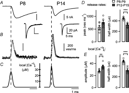
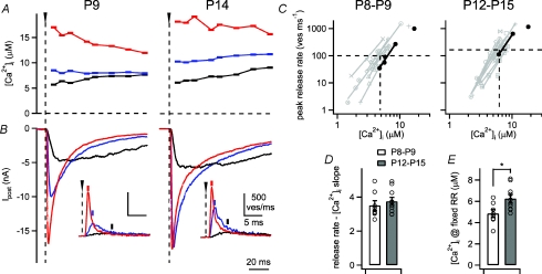
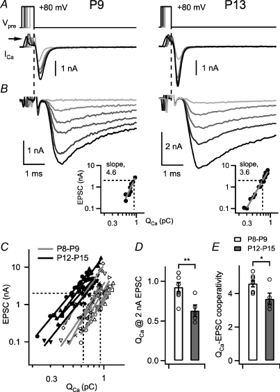
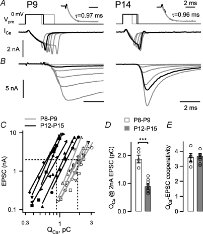
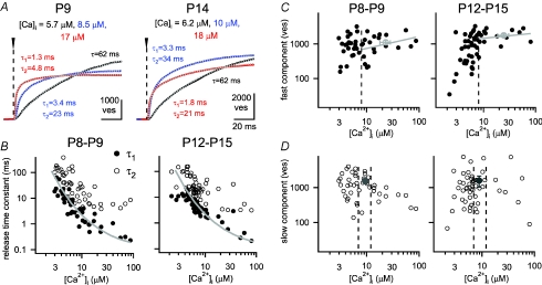
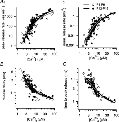
Developmental refinement of synaptic transmission can occur via changes in several pre- and postsynaptic factors, but it has been unknown whether the intrinsic Ca2+ sensitivity of vesicle fusion in the nerve terminal can be regulated during development. Using the calyx of Held, a giant synapse in the auditory pathway, we studied the presynaptic mechanisms underlying the developmental regulation of Ca2+-secretion coupling, comparing a time period before, and shortly after the onset of hearing in rats. We found an approximately 2-fold leftward shift in the relationship between EPSC amplitude and presynaptic Ca2+ current charge (QCa), indicating that brief presynaptic Ca2+ currents become significantly more efficient in driving release. Using a Ca2+ tail current protocol, we also found that the high cooperativity between EPSC amplitude and QCa was slightly reduced with development. In contrast, in presynaptic Ca2+ uncaging experiments, the intrinsic Ca2+ cooperativity of vesicle fusion was identical, and the intrinsic Ca2+ sensitivity was slightly reduced with development. This indicates that the significantly enhanced release efficiency of brief Ca2+ currents must be caused by a tighter co-localization of Ca2+ channels and readily releasable vesicles, but not by changes in the intrinsic properties of Ca2+-dependent release. Using the parameters of the intrinsic Ca2+ sensitivity measured at each developmental stage, we estimate that during a presynaptic action potential (AP), a given readily releasable vesicle experiences an about 1.3-fold higher 'local' intracellular Ca2+ concentration ([Ca2+]i) signal with development. Thus, the data indicate a tightening in the Ca2+ channel-vesicle co-localization during development, without a major change in the intrinsic Ca2+ sensitivity of vesicle fusion.


Figures

Figure 6
Figure 6. The ‘local’[Ca2+]i signal experienced by readily releasable vesicles during AP-evoked release increases with development
A, EPSCs (average of n= 5 traces each) evoked by afferent fibre stimulation in a P8 and a P14 synapse, in the presence of cyclothiazide (CTZ). The average mEPSC waveform recorded in each cell is also shown (lower trace; note different scales). B, transmitter release rates (average of n= 5 single sweeps; black data points) for the EPSCs shown in A, as obtained by deconvolution of the evoked EPSC with the mEPSC waveform (see Methods). The transmitter release rates predicted by the ‘local’[Ca2+]i waveforms shown in C are superimposed (grey traces). C, the iteratively refined local [Ca2+]i waveforms which best predicted the observed transmitter release rates shown in B. D, average peak transmitter release rates (left) and their half-widths (right) for each developmental stage. Note the highly significant (P < 0.001) reduction in the half-width of AP-evoked transmitter release with development (right panel). E, average amplitude (left) and half-widths (right) of the back-calculated local [Ca2+]i transients for both developmental stages. Note that the local [Ca2+]i signal is significantly higher (P= 0.009, left), and at the same time briefer in more mature animals (P < 0.001, right).
Figure 3
Figure 3. Ca2+ uncaging experiments measure the intracellular Ca2+ sensitivity and Ca2+ cooperativity of vesicle fusion at two developmental stages
A and B, presynaptic [Ca2+]i steps produced by Ca2+ uncaging with different UV light intensities (A) and the resulting EPSCs in the postsynaptic cell (B), for recordings in a P9 rat (left) and a P14 rat (right). The insets in B show transmitter release rates as obtained by EPSC deconvolution. C, plot of the peak release rates as a function of the corresponding [Ca2+]i steps on double-logarithmic coordinates. The values for [Ca2+]i steps below 10 μm amplitude were fitted by lines. The data obtained from the example cells (A and B) are shown by black symbols and lines; data points and fits for all other cells obtained with 2 mm DM-nitrophen are shown by grey symbols and lines. D, average slope values derived from fitting the peak release rate–[Ca2+]i relation (see C). There was no significant change (P= 0.49), indicating that the intrinsic Ca2+ cooperativity of release was unchanged during development. E, plot of the average interpolated [Ca2+]i value needed to evoke a peak transmitter release rate of 100 ves ms−1 (at P8–P9), or a pool-corrected value of 160 ves ms−1 at P12–P15 group (dashed lines in C). There was a 1.29-fold increase of this [Ca2+]i value, indicating a slightly lower Ca2+ sensitivity with development (P= 0.012).
Figure 2
Figure 2. A slight decrease in the Ca2+ current–release cooperativity during developmental maturation
A and B, presynaptic Ca2+ tail currents (A) and the corresponding postsynaptic EPSCs (B) in a P9 (left) and a P13 calyx of Held synapse (right), with presynaptic depolarizations to +80 mV of varying durations. The insets in B show plots of the EPSC amplitude versus QCa for the corresponding cells on the same scales, with slope values as indicated. C, plot of the EPSC amplitudes as a function of the presynaptic Ca2+ charge for n= 8 cells at P8–P9 (open symbols) and for n= 5 cells at P12–P15 (filled symbols). Each logarithmized data set was fitted with a line to yield the individual slope values. Note the leftward shift, and the trend towards shallower slopes in the more mature age group (P12–P15). D, average and individual values of the presynaptic Ca2+ charge needed to evoke an EPSC of 2 nA. Note the significant difference between age groups (P= 0.007). E, the slope values obtained from line fits to log–log plots of EPSC amplitude versus QCa. Note the slight, but statistically significant (P= 0.04) reduction of the Ca2+ current–release cooperativity with development.
Figure 1
Figure 1. A pronounced leftward shift in the EPSC–presynaptic Ca2+ charge relation during developmental maturation
A and B, presynaptic Ca2+ currents (A) and the corresponding postsynaptic EPSCs (B) in a P9 (left) and a P14 calyx of Held synapse, with presynaptic depolarizations to 0 mV of increasing lengths. The insets in A show an exponential fit (grey line) to the rising phase of the Ca2+ currents, with time constants as indicated. The traces highlighted by black lines show presynaptic Ca2+ currents with similar charge transfer, which evoke an ∼9-fold larger EPSC in the P14 synapse. C, plot of the EPSC amplitudes as a function of the presynaptic Ca2+ charge, for n= 7 cells at P8–P9 (open symbols) and for n= 6 cells at P12–P15 (filled symbols). The logarithmized data set of each cell was fitted with a line, which is superimposed. Note that the data from the older age group is clearly leftward shifted. D, average and individual values of the presynaptic Ca2+ charge needed to evoke an EPSC of 2 nA. E, the slope values of the line fits to the EPSC amplitude versus QCa data in double-logarithmic coordinates (‘Ca2+ current–release cooperativity’). Note the unchanged value between the two age groups (P= 0.7).
Figure 4
Figure 4. Ca2+ uncaging evokes a fast and a slow component of transmitter release at both developmental stages
A, integrated release rate traces for a P9 (left) and a P14 (right) synapse evoked by step-like elevations of [Ca2+]i (traces are from the same cells and in colours corresponding to the examples shown in Fig. 3A and B). The post-flash [Ca2+]i value for each response is indicated. The cumulative release rate traces were fitted with various exponential fit functions (see Methods), and the best-fit traces are shown superimposed (grey dotted lines): single exponentials for the lowest [Ca2+]i steps (black traces, time constant τ indicated), or double exponentials, with the indicated values for τ1 and τ2. B, the two time constants (τ1 and τ2) of the fast and the slow release component were plotted against the corresponding post-flash [Ca2+]i values at P8–P9 (left panel) and at P12–P15 (right panel). The prediction of the five-site model of Ca2+ binding and vesicle fusion, which effectively models the fast release component, is superimposed (grey line; parameters as given for the P8–P9 age group in the legend to Fig. 5). Note that this data set, and the data shown in C and D also include experiments done with 5 mm DM-nitrophen, which allowed us to obtain higher post-flash [Ca2+]i values of up to 90 μm. C and D, the number of vesicles released in each kinetic component of release as a function of post-flash [Ca2+]i, plotted separately for P8–P9 (left panels) and for P12–P15 rats (right panels). The numbers were estimated by the amplitude values of the fast and the slow time constants in fits of the cumulative release rate traces (A and B), when the best-fit functions were either double-exponentials, double-exponentials plus line, or triple exponential functions. The average number of vesicles released in the fast component by [Ca2+]i steps above 8 μm is indicated by the grey average symbol. For [Ca2+]i > 8 μm, the data for the fast release component were fitted by lines (grey lines in C). The average number of slowly released vesicles was estimated in the [Ca2+]i range of 7–12 μm (see dotted lines and grey average symbols in D), because the number of slowly released vesicles showed a tendency to decrease with [Ca2+]i steps > 15 μm.
Figure 5
Figure 5. The intrinsic Ca2+ sensitivity of transmitter release is largely unchanged during developmental refinement of synaptic transmission
A–C, plots of the peak transmitter release rates (Aa), the pool-corrected peak release rate (Ab), the minimal release delay (B), and the time to the peak of the transmitter release rate (C) as a function of presynaptic [Ca2+]i reached after flashes for both developmental groups (P8–P9, open symbols; P12–P15, filled symbols). In Ab, the peak release rates were normalized by the pool size estimate (FRP) of each individual cell. The data points for the two developmental groups largely overlap for all analysed parameters. The data sets in Aa, B and C were simultaneously fitted with the five-site model of cooperative Ca2+ binding and vesicle fusion (Schneggenburger & Neher, 2000), yielding the following parameters for each age group: kon= 1.21 × 10⁸m−1 s−1, koff= 6500 s−1, b= 0.26, γ= 6960 s−1 (P8–P9 age group; grey fit lines); and kon= 1.15 × 10⁸m−1 s−1, koff= 7900 s−1, b= 0.26, γ= 6960 s−1 (P12–P15 age group; black fit lines). In Ab, the fit predictions were normalized by the average FRP pool size values obtained in young synapses (1109 vesicles; P8–P9) and in more mature synapses (1767 vesicles; see Results). A small, constant delay (0.4 ms) was added to the model predictions of release delay (B) and time to peak release (C), indicating a time delay not explained by the model (see also Bollmann et al. 2000; Felmy et al. 2003).

Similar articles

Cited by

References

    1. Augustine GJ, Adler EM, Charlton MP. The calcium signal for transmitter secretion from presynaptic nerve terminals. Ann N Y Acad Sci. 1991;635:365–381. - PubMed
    1. Bollmann JH, Sakmann B. Control of synaptic strength and timing by the release-site Ca2+ signal. Nat Neurosci. 2005;8:426–434. - PubMed
    1. Bollmann JH, Sakmann B, Borst JG. Calcium sensitivity of glutamate release in a calyx-type terminal. Science. 2000;289:953–957. - PubMed
    1. Borst JG, Sakmann B. Calcium current during a single action potential in a large presynaptic terminal of the rat brainstem. J Physiol. 1998;506:143–157. - PMC - PubMed
    1. Borst JG, Sakmann B. Effect of changes in action potential shape on calcium currents and transmitter release in a calyx-type synapse of the rat auditory brainstem. Philos Trans R Soc Lond B Biol Sci. 1999;354:347–355. - PMC - PubMed

Publication types

MeSH terms

LinkOut - more resources